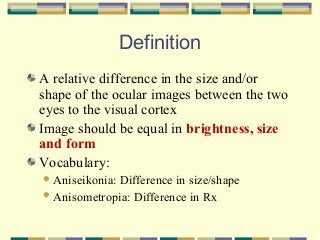
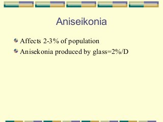
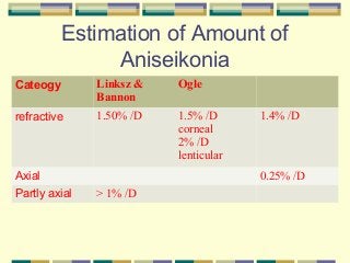
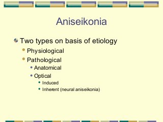
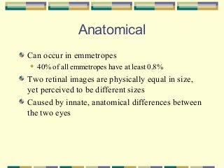
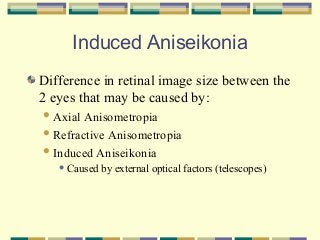
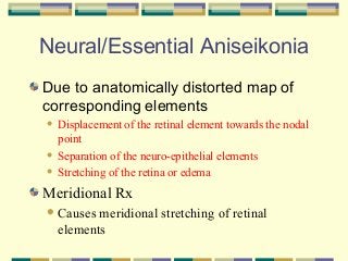
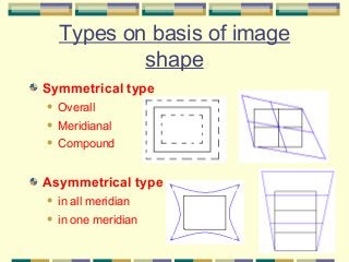
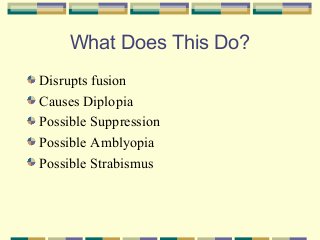
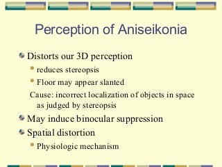
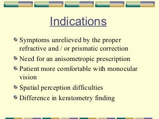
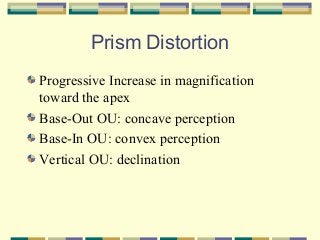
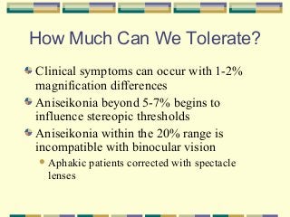
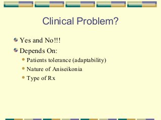
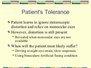
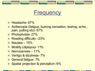
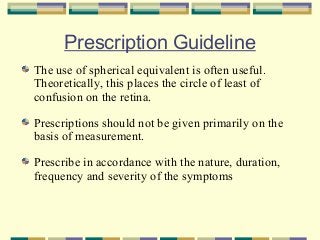
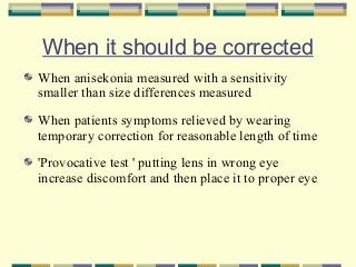
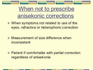
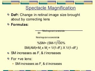
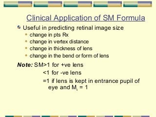
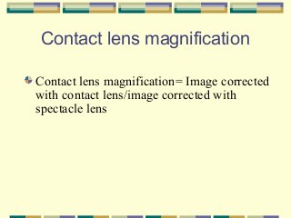
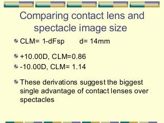
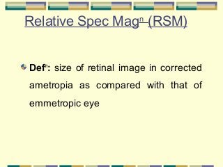
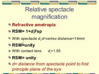
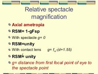
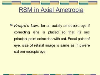
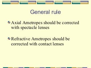
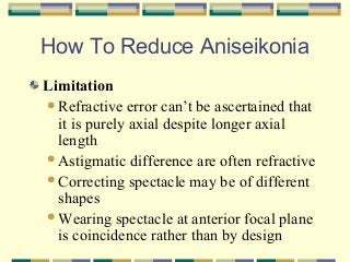
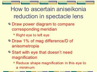
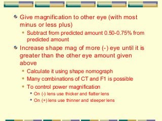
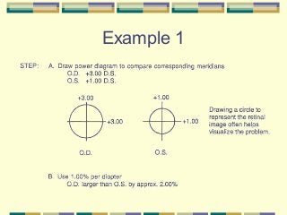
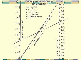
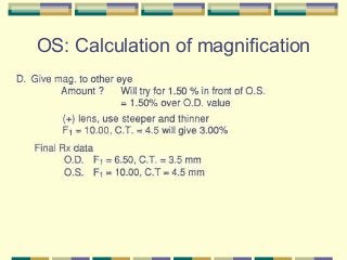
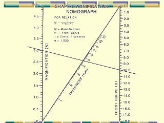
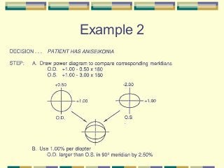
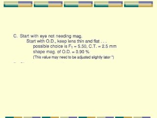
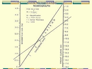
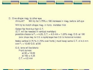
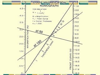
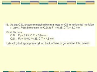
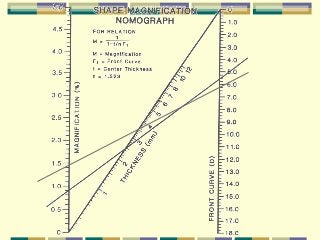
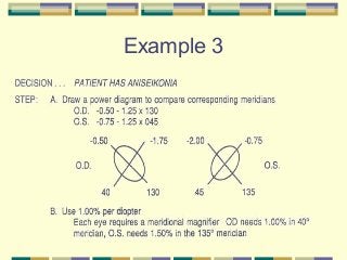
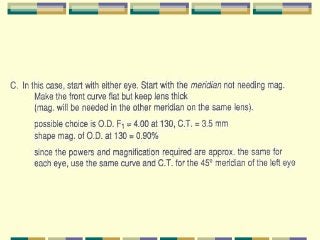
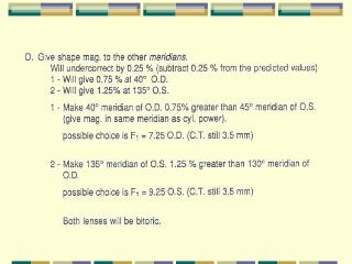
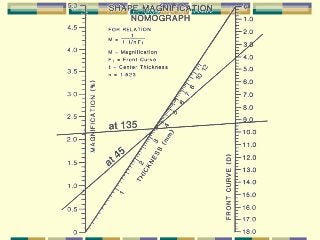
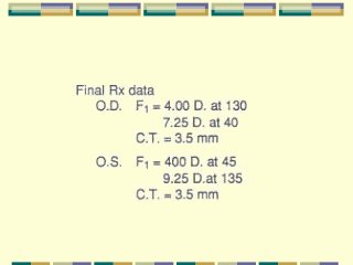
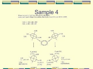
Aniseikonia
Gauri Shankar Shrestha,
M.Optom, FIACLE
Lecturer
BPKLCOS, IOM, TU
It’s true, aniseikonia
Is less lethal than pneumonia.
Doctors, nonetheless, if choosing,
Would find pneumonia less confusing
The fine art of prescribing glasses
Definition
A relative difference in the size and/or
shape of the ocular images between the two
eyes to the visual cortex
Image should be equal in brightness, size
and form
Vocabulary:
 Aniseikonia:

Difference in size/shape
 Anisometropia: Difference in Rx
Aniseikonia
Affects 2-3% of population
Anisekonia produced by glass=2%/D
Estimation of Amount of
Aniseikonia
Cateogy

Linksz &
Bannon

Ogle

refractive

1.50% /D

1.5% /D
corneal
2% /D
lenticular

0.25% /D

Axial
Partly axial

1.4% /D

> 1% /D
Aniseikonia
Two types on basis of etiology
 Physiological
 Pathological
 Anatomical
 Optical

Induced
 Inherent (neural aniseikonia)

Anatomical
Can occur in emmetropes


40% of all emmetropes have at least 0.8%

Two retinal images are physically equal in size,
yet perceived to be different sizes
Caused by innate, anatomical differences between
the two eyes
Induced Aniseikonia
Difference in retinal image size between the
2 eyes that may be caused by:
 Axial

Anisometropia
 Refractive Anisometropia
 Induced Aniseikonia
 Caused

by external optical factors (telescopes)
Neural/Essential Aniseikonia
Due to anatomically distorted map of
corresponding elements
Displacement of the retinal element towards the nodal
point
 Separation of the neuro-epithelial elements
 Stretching of the retina or edema


Meridional Rx
 Causes

meridional stretching of retinal
elements
Types on basis of image
shape
Symmetrical type


Overall



Meridianal



Compound

Asymmetrical type


in all meridian



in one meridian
What Does This Do?
Disrupts fusion
Causes Diplopia
Possible Suppression
Possible Amblyopia
Possible Strabismus
Perception of Aniseikonia
Distorts our 3D perception
 reduces

stereopsis
 Floor may appear slanted
Cause: incorrect localization of objects in space
as judged by stereopsis

May induce binocular suppression
Spatial distortion
 Physiologic

mechanism
Indications
Symptoms unrelieved by the proper
refractive and / or prismatic correction
Need for an anisometropic prescription
Patient more comfortable with monocular
vision
Spatial perception difficulties
Difference in keratometry finding
Prism Distortion
Progressive Increase in magnification
toward the apex
Base-Out OU: concave perception
Base-In OU: convex perception
Vertical OU: declination
How Much Can We Tolerate?
Clinical symptoms can occur with 1-2%
magnification differences
Aniseikonia beyond 5-7% begins to
influence stereopic thresholds
Aniseikonia within the 20% range is
incompatible with binocular vision
 Aphakic

lenses

patients corrected with spectacle
Clinical Problem?
Yes and No!!!
Depends On:
 Patients

tolerance (adaptability)
 Nature of Aniseikonia
 Type of Rx
Patient’s Tolerance
Patient learns to ignore stereoscopic
distortion and relies on monocular cues
However, distortion is still present
 Revealed

available

when monocular cues are not

When will the patient most likely suffer?
 Driving

at night-eye strain, slow responses
 Using binoculars-Artificial fusing condition
Frequency
o
o
o
o
o
o
o
o
o
o

Headache- 67%
Asthenopia (fatigue, burning sensation, tearing, ache ,
pain, pulling etc)- 67%
Photophobia- 27%
Reading difficulty –23%
Nausea – 15%
Motility (diplopia)- 11%
Nervousness – 11%
Vertigo & dizziness- 7%
General fatigue- 7%
Spatial projection & perception- 6%
Prescription Guideline
The use of spherical equivalent is often useful.
Theoretically, this places the circle of least of
confusion on the retina.
Prescriptions should not be given primarily on the
basis of measurement.
Prescribe in accordance with the nature, duration,
frequency and severity of the symptoms
When it should be corrected
When anisekonia measured with a sensitivity
smaller than size differences measured
When patients symptoms relieved by wearing
temporary correction for reasonable length of time
'Provocative test ' putting lens in wrong eye
increase discomfort and then place it to proper eye
When not to prescribe
anisekonic corrections
o

When symptoms not related to use of the
eyes, refractive or heterophoric correction

o

Measurement of size difference when
inconsistent

o

Patient if comfortable with partial correction
regardless of anisekonia
Spectacle Magnification
Defn: Change in retinal image size brought
about by correcting lens
Formulas:
.

SM=

Retinal image size in corrected state

Retinal image size in uncorrected state

%SM= (SM-1)100%
SM(AM)=Ms x Mp = 1/(1-tF1) X 1/(1-dFv)

SM increases as F1 & t increases

For +ve lens:


SM increases as Fv & d increases
Clinical Application of SM Formula
Useful in predicting retinal image size
change in pts Rx
 change in vertex distance
 change in thickness of lens
 change in the bend or form of lens


.

Note: SM>1 for +ve lens
<1 for -ve lens
=1 if lens is kept in entrance pupil of
eye and Ms = 1
Contact lens magnification
Contact lens magnification= Image corrected
with contact lens/image corrected with
spectacle lens
Comparing contact lens and
spectacle image size
CLM= 1-dFsp

d= 14mm

+10.00D, CLM=0.86
-10.00D, CLM= 1.14
These derivations suggest the biggest
single advantage of contact lenses over
spectacles
Relative Spec Magn (RSM)
Defn: size of retinal image in corrected
ametropia as compared with that of
emmetropic eye
Relative spectacle
magnification
Refractive ametropia
RSM= 1+d2Fsp
With spectacle d2=d=vertex distance=14mm

RSM=unity
With contact lens

d2=1.55

RSM= unity
d= distance from spectacle point to first
principle plane of the eye
Relative spectacle
magnification
Axial ametropia
RSM= 1-gFsp
With spectacle g= 0

RSM=unity
With contact lens

g= feye-(d+1.55)

RSM= unity
g= distance from first focal point of eye to
the spectacle point
RSM in Axial Ametropia
Knapp’s Law: for an axially ametropic eye if
correcting lens is placed so that its sec
principal point coincides with ant. Focal point of
eye, size of retinal image is same as if it were
std emmetropic eye
General rule
Axial Ametropes should be corrected
with spectacle lenses
Refractive Ametropes should be
corrected with contact lenses
How To Reduce Aniseikonia
Limitation
Refractive error can’t be ascertained that
it is purely axial despite longer axial
length
Astigmatic difference are often refractive
Correcting spectacle may be of different
shapes
Wearing spectacle at anterior focal plane
is coincidence rather than by design
How to ascertain aniseikonia
reduction in spectacle lens
Draw power diagram to compare
corresponding meridian
 Right

eye to left eye

Draw 1% of mag difference/D of
anisometropia
Start with eye that doesn’t need
magnification
 Reduce

shape magnification in this eye to
a minimum
Give magnification to other eye (with most
minus or less plus)


Subtract from predicted amount 0.50-0.75% from
predicted amount

Increase shape mag of more (-) eye until it is
greater than the other eye amount given
above
Calculate it using shape nomograph
 Many combinations of CT and F1 is possible
 To control power magnification


On (-) lens use thicker and flatter lens
 On (+) lens use thinner and steeper lens

Example 1
OS: Calculation of magnification
Example 2
Example 3
Sample 4
Frame selection
References
Optics of contact lens, IACLE module II
Milder B, Rubin ML. Anisometropia,
Fine art of prescribing glasses.
Fannin TE, Grosvenor TP. Clinical
optics
Polasky M. Instructional media center,
College of optometry, The Ohio State
University
Thank you

Aniseikonia

  • 1.
    Aniseikonia Gauri Shankar Shrestha, M.Optom,FIACLE Lecturer BPKLCOS, IOM, TU
  • 2.
    It’s true, aniseikonia Isless lethal than pneumonia. Doctors, nonetheless, if choosing, Would find pneumonia less confusing The fine art of prescribing glasses
  • 3.
    Definition A relative differencein the size and/or shape of the ocular images between the two eyes to the visual cortex Image should be equal in brightness, size and form Vocabulary:  Aniseikonia: Difference in size/shape  Anisometropia: Difference in Rx
  • 4.
    Aniseikonia Affects 2-3% ofpopulation Anisekonia produced by glass=2%/D
  • 5.
    Estimation of Amountof Aniseikonia Cateogy Linksz & Bannon Ogle refractive 1.50% /D 1.5% /D corneal 2% /D lenticular 0.25% /D Axial Partly axial 1.4% /D > 1% /D
  • 6.
    Aniseikonia Two types onbasis of etiology  Physiological  Pathological  Anatomical  Optical Induced  Inherent (neural aniseikonia) 
  • 7.
    Anatomical Can occur inemmetropes  40% of all emmetropes have at least 0.8% Two retinal images are physically equal in size, yet perceived to be different sizes Caused by innate, anatomical differences between the two eyes
  • 8.
    Induced Aniseikonia Difference inretinal image size between the 2 eyes that may be caused by:  Axial Anisometropia  Refractive Anisometropia  Induced Aniseikonia  Caused by external optical factors (telescopes)
  • 9.
    Neural/Essential Aniseikonia Due toanatomically distorted map of corresponding elements Displacement of the retinal element towards the nodal point  Separation of the neuro-epithelial elements  Stretching of the retina or edema  Meridional Rx  Causes meridional stretching of retinal elements
  • 10.
    Types on basisof image shape Symmetrical type  Overall  Meridianal  Compound Asymmetrical type  in all meridian  in one meridian
  • 11.
    What Does ThisDo? Disrupts fusion Causes Diplopia Possible Suppression Possible Amblyopia Possible Strabismus
  • 12.
    Perception of Aniseikonia Distortsour 3D perception  reduces stereopsis  Floor may appear slanted Cause: incorrect localization of objects in space as judged by stereopsis May induce binocular suppression Spatial distortion  Physiologic mechanism
  • 13.
    Indications Symptoms unrelieved bythe proper refractive and / or prismatic correction Need for an anisometropic prescription Patient more comfortable with monocular vision Spatial perception difficulties Difference in keratometry finding
  • 14.
    Prism Distortion Progressive Increasein magnification toward the apex Base-Out OU: concave perception Base-In OU: convex perception Vertical OU: declination
  • 15.
    How Much CanWe Tolerate? Clinical symptoms can occur with 1-2% magnification differences Aniseikonia beyond 5-7% begins to influence stereopic thresholds Aniseikonia within the 20% range is incompatible with binocular vision  Aphakic lenses patients corrected with spectacle
  • 16.
    Clinical Problem? Yes andNo!!! Depends On:  Patients tolerance (adaptability)  Nature of Aniseikonia  Type of Rx
  • 17.
    Patient’s Tolerance Patient learnsto ignore stereoscopic distortion and relies on monocular cues However, distortion is still present  Revealed available when monocular cues are not When will the patient most likely suffer?  Driving at night-eye strain, slow responses  Using binoculars-Artificial fusing condition
  • 18.
    Frequency o o o o o o o o o o Headache- 67% Asthenopia (fatigue,burning sensation, tearing, ache , pain, pulling etc)- 67% Photophobia- 27% Reading difficulty –23% Nausea – 15% Motility (diplopia)- 11% Nervousness – 11% Vertigo & dizziness- 7% General fatigue- 7% Spatial projection & perception- 6%
  • 19.
    Prescription Guideline The useof spherical equivalent is often useful. Theoretically, this places the circle of least of confusion on the retina. Prescriptions should not be given primarily on the basis of measurement. Prescribe in accordance with the nature, duration, frequency and severity of the symptoms
  • 20.
    When it shouldbe corrected When anisekonia measured with a sensitivity smaller than size differences measured When patients symptoms relieved by wearing temporary correction for reasonable length of time 'Provocative test ' putting lens in wrong eye increase discomfort and then place it to proper eye
  • 21.
    When not toprescribe anisekonic corrections o When symptoms not related to use of the eyes, refractive or heterophoric correction o Measurement of size difference when inconsistent o Patient if comfortable with partial correction regardless of anisekonia
  • 22.
    Spectacle Magnification Defn: Changein retinal image size brought about by correcting lens Formulas: . SM= Retinal image size in corrected state Retinal image size in uncorrected state %SM= (SM-1)100% SM(AM)=Ms x Mp = 1/(1-tF1) X 1/(1-dFv) SM increases as F1 & t increases For +ve lens:  SM increases as Fv & d increases
  • 23.
    Clinical Application ofSM Formula Useful in predicting retinal image size change in pts Rx  change in vertex distance  change in thickness of lens  change in the bend or form of lens  . Note: SM>1 for +ve lens <1 for -ve lens =1 if lens is kept in entrance pupil of eye and Ms = 1
  • 24.
    Contact lens magnification Contactlens magnification= Image corrected with contact lens/image corrected with spectacle lens
  • 25.
    Comparing contact lensand spectacle image size CLM= 1-dFsp d= 14mm +10.00D, CLM=0.86 -10.00D, CLM= 1.14 These derivations suggest the biggest single advantage of contact lenses over spectacles
  • 26.
    Relative Spec Magn(RSM) Defn: size of retinal image in corrected ametropia as compared with that of emmetropic eye
  • 27.
    Relative spectacle magnification Refractive ametropia RSM=1+d2Fsp With spectacle d2=d=vertex distance=14mm RSM=unity With contact lens d2=1.55 RSM= unity d= distance from spectacle point to first principle plane of the eye
  • 28.
    Relative spectacle magnification Axial ametropia RSM=1-gFsp With spectacle g= 0 RSM=unity With contact lens g= feye-(d+1.55) RSM= unity g= distance from first focal point of eye to the spectacle point
  • 29.
    RSM in AxialAmetropia Knapp’s Law: for an axially ametropic eye if correcting lens is placed so that its sec principal point coincides with ant. Focal point of eye, size of retinal image is same as if it were std emmetropic eye
  • 30.
    General rule Axial Ametropesshould be corrected with spectacle lenses Refractive Ametropes should be corrected with contact lenses
  • 31.
    How To ReduceAniseikonia Limitation Refractive error can’t be ascertained that it is purely axial despite longer axial length Astigmatic difference are often refractive Correcting spectacle may be of different shapes Wearing spectacle at anterior focal plane is coincidence rather than by design
  • 32.
    How to ascertainaniseikonia reduction in spectacle lens Draw power diagram to compare corresponding meridian  Right eye to left eye Draw 1% of mag difference/D of anisometropia Start with eye that doesn’t need magnification  Reduce shape magnification in this eye to a minimum
  • 33.
    Give magnification toother eye (with most minus or less plus)  Subtract from predicted amount 0.50-0.75% from predicted amount Increase shape mag of more (-) eye until it is greater than the other eye amount given above Calculate it using shape nomograph  Many combinations of CT and F1 is possible  To control power magnification  On (-) lens use thicker and flatter lens  On (+) lens use thinner and steeper lens 
  • 34.
  • 36.
    OS: Calculation ofmagnification
  • 38.
  • 45.
  • 51.
  • 52.
  • 53.
    References Optics of contactlens, IACLE module II Milder B, Rubin ML. Anisometropia, Fine art of prescribing glasses. Fannin TE, Grosvenor TP. Clinical optics Polasky M. Instructional media center, College of optometry, The Ohio State University
  • 54.

Editor's Notes

  • #4 Ocular image is determined not only by the dioptric image on the retina but also on the distribution of retinal elements and the physiological and psychological modifications of the dioptric image in the process of reaching the higher centers. The normal image size differences, called disparities, are the basis of stereopsis.
  • #8 . .
  • #10 Displacement of the retinal element towards the nodal point. Separation of the neuro-epithelial elements. Stretching of the retina or edema
  • #13 Since anisekonia is a binocular anomaly, it adds further disparity to normal disparities which causes incorrect localization of objects in space as judged by stereopsis
  • #16 With large amount of anisekonia(&amp;gt;5%), it becomes impossible to achieve fusion and hence prevents central fusion.