Downloaded 171 times


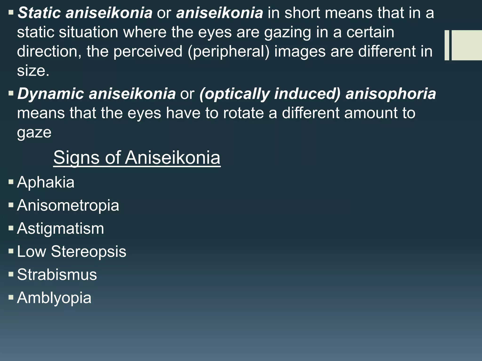


This document discusses aniseikonia, which is a condition where the two eyes perceive images as being different in size. It can be caused by differences in the dioptric images formed by the retina or differences in retinal element distribution. The document describes static aniseikonia, where peripheral images differ in size with static gaze, and dynamic aniseikonia, where the eyes must rotate different amounts to gaze. Signs include aphakia, anisometropia, astigmatism, low stereopsis, and strabismus. Symptoms include headaches, asthenopia, reading difficulty, photophobia, nausea, vertigo, and fatigue. Aniseikonia is measured using an e


![Aniseikonia [ophthalmology description for medical students ]](https://cdn.slidesharecdn.com/ss_thumbnails/aniseikonia-201008163906-thumbnail.jpg?width=640&height=640&fit=bounds)


![Apporach to lung biopsy [Auto-saved].pptx latest](https://cdn.slidesharecdn.com/ss_thumbnails/apporachtolungbiopsyauto-saved-251211225655-93258539-thumbnail.jpg?width=640&height=640&fit=bounds)


