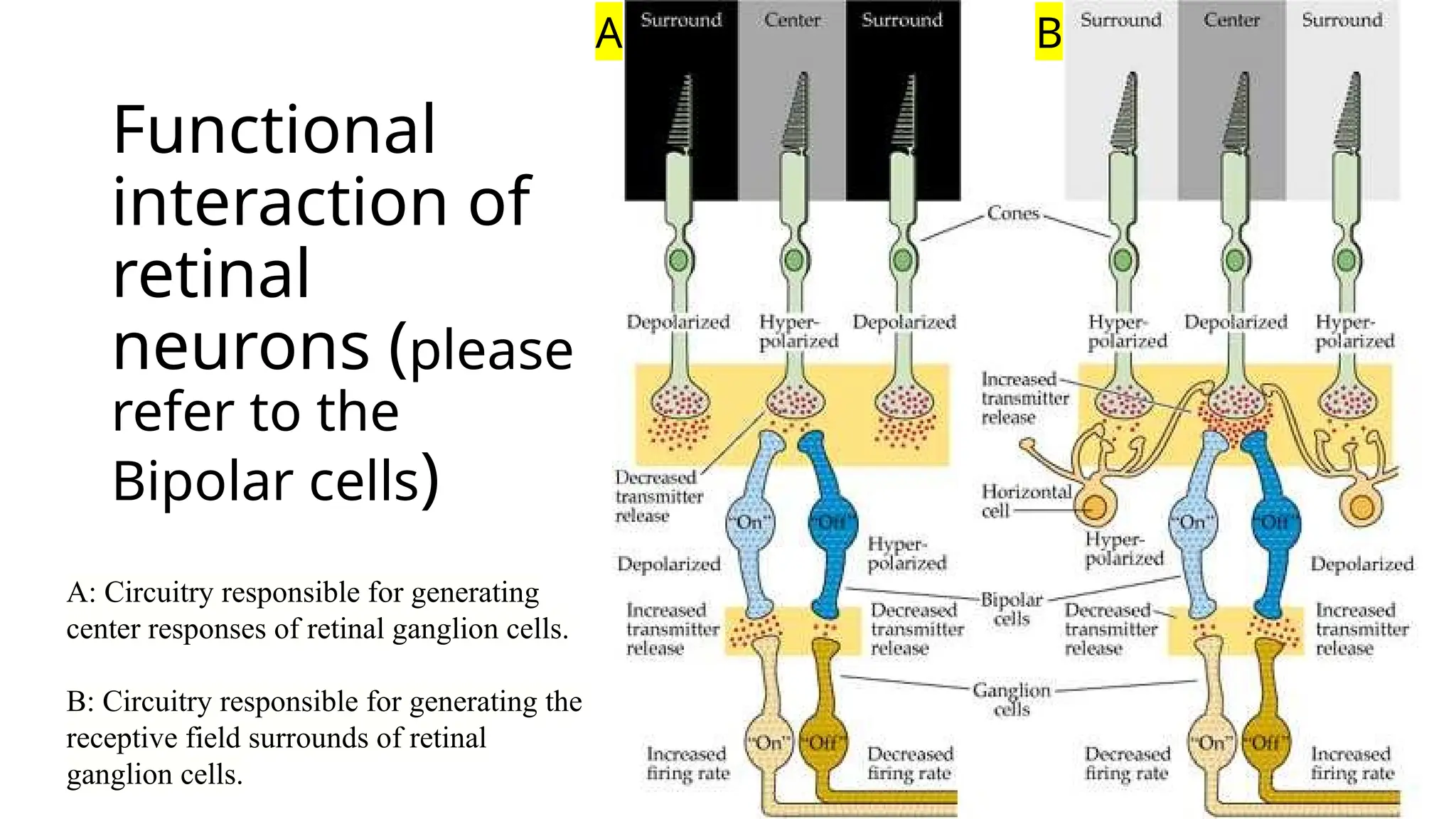
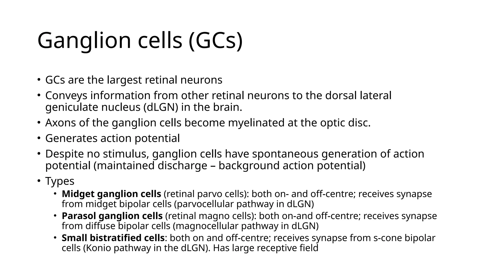
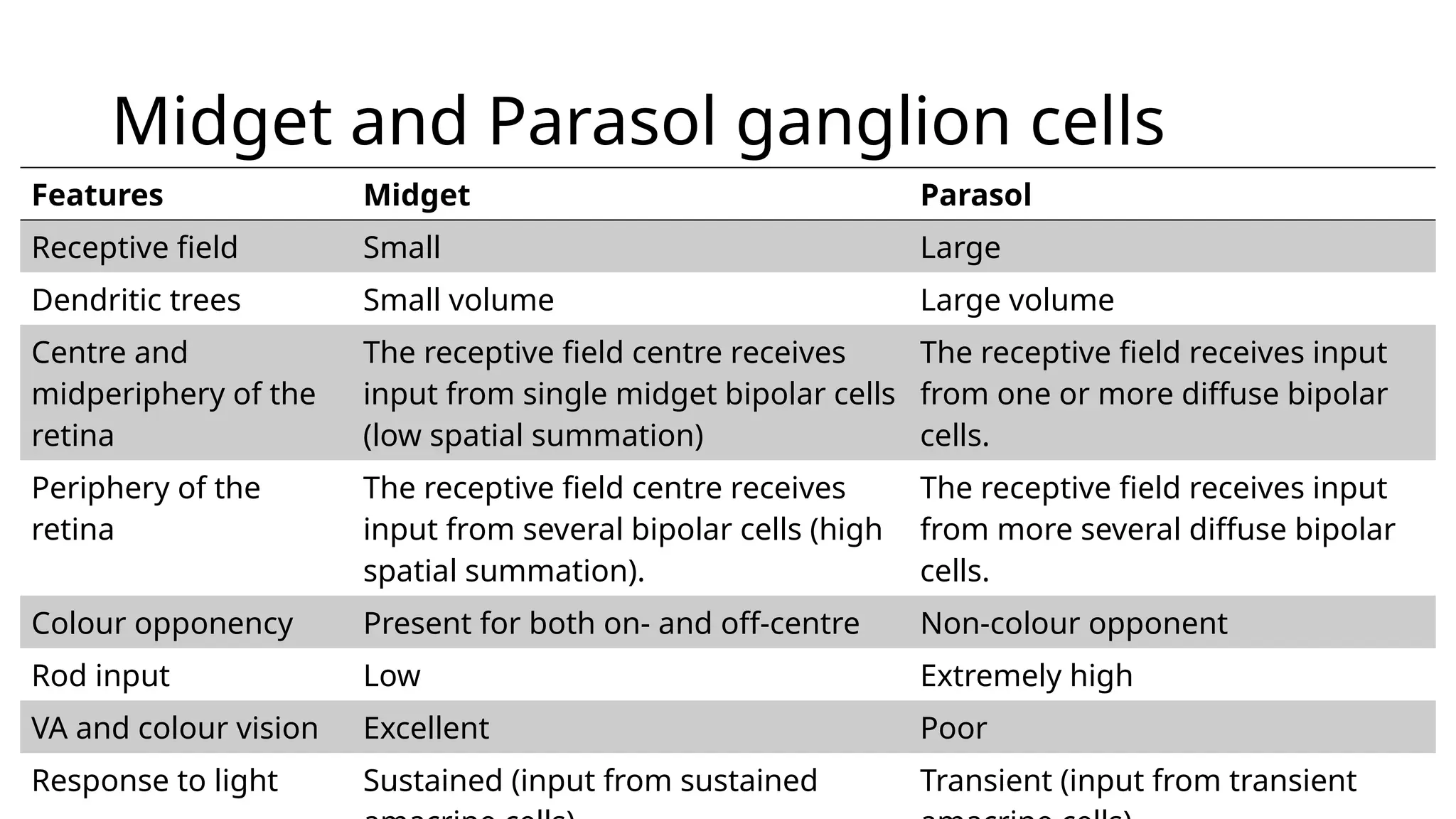
Physiologic
Model of
Spatial Vision
and Retinal
Sampling
Dr Gauri Sr Shrestha
MMC, IOM, TU
Course
overview
Learning outcomes
• Understand basic organisation of retinal neural cells and
their processing.
• Understand the spatial summation and resolution
• Understand spatial frequency tunning and neural channels
at the retinal level
• Understand the receptive field characteristics of the neurons
that represents retinal processing of visual information
(photoreceptors, horizontal cells, bipolar cells, amacrine
cells and ganglion cells)
Organisatio
n of neural
cells in the
retina
Retinal photoreceptor
cells
Rod spherule Cone pedicle
Ellipsoid
Myoid
Outer rod
fibres
Inner rod
fibres
Ellipsoid
Myoid
Inner cone
fibres
Rod cells
• 40 – 60 µm long
• Rod outer segment
• is cylindrical, highly refractile and contains visual pigments (visual purples)
• Consists of lipid protein lamellar discs stacked one over the other (600 – 1000
discs/ rod) and are surrounded by cell membrane.
• Rod inner segment
• Is thicker than the outer segment and has two regions.
• Ellipsoid (the outer portion): contains abundant number of mitochondria.
• Myoid (the inner portion): contains the glycogen and usual organelles.
Rod cells
• Peak density of rods occurs 20o
from fovea: 150,000 rods/mm2
• No rods are present at the fovea (0.35 mm, 1.25o
of VF
• Total Number of rods in the retina are 120 million
• Visual purple: Rhodopsin
• Rhodopsin most readily absorbs wavelengths of 507 nm.
• One molecule of rhodopsin absorbs one quanta of light, it is
‘bleached’
• Bleached: the molecule is not capable of capturing another
quantum
• Spontaneously become ‘unbleached’
• 50% recover within 5 minutes
Cone cells
• 40 – 80 µm long (longest at the fovea and shortest at the periphery)
• Cone outer segment
• is conical in shape and contains iodopsin
• Consists of lipid protein lamellar discs stacked one over the other (1000 –
1200 discs/ cone) and are surrounded by cell membrane on the one side only.
• Cone inner segment
• Is similar to the rods and has two regions.
• Ellipsoid (the outer portion): is very lumpy and contains abundant number of
mitochondria.
• However, outer fibre is absent and has a stout inner fibre.
Cone cells
• Cones are most densely packed at the fovea: 150,000 cones/mm2
• Only 4% of total cones are foveal
• Total Number of cones in the retina are 6 million
• 3 types of cone photopigments:
• Erythrolabe: maximum absorption at 565 nm (L-cones, red-light sensitive cones)
• Chlorolabe: maximum absorption at 535 nm (M-cones, green-light sensitive
cones)
• Cyanolabe: maximum absorption at 430 nm (S-cones, blue-light sensitive cones)
• Recover from bleaching more rapidly than rhodopsin
• 50% of cones will recover within 1.5 minutes
Distributio
n of rods
and cones
in the
retina
Recording of electrical activity in the
retina
• The retina analyses the optical images fell on it and encodes
them into a complex neural signal to transmit the signal to
higher visual centres.
• Isolated electrical activity is recorded through
• Extracellular recording (in-vitro) – for both action potential and
graded (slow potential)
• Intracellular recording (in-vivo) – for graded (slow potential)
Receptive field: area that influences the neural activity of the cell.
Excitation as an increase in the frequency of action potentials (indicated by plus sign)
Inhibition as a decrease in the frequency of action potential (indicated by minus sign)
Graded potential versus Action
potential
Features Graded Action
Type of signal • Input signal
• Depolarising or hyperpolarising
• Output signal
• depolarising
Travel Conveys over small distance
(membrane potential decreases
with distance from the stimulation)
Transfer over long distance
(propagate along entire membrane
surface without decreasing its
strength
Strength of signal Variable, depends on the
magnitude of stimulus
Always same (all or none)
Threshold No threshold is required to initiate Stimulus reaches threshold level to
generate AP
Site Dendrites and cell body Axon
Receptive field of retinal
nerve cells
• The various retinal cells show different
receptive field properties (refer to the
picture on the right)
• Photoreceptor and horizontal cells have diffused
receptive field
• Bipolar, amacrine and ganglion cells have centre-
surround organisation in their receptive field.
Receptive
field
Cell
type
Receptive field Stimulus
Photoreceptor diffused light
Horizontal cell a spot of light
Bipolar cells A linear light (a bar of light)
Ganglion cell spatial gratings (alternate light and dark
Receptive field of retinal nerve
cells
• In centre surround
organisation, the electric
potential generated is
either excitatory or
inhibitory.
• Light falling on the
receptive field centre has
the opposite effect of light
falling on the surrounding
area of the receptive field.
Ganglion
cell
receptive
field
Spatial resolution and spatial
summation
• Dark band of stimulus
as well as light band of
stimulus should
stimulate ganglion
cells separately.
• Spatial summation
describes the eye’s
ability to summate
quanta over a certain
area (critical diameter).
Ricco’s law
• For small stimuli (below a critical
diameter), the total luminous energy
(intensity × area) must reach a
constant threshold for detection (k).
• A dim but large stimulus can be
detected just as easily as a small
but bright one
• In parafoveal area (4 – 7°), critical
diameter = 30 mins of arc
• At an eccentricity of 30°, the critical
dimeter = 2° of arc
L.An
= k
n = 1, total spatial summation
n = 0, no spatial summation
Spatial tuning
• Spatial tuning refers to the ability of
neurons in the visual system to respond
selectively to stimuli of specific sizes,
shapes, or locations within the visual
field
• When a grating is presented to the
ganglion cells, the spatial tunning will be
maximum when
• The dark area fall on the inhibitory surround
• The light areas fall on the excitatory centre
• The response will be very strong.
a more optimal stimulus
for the ganglion cell
Spatial tunning
Spatial
tunning
Spatial
antagonism
• Bipolar cell is the first cell
to show spatial antagonism
to a bar of light
• A spatial grating is a strong
stimulus for a ganglion cell.
• Light bar falls on excitatory
centre and
• Dark bar falls on the
inhibitory surround
a more optimal stimulus
for the ganglion cell
the large bright bar of the low-frequency
grating falls both on the receptive field’s
centre and surround causing lateral inhibition
Mach Bands
• Refers to an optical phenomenon
(sometimes called optical illusion) from
EDGE ENHANCEMENT (a relative
enhancement of high spatial
frequencies) at the borders of gradual
physical transition.
• The edges of dark objects next to light
objects appear darker and vice versa at
the junction of a gradual change of
bright to dark regions.
• Gradual transition consists of low
frequencies
• We have a low sensitivity to these
frequencies (lateral inhibition)
The Visual System as
a Fourier Analyzer
• The contrast sensitivity function
of our visual system consists of
several independent narrower
spatial frequency channels.
• breaks the visual world into
spatial frequency components
• consists several narrower channel
(no single channel exists)
• again, reassembles components
and forms our spatial perception
The Visual
System as a
Fourier Analyzer
• e.g., A subject is
adapted to a
sinusoidal grating (6
cycle/ degree) for
about a minute prior
to determination of
the CSF.
• There is a discrete
reduction in
sensitivity at the
adapted frequency (6
cycle/ degree)
The Visual System
as a Fourier
Analyzer
• Now the subject is adapted
to a square wave grating
of 6 cycle/ degree.
• Our visual system breaks
the Complex stimulus
(square-wave) into its
components (fundamental
and harmonic frequencies)
• There would be decrease in
sensitivity at those
frequencies.
Blakemore and Campbell (1969)
6 cycle/degree
18 cycle/degree
Photoreceptors
• Photoreceptors convert light energy into
electrical activity.
• Photoreceptors are in the state of
depolarisation (-50mV) in the dark relative
to other neuron cells.
• When exposed to light, they hyperpolarise
(-70mV).
• Potential produced by photoreceptors are
graded.
• 10% rhodopsin bleaching closes Na+
channel, no rhodopsin further
hyperpolarises (rod saturation)
Dark
current
From light reception to
receptor potential
Horizontal cells (HCs)
• Several photoreceptors (rods and
cones) distributed over a large area
of the retina synapse with the widely
dispersed dendrites of a single
horizontal cell.
• This interconnection contributes to
the spatial summation.
• HCs shows sign-conserving synapses
(Invaginating synapse) with
photoreceptors.
Horizontal cells (HCs)
• HCs hyperpolarise in
response to the light.
• HCs produce graded
potential (no action
potential).
• Process luminosity.
• Two types of horizontal cells
• H1: primary input from the M-
and L-cones, and little input
from S-cones
• H2: Strong input from S-cones
and also receives input from
M- and L-cones.
Spatial summation and lateral
inhibition facilitated by HCs
A. HCs collect information from
photoreceptors in the receptive
field surround (or centre) and
feedback on to photoreceptors
in the receptive field centre (or
surround) to generate the
antagonistic receptive field
surround (or centre) of the
bipolar cells
B. The reciprocal synapse between
cones and horizontal cells
mediates negative feedback
Bipolar (BP) cells
• Bipolar cells are the FIRST retinal cells
to mediate spatial antagonism
• Bipolar cells mediates graded potential
(no action potential)
• S-cones receive input from a distinct
class of bipolar cells (S-cone bipolar
cells)
• Dendrites of BP cells synapse with
photoreceptors and horizontal cells
• Axons of BP cells synapse with
ganglion and amacrine cells
• Neurotransmitter released by BP cells -
glutamate
Sign
conserving
(invaginating)
synapse
Conventional
flat synapse
Functional
interaction of
retinal
neurons (please
refer to the
Bipolar cells)
A: Circuitry responsible for generating
center responses of retinal ganglion cells.
B: Circuitry responsible for generating the
receptive field surrounds of retinal
ganglion cells.
A B
On- and Off-centre bipolar cells
Features On-centre Off-centre
Synapses with
Photoreceptors
Sign conserving Flat
Light on centre of the
receptive field
Excitation (depolarisation) Inhibition
(hyperpolarisation)
Light on surround of the
receptive field
Inhibition (hyperpolarisation) Excitation (depolarisation)
Synapses Inner sublayer of IPL Outer sublayer of IPL
Glutamate released by
photoreceptors
Inhibitory (reduction in
glutamate released by
photoreceptors causes
depolarisation (excitation)
Excitatory (reduction in
glutamate released by
photoreceptors causes
hyperpolarisation
Midget and diffuse bipolar cells
Features Midget Diffuse
Morphology Less tissue volume and less
dendritic branches
More tissue volume and more
dendritic branches
Centre and
midperiphery of the
retina
The receptive field centre receives
input from single M- or L-cones
(high VA, colour opponent)
The receptive field receives input
from 5 – 10 cones (more than
one cone type (M or L).
Periphery of the retina The receptive field centre receives
input from several photoreceptors
(Low VA).
Receptive field
surround
Receives input from single H1 HCs Receives input from several H1
HCs
Colour opponency Present for both on- and off-centre Non-colour opponent
Organisati
on of the
primate
retina
Amacrine cells
• Location: between INL and GCL of the retina
• Presynaptic connection: Bipolar cells
• Post-synaptic connection: Ganglion cells
• Shows centre surround organisation
• Is the FIRST cell to display action potential.
• Shows time-related characteristics of neural
response
• Responds briefly and transiently at the
stimulus onset and off-set (helps ganglion
cells to detect change in light intensity)
Amacrine cells
• Functions of Amacrine cells:
• Plays a role in coding movement
• Bridge the connection between rods and cones in scotopic vision for high sensitivity
• Adjust light sensitivity for photopic and scotopic vision
• Provides synaptic feedback to BP cells
• Types by response to the light
1. Sustained amacrine cells
2. Transient amacrine cells
• Types by cell types:
1. Sublaminar cell A: contains bipolar axons and off-centre ganglion cell connections
(hyperpolarising response to light)
2. Sublaminar cell B: contains bipolar axons and on-centre ganglion cell connections
(depolarising response to light)
Ganglion cells (GCs)
• GCs are the largest retinal neurons
• Conveys information from other retinal neurons to the dorsal lateral
geniculate nucleus (dLGN) in the brain.
• Axons of the ganglion cells become myelinated at the optic disc.
• Generates action potential
• Despite no stimulus, ganglion cells have spontaneous generation of action
potential (maintained discharge – background action potential)
• Types
• Midget ganglion cells (retinal parvo cells): both on- and off-centre; receives synapse
from midget bipolar cells (parvocellular pathway in dLGN)
• Parasol ganglion cells (retinal magno cells): both on-and off-centre; receives synapse
from diffuse bipolar cells (magnocellular pathway in dLGN)
• Small bistratified cells: both on and off-centre; receives synapse from s-cone bipolar
cells (Konio pathway in the dLGN). Has large receptive field
Midget and Parasol ganglion cells
Features Midget Parasol
Receptive field Small Large
Dendritic trees Small volume Large volume
Centre and
midperiphery of the
retina
The receptive field centre receives
input from single midget bipolar cells
(low spatial summation)
The receptive field receives input
from one or more diffuse bipolar
cells.
Periphery of the
retina
The receptive field centre receives
input from several bipolar cells (high
spatial summation).
The receptive field receives input
from more several diffuse bipolar
cells.
Colour opponency Present for both on- and off-centre Non-colour opponent
Rod input Low Extremely high
VA and colour vision Excellent Poor
Response to light Sustained (input from sustained Transient (input from transient
Receptive field organisation of the
ganglion cells
• The receptive field
of the ganglion
cells overlap.
• A small spot of light
can excite or inhibit
many ganglion cells
Other types of ganglion cells
• Ganglion cells projecting to the superior colliculus
(midbrain): controls eye movements
• Melanopsin containing ganglion cells: projects to the
suprachiasmatic nucleus of the hypothalamus and is
responsible for the circadian rhythm.
• Intrinsically photosensitive (retino-hypothalamic track)
• Ganglion cells projecting to pulvinar region: visual attention
and motion processing.
Linearity of ganglion
cell receptive field
• Ganglion cells act like shift-
invariant linear systems.
• The input is stimulus
contrast. The output is firing
rate.
• Add light intensities at each
point weighted by positive or
negative centre and surround
subregions of the receptive
field, their response changes
to the stimulus linearly.
Summary
• The retinal neural cells transfer signals vertically from photoreceptors – bipolar
cell – ganglion cells.
• The lateral communication between retinal neural cells is mediated by horizontal
and amacrine cells.
• The spatial summation takes place within the critical diameter of the retina and
also the area of summation varies with the retinal location.
• Various retinal cell exhibits various types of receptive fields. The first cells to
exhibit lateral inhibition is bipolar cells and to generate action potential is the
amacrine cells.
• Our visual system shows several independent narrower spatial frequency
channels.
• Neural convergence is mandatory (Photoreceptors = 126 million, ganglion cells =
1 million)
Thank you
• Further reading
• neurotransmitters in the retina
(https://www.ncbi.nlm.nih.gov/books/NBK11546/).

1. Physiologic Model of Spatial Vision and Retinal Sampling.pptx

  • 1.
    Physiologic Model of Spatial Vision andRetinal Sampling Dr Gauri Sr Shrestha MMC, IOM, TU
  • 2.
  • 3.
    Learning outcomes • Understandbasic organisation of retinal neural cells and their processing. • Understand the spatial summation and resolution • Understand spatial frequency tunning and neural channels at the retinal level • Understand the receptive field characteristics of the neurons that represents retinal processing of visual information (photoreceptors, horizontal cells, bipolar cells, amacrine cells and ganglion cells)
  • 4.
  • 5.
    Retinal photoreceptor cells Rod spheruleCone pedicle Ellipsoid Myoid Outer rod fibres Inner rod fibres Ellipsoid Myoid Inner cone fibres
  • 6.
    Rod cells • 40– 60 µm long • Rod outer segment • is cylindrical, highly refractile and contains visual pigments (visual purples) • Consists of lipid protein lamellar discs stacked one over the other (600 – 1000 discs/ rod) and are surrounded by cell membrane. • Rod inner segment • Is thicker than the outer segment and has two regions. • Ellipsoid (the outer portion): contains abundant number of mitochondria. • Myoid (the inner portion): contains the glycogen and usual organelles.
  • 7.
    Rod cells • Peakdensity of rods occurs 20o from fovea: 150,000 rods/mm2 • No rods are present at the fovea (0.35 mm, 1.25o of VF • Total Number of rods in the retina are 120 million • Visual purple: Rhodopsin • Rhodopsin most readily absorbs wavelengths of 507 nm. • One molecule of rhodopsin absorbs one quanta of light, it is ‘bleached’ • Bleached: the molecule is not capable of capturing another quantum • Spontaneously become ‘unbleached’ • 50% recover within 5 minutes
  • 8.
    Cone cells • 40– 80 µm long (longest at the fovea and shortest at the periphery) • Cone outer segment • is conical in shape and contains iodopsin • Consists of lipid protein lamellar discs stacked one over the other (1000 – 1200 discs/ cone) and are surrounded by cell membrane on the one side only. • Cone inner segment • Is similar to the rods and has two regions. • Ellipsoid (the outer portion): is very lumpy and contains abundant number of mitochondria. • However, outer fibre is absent and has a stout inner fibre.
  • 9.
    Cone cells • Conesare most densely packed at the fovea: 150,000 cones/mm2 • Only 4% of total cones are foveal • Total Number of cones in the retina are 6 million • 3 types of cone photopigments: • Erythrolabe: maximum absorption at 565 nm (L-cones, red-light sensitive cones) • Chlorolabe: maximum absorption at 535 nm (M-cones, green-light sensitive cones) • Cyanolabe: maximum absorption at 430 nm (S-cones, blue-light sensitive cones) • Recover from bleaching more rapidly than rhodopsin • 50% of cones will recover within 1.5 minutes
  • 10.
    Distributio n of rods andcones in the retina
  • 11.
    Recording of electricalactivity in the retina • The retina analyses the optical images fell on it and encodes them into a complex neural signal to transmit the signal to higher visual centres. • Isolated electrical activity is recorded through • Extracellular recording (in-vitro) – for both action potential and graded (slow potential) • Intracellular recording (in-vivo) – for graded (slow potential) Receptive field: area that influences the neural activity of the cell. Excitation as an increase in the frequency of action potentials (indicated by plus sign) Inhibition as a decrease in the frequency of action potential (indicated by minus sign)
  • 12.
    Graded potential versusAction potential Features Graded Action Type of signal • Input signal • Depolarising or hyperpolarising • Output signal • depolarising Travel Conveys over small distance (membrane potential decreases with distance from the stimulation) Transfer over long distance (propagate along entire membrane surface without decreasing its strength Strength of signal Variable, depends on the magnitude of stimulus Always same (all or none) Threshold No threshold is required to initiate Stimulus reaches threshold level to generate AP Site Dendrites and cell body Axon
  • 13.
    Receptive field ofretinal nerve cells • The various retinal cells show different receptive field properties (refer to the picture on the right) • Photoreceptor and horizontal cells have diffused receptive field • Bipolar, amacrine and ganglion cells have centre- surround organisation in their receptive field. Receptive field Cell type Receptive field Stimulus Photoreceptor diffused light Horizontal cell a spot of light Bipolar cells A linear light (a bar of light) Ganglion cell spatial gratings (alternate light and dark
  • 14.
    Receptive field ofretinal nerve cells • In centre surround organisation, the electric potential generated is either excitatory or inhibitory. • Light falling on the receptive field centre has the opposite effect of light falling on the surrounding area of the receptive field.
  • 15.
  • 16.
    Spatial resolution andspatial summation • Dark band of stimulus as well as light band of stimulus should stimulate ganglion cells separately. • Spatial summation describes the eye’s ability to summate quanta over a certain area (critical diameter).
  • 17.
    Ricco’s law • Forsmall stimuli (below a critical diameter), the total luminous energy (intensity × area) must reach a constant threshold for detection (k). • A dim but large stimulus can be detected just as easily as a small but bright one • In parafoveal area (4 – 7°), critical diameter = 30 mins of arc • At an eccentricity of 30°, the critical dimeter = 2° of arc L.An = k n = 1, total spatial summation n = 0, no spatial summation
  • 18.
    Spatial tuning • Spatialtuning refers to the ability of neurons in the visual system to respond selectively to stimuli of specific sizes, shapes, or locations within the visual field • When a grating is presented to the ganglion cells, the spatial tunning will be maximum when • The dark area fall on the inhibitory surround • The light areas fall on the excitatory centre • The response will be very strong. a more optimal stimulus for the ganglion cell
  • 19.
  • 20.
  • 21.
    Spatial antagonism • Bipolar cellis the first cell to show spatial antagonism to a bar of light • A spatial grating is a strong stimulus for a ganglion cell. • Light bar falls on excitatory centre and • Dark bar falls on the inhibitory surround a more optimal stimulus for the ganglion cell the large bright bar of the low-frequency grating falls both on the receptive field’s centre and surround causing lateral inhibition
  • 22.
    Mach Bands • Refersto an optical phenomenon (sometimes called optical illusion) from EDGE ENHANCEMENT (a relative enhancement of high spatial frequencies) at the borders of gradual physical transition. • The edges of dark objects next to light objects appear darker and vice versa at the junction of a gradual change of bright to dark regions. • Gradual transition consists of low frequencies • We have a low sensitivity to these frequencies (lateral inhibition)
  • 23.
    The Visual Systemas a Fourier Analyzer • The contrast sensitivity function of our visual system consists of several independent narrower spatial frequency channels. • breaks the visual world into spatial frequency components • consists several narrower channel (no single channel exists) • again, reassembles components and forms our spatial perception
  • 24.
    The Visual System asa Fourier Analyzer • e.g., A subject is adapted to a sinusoidal grating (6 cycle/ degree) for about a minute prior to determination of the CSF. • There is a discrete reduction in sensitivity at the adapted frequency (6 cycle/ degree)
  • 25.
    The Visual System asa Fourier Analyzer • Now the subject is adapted to a square wave grating of 6 cycle/ degree. • Our visual system breaks the Complex stimulus (square-wave) into its components (fundamental and harmonic frequencies) • There would be decrease in sensitivity at those frequencies. Blakemore and Campbell (1969) 6 cycle/degree 18 cycle/degree
  • 26.
    Photoreceptors • Photoreceptors convertlight energy into electrical activity. • Photoreceptors are in the state of depolarisation (-50mV) in the dark relative to other neuron cells. • When exposed to light, they hyperpolarise (-70mV). • Potential produced by photoreceptors are graded. • 10% rhodopsin bleaching closes Na+ channel, no rhodopsin further hyperpolarises (rod saturation) Dark current
  • 27.
    From light receptionto receptor potential
  • 28.
    Horizontal cells (HCs) •Several photoreceptors (rods and cones) distributed over a large area of the retina synapse with the widely dispersed dendrites of a single horizontal cell. • This interconnection contributes to the spatial summation. • HCs shows sign-conserving synapses (Invaginating synapse) with photoreceptors.
  • 29.
    Horizontal cells (HCs) •HCs hyperpolarise in response to the light. • HCs produce graded potential (no action potential). • Process luminosity. • Two types of horizontal cells • H1: primary input from the M- and L-cones, and little input from S-cones • H2: Strong input from S-cones and also receives input from M- and L-cones.
  • 30.
    Spatial summation andlateral inhibition facilitated by HCs A. HCs collect information from photoreceptors in the receptive field surround (or centre) and feedback on to photoreceptors in the receptive field centre (or surround) to generate the antagonistic receptive field surround (or centre) of the bipolar cells B. The reciprocal synapse between cones and horizontal cells mediates negative feedback
  • 31.
    Bipolar (BP) cells •Bipolar cells are the FIRST retinal cells to mediate spatial antagonism • Bipolar cells mediates graded potential (no action potential) • S-cones receive input from a distinct class of bipolar cells (S-cone bipolar cells) • Dendrites of BP cells synapse with photoreceptors and horizontal cells • Axons of BP cells synapse with ganglion and amacrine cells • Neurotransmitter released by BP cells - glutamate Sign conserving (invaginating) synapse Conventional flat synapse
  • 32.
    Functional interaction of retinal neurons (please referto the Bipolar cells) A: Circuitry responsible for generating center responses of retinal ganglion cells. B: Circuitry responsible for generating the receptive field surrounds of retinal ganglion cells. A B
  • 33.
    On- and Off-centrebipolar cells Features On-centre Off-centre Synapses with Photoreceptors Sign conserving Flat Light on centre of the receptive field Excitation (depolarisation) Inhibition (hyperpolarisation) Light on surround of the receptive field Inhibition (hyperpolarisation) Excitation (depolarisation) Synapses Inner sublayer of IPL Outer sublayer of IPL Glutamate released by photoreceptors Inhibitory (reduction in glutamate released by photoreceptors causes depolarisation (excitation) Excitatory (reduction in glutamate released by photoreceptors causes hyperpolarisation
  • 34.
    Midget and diffusebipolar cells Features Midget Diffuse Morphology Less tissue volume and less dendritic branches More tissue volume and more dendritic branches Centre and midperiphery of the retina The receptive field centre receives input from single M- or L-cones (high VA, colour opponent) The receptive field receives input from 5 – 10 cones (more than one cone type (M or L). Periphery of the retina The receptive field centre receives input from several photoreceptors (Low VA). Receptive field surround Receives input from single H1 HCs Receives input from several H1 HCs Colour opponency Present for both on- and off-centre Non-colour opponent
  • 35.
  • 36.
    Amacrine cells • Location:between INL and GCL of the retina • Presynaptic connection: Bipolar cells • Post-synaptic connection: Ganglion cells • Shows centre surround organisation • Is the FIRST cell to display action potential. • Shows time-related characteristics of neural response • Responds briefly and transiently at the stimulus onset and off-set (helps ganglion cells to detect change in light intensity)
  • 37.
    Amacrine cells • Functionsof Amacrine cells: • Plays a role in coding movement • Bridge the connection between rods and cones in scotopic vision for high sensitivity • Adjust light sensitivity for photopic and scotopic vision • Provides synaptic feedback to BP cells • Types by response to the light 1. Sustained amacrine cells 2. Transient amacrine cells • Types by cell types: 1. Sublaminar cell A: contains bipolar axons and off-centre ganglion cell connections (hyperpolarising response to light) 2. Sublaminar cell B: contains bipolar axons and on-centre ganglion cell connections (depolarising response to light)
  • 38.
    Ganglion cells (GCs) •GCs are the largest retinal neurons • Conveys information from other retinal neurons to the dorsal lateral geniculate nucleus (dLGN) in the brain. • Axons of the ganglion cells become myelinated at the optic disc. • Generates action potential • Despite no stimulus, ganglion cells have spontaneous generation of action potential (maintained discharge – background action potential) • Types • Midget ganglion cells (retinal parvo cells): both on- and off-centre; receives synapse from midget bipolar cells (parvocellular pathway in dLGN) • Parasol ganglion cells (retinal magno cells): both on-and off-centre; receives synapse from diffuse bipolar cells (magnocellular pathway in dLGN) • Small bistratified cells: both on and off-centre; receives synapse from s-cone bipolar cells (Konio pathway in the dLGN). Has large receptive field
  • 39.
    Midget and Parasolganglion cells Features Midget Parasol Receptive field Small Large Dendritic trees Small volume Large volume Centre and midperiphery of the retina The receptive field centre receives input from single midget bipolar cells (low spatial summation) The receptive field receives input from one or more diffuse bipolar cells. Periphery of the retina The receptive field centre receives input from several bipolar cells (high spatial summation). The receptive field receives input from more several diffuse bipolar cells. Colour opponency Present for both on- and off-centre Non-colour opponent Rod input Low Extremely high VA and colour vision Excellent Poor Response to light Sustained (input from sustained Transient (input from transient
  • 40.
    Receptive field organisationof the ganglion cells • The receptive field of the ganglion cells overlap. • A small spot of light can excite or inhibit many ganglion cells
  • 41.
    Other types ofganglion cells • Ganglion cells projecting to the superior colliculus (midbrain): controls eye movements • Melanopsin containing ganglion cells: projects to the suprachiasmatic nucleus of the hypothalamus and is responsible for the circadian rhythm. • Intrinsically photosensitive (retino-hypothalamic track) • Ganglion cells projecting to pulvinar region: visual attention and motion processing.
  • 42.
    Linearity of ganglion cellreceptive field • Ganglion cells act like shift- invariant linear systems. • The input is stimulus contrast. The output is firing rate. • Add light intensities at each point weighted by positive or negative centre and surround subregions of the receptive field, their response changes to the stimulus linearly.
  • 43.
    Summary • The retinalneural cells transfer signals vertically from photoreceptors – bipolar cell – ganglion cells. • The lateral communication between retinal neural cells is mediated by horizontal and amacrine cells. • The spatial summation takes place within the critical diameter of the retina and also the area of summation varies with the retinal location. • Various retinal cell exhibits various types of receptive fields. The first cells to exhibit lateral inhibition is bipolar cells and to generate action potential is the amacrine cells. • Our visual system shows several independent narrower spatial frequency channels. • Neural convergence is mandatory (Photoreceptors = 126 million, ganglion cells = 1 million)
  • 44.
    Thank you • Furtherreading • neurotransmitters in the retina (https://www.ncbi.nlm.nih.gov/books/NBK11546/).

Editor's Notes

  • #5 Membrane bound discs. Light sensitive proteins embedded in the discs. Inner and outer segments (rods and cone discs) Cell body and nucleus (ONL) A cell process or stalk (OPL)
  • #6 https://www.britannica.com/science/photoreception/Structure-and-function-of-photoreceptors
  • #17 https://www.britannica.com/science/human-eye/Plexiform-layers This area over which spatial summation operates is called the critical diameter The threshold luminance of a test patch (to be just visible) depends on its size within the critical diameter a larger patch requires a lower luminance and vice versa With in the area of spatial summation, 100 quanta of light falling on a single rod are as effective as one quanta falling simultaneously on 100 rods.
  • #22 We perceive bright and dark bands at the junctions of dark and bright regions A relative enhancement of high spatial frequencies which results in the perception of enhanced boundaries, represented by Mach Band, at the borders of gradual physical transition.
  • #24 In single channel: there will be overall reduction in CSF for all frequencies.
  • #34 Assignment task: why midget bipolar cells have the excellent VA and colour vision?