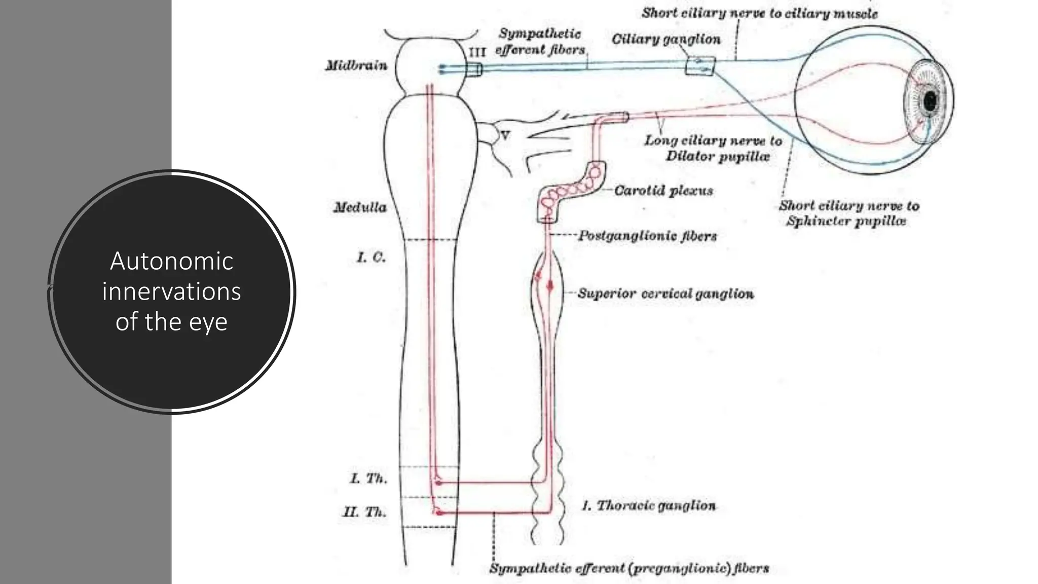
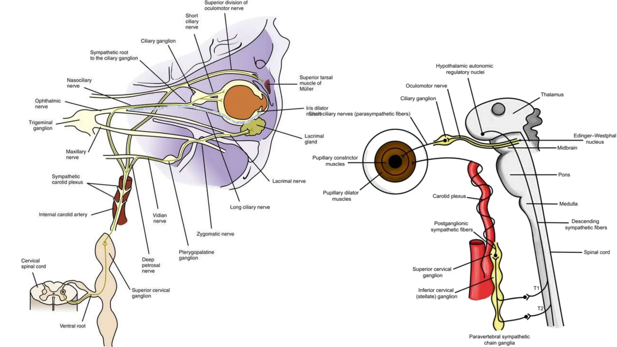
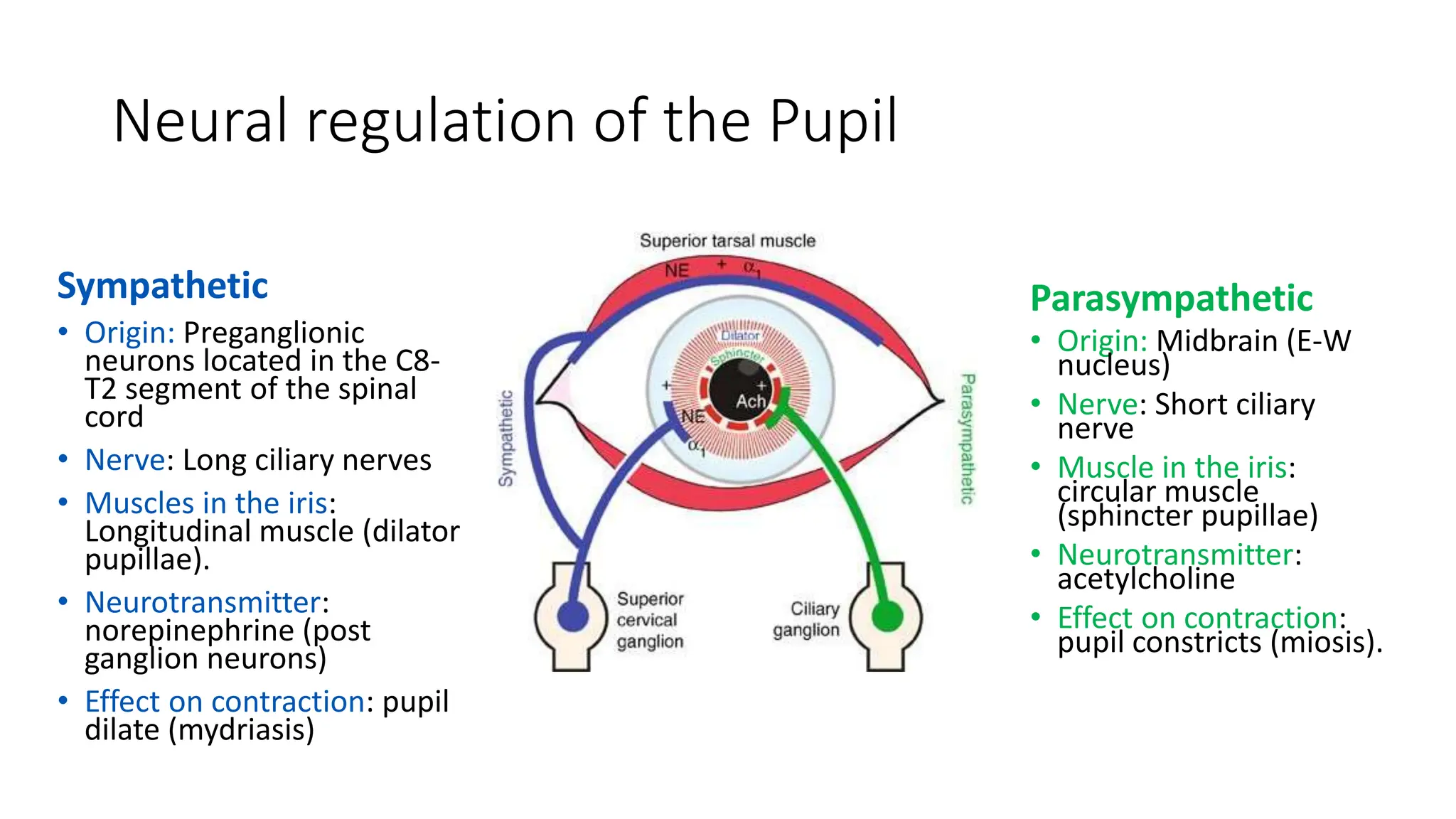
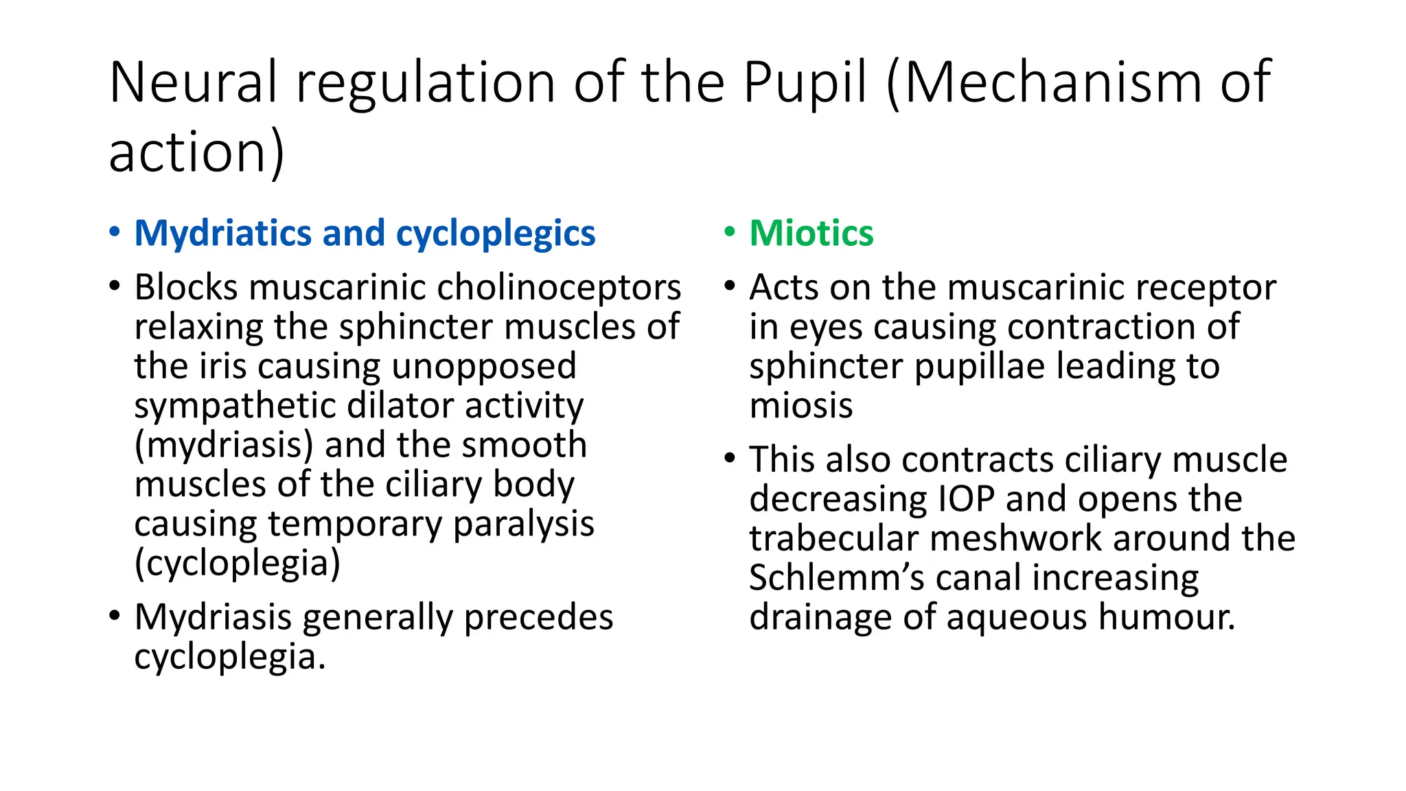
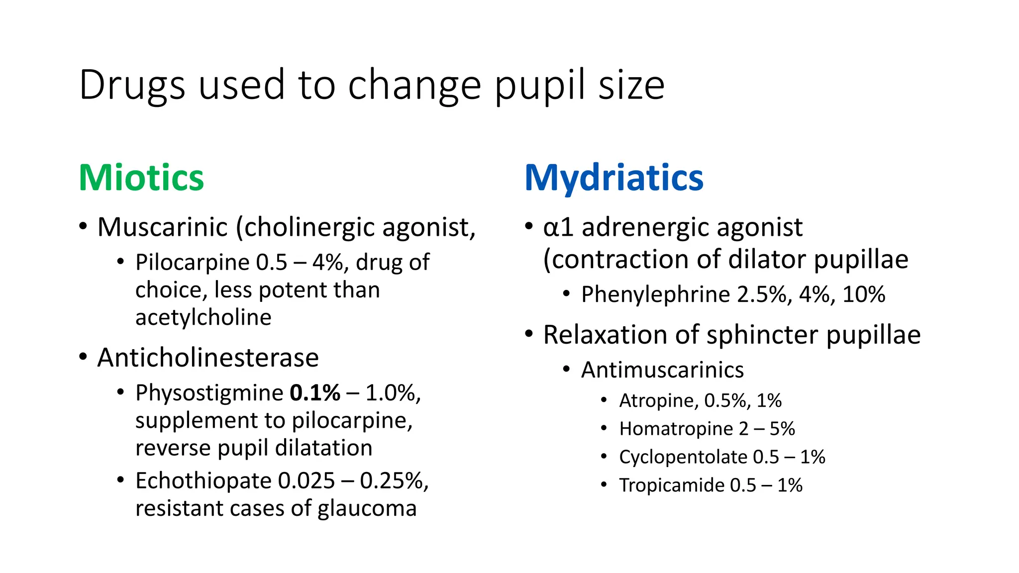
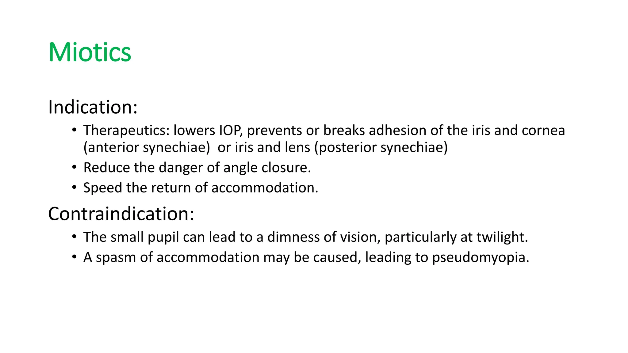
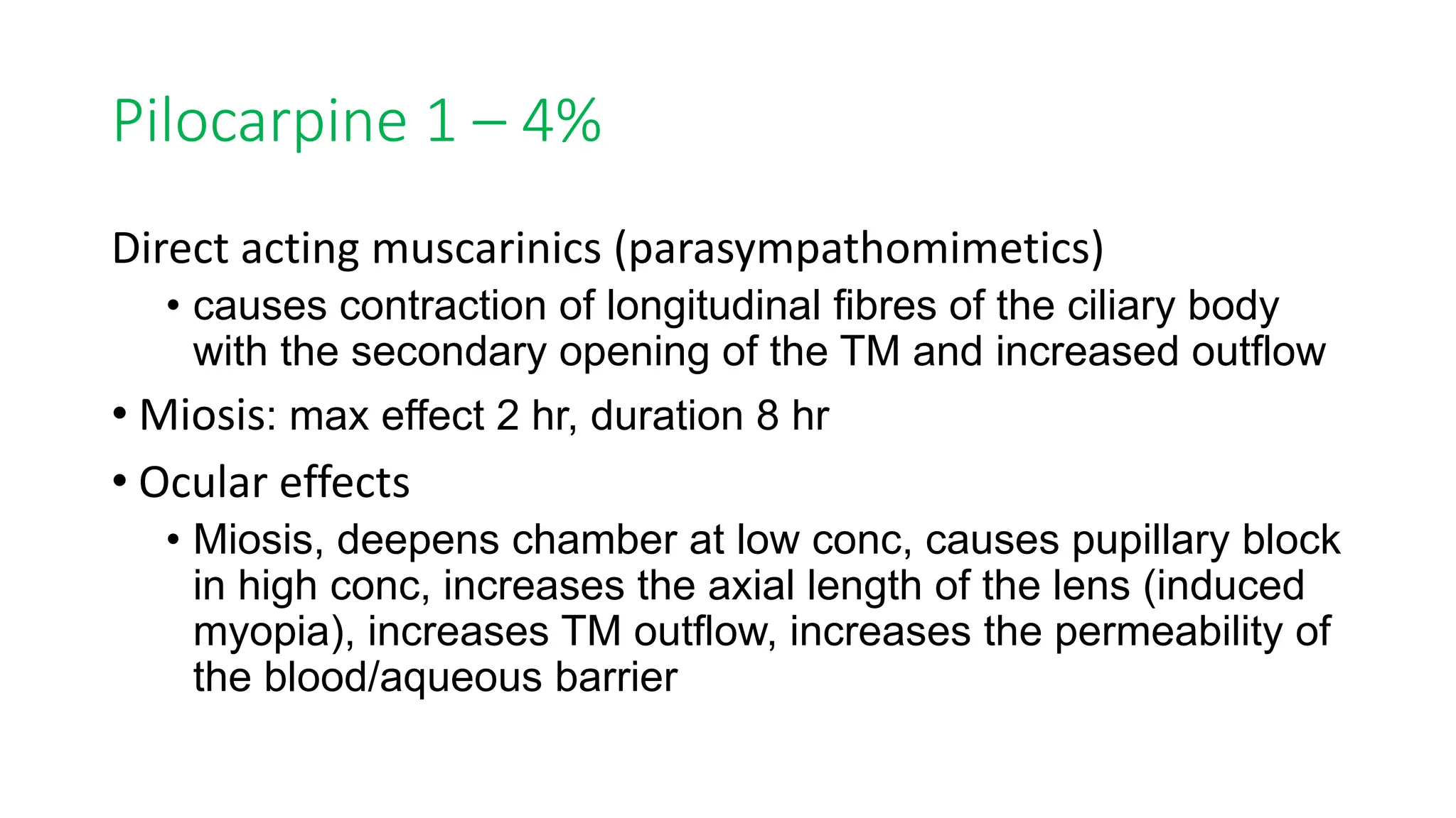
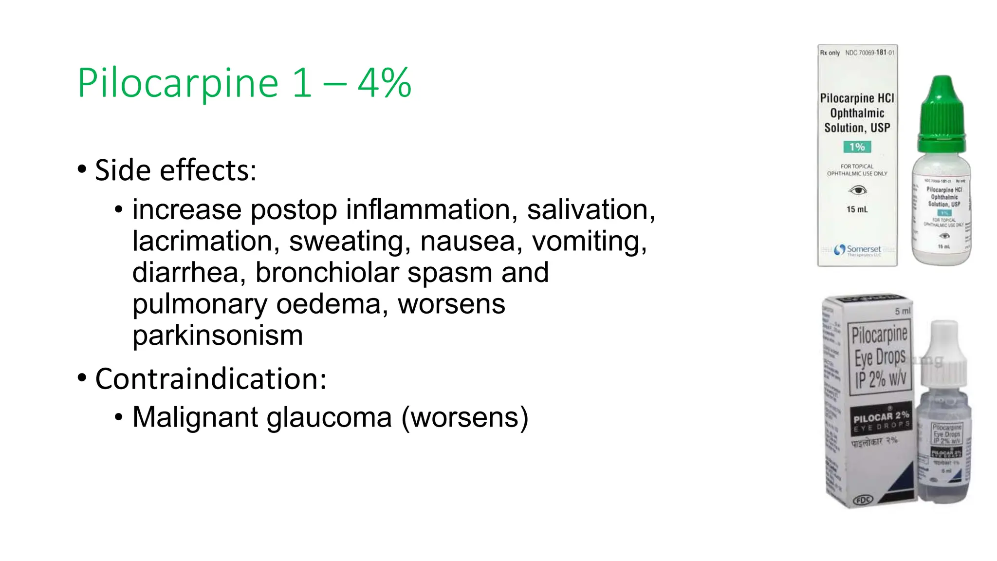
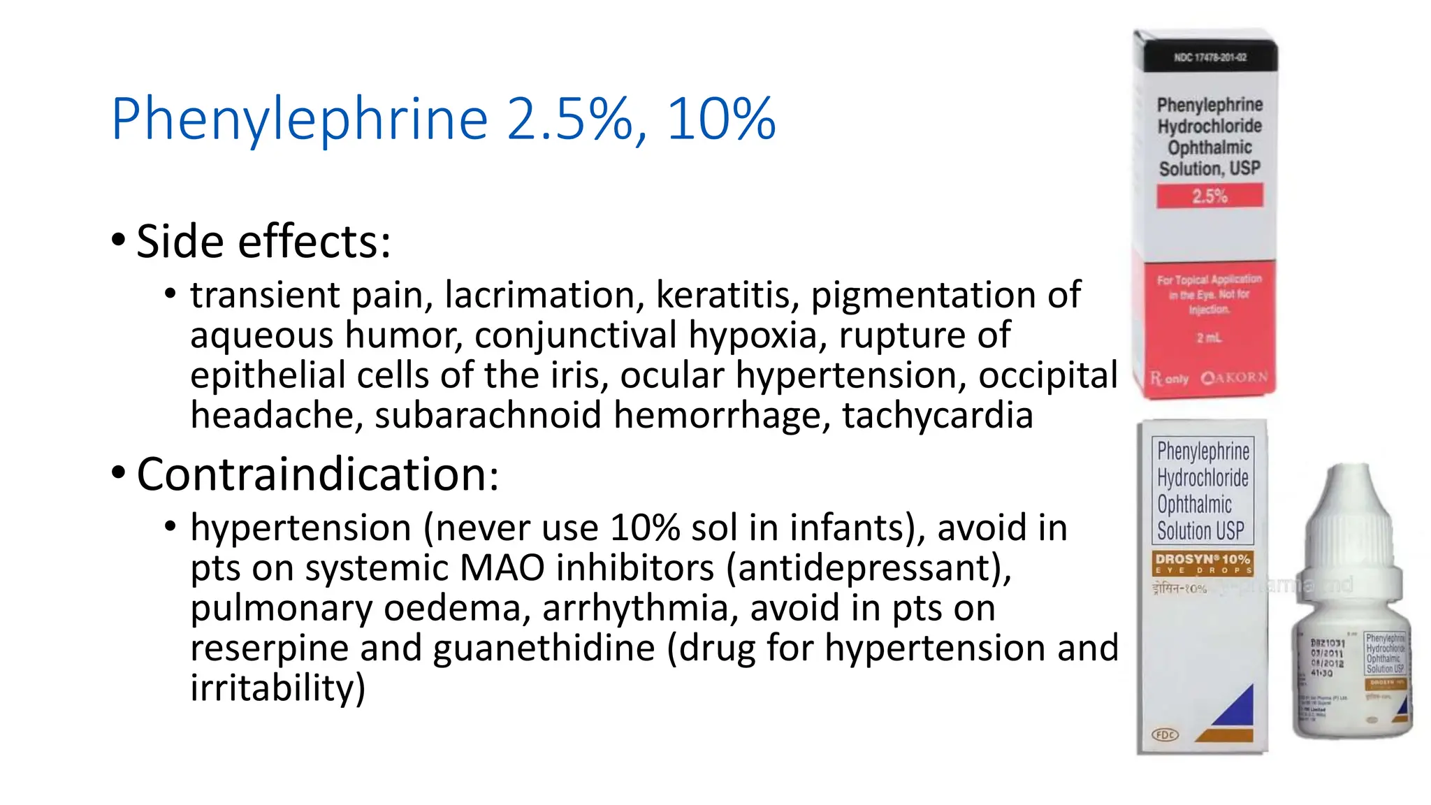
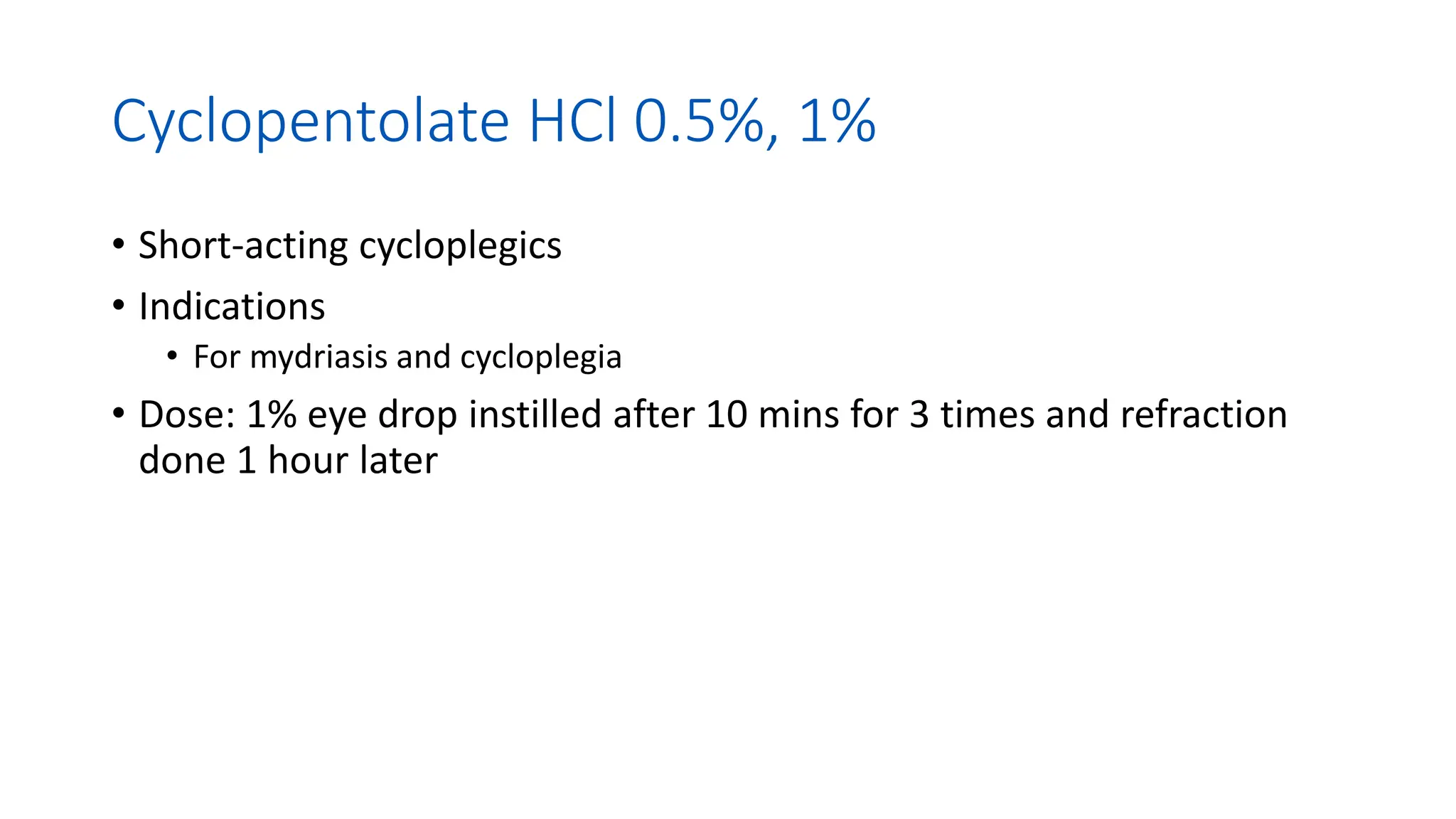
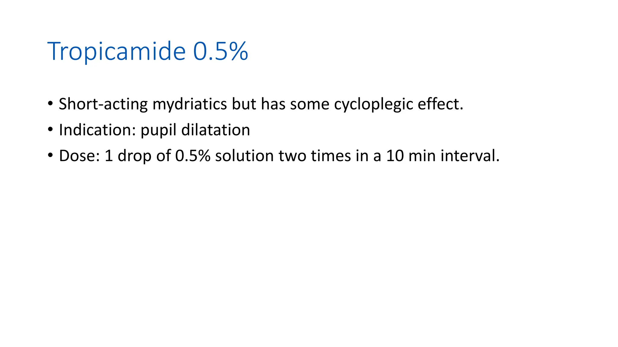
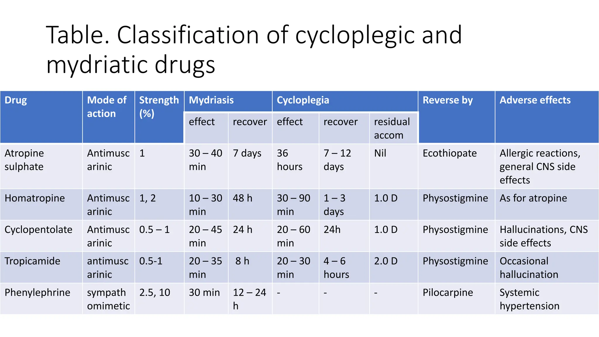
4. Neural regulation of the Pupil
Parasympathetic
• Origin: Midbrain (E-W
nucleus)
• Nerve: Short ciliary
nerve
• Muscle in the iris:
circular muscle
(sphincter pupillae)
• Neurotransmitter:
acetylcholine
• Effect on contraction:
pupil constricts (miosis).
Sympathetic
• Origin: Preganglionic
neurons located in the C8-
T2 segment of the spinal
cord
• Nerve: Long ciliary nerves
• Muscles in the iris:
Longitudinal muscle (dilator
pupillae).
• Neurotransmitter:
norepinephrine (post
ganglion neurons)
• Effect on contraction: pupil
dilate (mydriasis)
5. Neural regulation of the Pupil (Mechanism of
action)
• Mydriatics and cycloplegics
• Blocks muscarinic cholinoceptors
relaxing the sphincter muscles of
the iris causing unopposed
sympathetic dilator activity
(mydriasis) and the smooth
muscles of the ciliary body
causing temporary paralysis
(cycloplegia)
• Mydriasis generally precedes
cycloplegia.
• Miotics
• Acts on the muscarinic receptor
in eyes causing contraction of
sphincter pupillae leading to
miosis
• This also contracts ciliary muscle
decreasing IOP and opens the
trabecular meshwork around the
Schlemm’s canal increasing
drainage of aqueous humour.
6. Drugs used to change pupil size
Miotics
• Muscarinic (cholinergic agonist,
• Pilocarpine 0.5 – 4%, drug of
choice, less potent than
acetylcholine
• Anticholinesterase
• Physostigmine 0.1% – 1.0%,
supplement to pilocarpine,
reverse pupil dilatation
• Echothiopate 0.025 – 0.25%,
resistant cases of glaucoma
Mydriatics
• α1 adrenergic agonist
(contraction of dilator pupillae
• Phenylephrine 2.5%, 4%, 10%
• Relaxation of sphincter pupillae
• Antimuscarinics
• Atropine, 0.5%, 1%
• Homatropine 2 – 5%
• Cyclopentolate 0.5 – 1%
• Tropicamide 0.5 – 1%
7. Miotics
Indication:
• Therapeutics: lowers IOP, prevents or breaks adhesion of the iris and cornea
(anterior synechiae) or iris and lens (posterior synechiae)
• Reduce the danger of angle closure.
• Speed the return of accommodation.
Contraindication:
• The small pupil can lead to a dimness of vision, particularly at twilight.
• A spasm of accommodation may be caused, leading to pseudomyopia.
8. Pilocarpine 1 – 4%
Direct acting muscarinics (parasympathomimetics)
• causes contraction of longitudinal fibres of the ciliary body
with the secondary opening of the TM and increased outflow
• Miosis: max effect 2 hr, duration 8 hr
• Ocular effects
• Miosis, deepens chamber at low conc, causes pupillary block
in high conc, increases the axial length of the lens (induced
myopia), increases TM outflow, increases the permeability of
the blood/aqueous barrier
10. Mydriatics
Indication
• Onset of vitreous opacities, with the symptoms of flashing light.
• Relatively sudden decrease in visual acuity.
• Unexplained loss of visual field.
• Unexplained ocular pain, unaccompanied by raised intraocular pressure.
• Redness of the eye with no infection, allergy, or raised IOP.
• After contusion, to exclude the presence of ocular damage.
• Difficulty in fundus examination due to media opacities
• Diabetic patients – annually as a general rule.
• Fundus photography
11. Mydriatics
Contraindication
• patients using pilocarpine for the treatment of glaucoma
• narrow-angle glaucoma
• abnormally shallow anterior chamber (due to the risk of angle-closure
glaucoma)
• dislocation of the crystalline, or an intraocular lens
• an intraocular lens of the anterior chamber or iris-supported type.
12. Phenylephrine 2.5%, 10%
• An alpha-receptor agonist
• A potent vasoconstrictor (Blanching test) and mydriatics
• Mydriasis - onset 30-60 min; for 2.5% solution half time 3
hours, for 10% solution half time 6 hours
• Side effect: rise in blood pressure
• Indication:
• decongestant and vasoconstrictor, pupil dilation, intraocular
surgeries
• Diagnosis of Horner syndrome
13. Phenylephrine 2.5%, 10%
• Side effects:
• transient pain, lacrimation, keratitis, pigmentation of
aqueous humor, conjunctival hypoxia, rupture of
epithelial cells of the iris, ocular hypertension, occipital
headache, subarachnoid hemorrhage, tachycardia
• Contraindication:
• hypertension (never use 10% sol in infants), avoid in
pts on systemic MAO inhibitors (antidepressant),
pulmonary oedema, arrhythmia, avoid in pts on
reserpine and guanethidine (drug for hypertension and
irritability)
14. Cycloplegics
Indication
• children with constant or intermittent esotropia
• children and young adults with asthenopia and esophoria, especially when a
latent refractive error is suspected
• when retinoscopy suggests that accommodation is fluctuating significantly
• when the retinoscopy findings differ significantly from the findings of subjective
refraction
• in cases of anomalies of accommodation such as accommodative insufficiency,
accommodative fatigue, accommodative inertia, and spasm of accommodation
• in cases where retinoscopy is very difficult due to a lack of patient cooperation
• candidates for refractive surgery.
16. Atropine sulphate 0.5%, 1.0%
• A natural alkaloid (an extract of the belladonna plant)
• Readily absorbs from the gastrointestinal tract, mucous membrane and the
eye
• Metabolise in the liver and excreted through urine.
• Indication:
• Cycloplegic refraction in children below age 4 years (dose: 1% ointment TID for 3
days for complete cycloplegia)
• Treatment of amblyopia (penalization)
• Relieves pain associated with eye inflammation by relaxing the ciliary muscle spasm,
prevents synechiae formation by pupil dilation, and reduces protein and cell
precipitation from inflamed vessels in the anterior chamber.
• Recently, indicated for myopia treatment to prevent myopia progression
• Atropine poisoning: physostigmaine salicylate 1 – 4 mg SQ or IV
17. Homatropine HBr 2%
• Long-acting cycloplegics
• Better than cyclopentolate eye drop
• Indication
• Cycloplegic refraction
• Doses: one drop instilled every 10 minutes for 3 times for cycloplegic
refraction
18. Cyclopentolate HCl 0.5%, 1%
• Short-acting cycloplegics
• Indications
• For mydriasis and cycloplegia
• Dose: 1% eye drop instilled after 10 mins for 3 times and refraction
done 1 hour later
19. Tropicamide 0.5%
• Short-acting mydriatics but has some cycloplegic effect.
• Indication: pupil dilatation
• Dose: 1 drop of 0.5% solution two times in a 10 min interval.
20. Table. Classification of cycloplegic and
mydriatic drugs
Drug Mode of
action
Strength
(%)
Mydriasis Cycloplegia Reverse by Adverse effects
effect recover effect recover residual
accom
Atropine
sulphate
Antimusc
arinic
1 30 – 40
min
7 days 36
hours
7 – 12
days
Nil Ecothiopate Allergic reactions,
general CNS side
effects
Homatropine Antimusc
arinic
1, 2 10 – 30
min
48 h 30 – 90
min
1 – 3
days
1.0 D Physostigmine As for atropine
Cyclopentolate Antimusc
arinic
0.5 – 1 20 – 45
min
24 h 20 – 60
min
24h 1.0 D Physostigmine Hallucinations, CNS
side effects
Tropicamide antimusc
arinic
0.5-1 20 – 35
min
8 h 20 – 30
min
4 – 6
hours
2.0 D Physostigmine Occasional
hallucination
Phenylephrine sympath
omimetic
2.5, 10 30 min 12 – 24
h
- - - Pilocarpine Systemic
hypertension
Miotics
Side effects:
accommodative spasm, sweating, salivation, bronchospasm, nausea and vomiting, CNS disturbances, decrease vision in low illumination
Precaution:
pregnancy, lactation, hypersensitivity reactions
Mydriatics and Cycloplegics
Side effects:
photophobia, blurring of near vision, increase IOP, contact dermatitis, dryness of mouth and skin, fever, rash, thirst, tachycardia, irritability, hyperactivity, confusion, urinary retention, restlessness, hallucination
Precaution
Narrow anterior angle, down’s syndrome, intermittent convergent strabismus, use in lactating mother, blond children, spastic paralysis, brain damage
It nevertheless introduces the danger of pupil block
Avoid photophobia, especially on a bright, sunny day.
Alternative: relaxation of dilator pupillae (α1 adrenergic antagonists)
It nevertheless introduces the danger of pupil block
Avoid photophobia, especially on a bright, sunny day.
Onset of vitreous opacities, accompanied by the symptoms of a flashing light.
Relatively sudden decrease in visual acuity.
Unexplained loss of visual field.
Unexplained ocular pain, unaccompanied by raised intraocular pressure.
Redness of the eye that cannot be attributed to infection, allergy, or raised IOP.
After contusion, to exclude the presence of ocular damage.
Difficulty in observing the fundus due to reduced transparency of the media.
Diabetic patients – annually as a general rule.
Fundus photography, although some cameras are now designed to be non-mydriatic.
Horner syndrome is a rare condition classically presenting with partial ptosis (drooping or falling of the upper eyelid), miosis (constricted pupil), and facial anhidrosis (absence of sweating) due to a disruption in the sympathetic nerve supply.