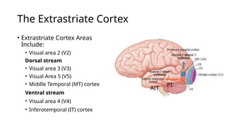
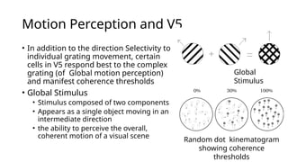
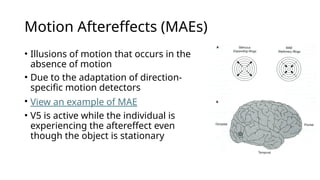
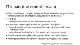
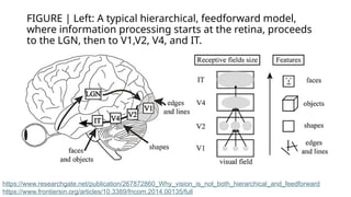
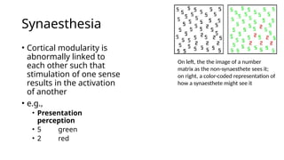
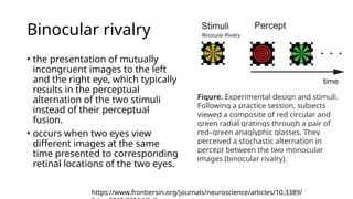
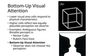
Extrastriate cortex
Dr Gauri Sr Shrestha
MMC, IOM, TU
Learning objectives
• Overview the extrastriate cortex.
• Understand the extrastriate cortex inputs and projections.
• Elaborate dorsal and ventral processing streams.
• Understand visual integration and attention associated with
the extrastriate cortex.
• Define the types of visual agnosia.
The Extrastriate cortex
• Attributes to analysis of specific visual stimuli and have a
degree of functional specificity relating to particular stimulus
attributes
• e.g., color, form, movement, texture, binocular disparity etc
• Human have around 20 cortical visual associated areas
• V1 and extrastriate cortices together occupy ~ 30–40% of the
cortex.
• V1 ~ 12 – 18 %
• Retinotopic organisation is cruder than that in V1
• No distinguished by Line of Gennari
Visual
processin
g in the
nervous
system
The Extrastriate Cortex
• Extrastriate Cortex Areas
Include:
• Visual area 2 (V2)
Dorsal stream
• Visual area 3 (V3)
• Visual Area 5 (V5)
• Middle Temporal (MT) cortex
Ventral stream
• Visual area 4 (V4)
• Inferotemporal (IT) cortex
AIT
PI
Processing Streams
Stream Function Input Origin
Passes
through
Destination Pathway
Dorsal
(parietal)
Spatial
location,
motion
perception
M and K
channels
V1
V2, V3,
MT (V5)
Parietal
cortex
Where
(Magno)
Ventral
(temporal)
Object
recognition
P, M,
and K
channels
V1 V2, V4
Temporal
cortex
What
(Parvo)
V2 inputs and projections
• V2 Inputs
• from the same V1 layers and is segregated
according to two pathways:
• (a)CO blobs to thin stripes
• (b)Interblob areas to pale and thick stripes
• V2 projections
• The neurons in the thin and pale
stripes project to V4
• Associated with a ventral stream for form
processing, color, and object recognition.
• The thick stripes project to V5, a dorsal
stream area
• associated with motion processing
• Some thick stripe neurons project to the SC
for controlling saccadic eye movement.
https://entokey.com/the-extrastriate-cortex/
V3 inputs and projections
(for supplementary reading)
• V3 is a narrow area of the cortex in front of V2.
• Its precise location and function are controversial.
• V3 dorsal and ventral halves represent the lower and upper
visual quadrants, respectively
• Dorsal half: direction of movement, stereopsis
• Ventral half: color, orientation
V5/ MT inputs and projections (the
Dorsal stream)
• V5/ MT inputs
• input from direction-selective cells in V1
• Input from the cells located in thick stripes in V2.
• V5 neurones are sélective for:
• orientation of elongated contours
• direction of movement
• binocular disparity
• wide-field motion contrast.
• V5 has connections with frontal eye fields important in generating smooth
pursuit movements
• V5 also receives inputs from the pulvinar and koniocellular LGN cells that
bypass V1.
Motion Perception and V5
• In addition to the direction Selectivity to
individual grating movement, certain
cells in V5 respond best to the complex
grating (of Global motion perception)
and manifest coherence thresholds
• Global Stimulus
• Stimulus composed of two components
• Appears as a single object moving in an
intermediate direction
• the ability to perceive the overall,
coherent motion of a visual scene
Global
Stimulus
Random dot kinematogram
showing coherence
thresholds
Motion Aftereffects (MAEs)
• Illusions of motion that occurs in the
absence of motion
• Due to the adaptation of direction-
specific motion detectors
• View an example of MAE
• V5 is active while the individual is
experiencing the aftereffect even
though the object is stationary
V4 inputs and projections (the ventral
stream)
• Receptive fields of V4 neurons are substantially larger than
those of V2 and V3 neurons
• V4 Inputs
• receives direct inputs from V1, V2 (thin stripes and pale stripes), and V3
(ventral half).
• Receives V1 inputs from P, M, and K channels arising from layer 3 CO
blobs and interblob regions.
• V4 projections
• projects to inferotemporal areas involved in detailed object form
analysis (object recognition)
• Cells in V4 are principally concerned with color sensitivity
IT inputs (the ventral stream)
• Cells have large, complex receptive fields selective for particular
combinations of orientation, size, texture, and color.
• IT Input
• receives input from V2, V4, and the prefrontal cortex
• is involved in perception and recognition of objects
• can display invariance to stimulus size, rotation, and visual
field location.
• can detect sophisticated forms (circles, squares, ovals)
• Fusiform face area (FFA)- recognizes faces not other objects
• Parahippocampal area (PPA)- recognizes objects and places
https://entokey.com/the-extrastriate-cortex/
Overview of the extrastriate cortex
Stream
Extrastriate
area
Function
V2
Thin dark stripes: color processing
Thick dark stripes: orientation selectivity and
binocular disparity
Pale stripes: form processing and object recognition
Dorsal
V5/MT
V3
Parietal areas
Direction of movement, binocular disparity
Dorsal V3: direction of movement
Ventral V3: color, orientation
Visuospatial perception and movement planning
Ventral
V4
Inferotempor
al areas (IT)
Color sensitivity, object recognition
Complex receptive field properties, e.g., form
perception and face recognition
FIGURE | Left: A typical hierarchical, feedforward model,
where information processing starts at the retina, proceeds
to the LGN, then to V1,V2, V4, and IT.
https://www.researchgate.net/publication/267872860_Why_vision_is_not_both_hierarchical_and_feedforward
https://www.frontiersin.org/articles/10.3389/fncom.2014.00135/full
Additional inputs to extrastriate cortex
• Some extrastriate cortical areas (e.g., area MT) receive
substantial direct input from dorsal thalamic nuclei
(the lateral geniculate nucleus (LGN) and the pulvinar).
• Laminae of the LGN (K layers) and regions of pulvinar which
project to the extrastriate cortices receive direct input from
the superficial layers of the superior colliculus (SC).
What happens with a lesion in the
cortex ?
• Striate Cortex
• Simple blind spots (scotomas)
• Extrastriate Cortex
• Ventral Processing Stream
• Visual Agnosia – inability to recognize objects**
• Dorsal Processing Stream (superior temporal lobe)
• Visual Neglect – patient ignores area of visual field
Visual agnosia
• Form and pattern
▫ Object agnosia inability to recognize real objects
▫ Agnosia for drawing inability to recognize drawing objects
▫ Prosopagnosia inability to recognize faces
• Color
▫ Color agnosia inability to associate colors with objects
▫ Color anomia inability to name colors
▫ Achromatopsia inability to distinguish hues
• Depth and movements
▫ Visual spatial agnosia inability to perceive stereoscopic vision and
topographical relations
▫ Akinetopsia inability to perceive motion
Prosopagnosia
Synaesthesia
• Cortical modularity is
abnormally linked to
each other such that
stimulation of one sense
results in the activation
of another
• e.g.,
• Presentation
perception
• 5 green
• 2 red
On left, the the image of a number
matrix as the non-synaesthete sees it;
on right, a color-coded representation of
how a synaesthete might see it
Grandmother cells (GC)
• GC is a hypothetical neuron that represents a complex but
specific concept or object
• Remembrance of visualized objects result in a pattern of
activity that is similar to that elicited by actually viewing the
object.
• It activates when a person "sees, hears, or sensibly
discriminates“ a specific entity, such as their grandmother.
• Visual signal is continuously processed until it reaches a
high-level cortical cell whose activity results in visual
consciousness
Visual
integration
• Information from various
cortical areas must be
integrated for our perception
• Example: Viewing a moving
red car
• Motion and position
information from dorsal
stream must be integrated
with color and form sense
from the ventral stream
• The integration occurs in the
Prefrontal cortex
Binocular rivalry
• the presentation of mutually
incongruent images to the left
and the right eye, which typically
results in the perceptual
alternation of the two stimuli
instead of their perceptual
fusion.
• occurs when two eyes view
different images at the same
time presented to corresponding
retinal locations of the two eyes.
Figure. Experimental design and stimuli.
Following a practice session, subjects
viewed a composite of red circular and
green radial gratings through a pair of
red–green anaglyphic glasses. They
perceived a stochastic alternation in
percept between the two monocular
images (binocular rivalry).
https://www.frontiersin.org/journals/neuroscience/articles/10.3389/
Binocular rivalry
• An observer is
presented with two
different images to the
two eyes. During
binocular rivalry, the
observer experiences
alternating perceptions
in a temporally
stochastic fashion,
rather than a constant
mixture of the two
images.
https://link.springer.com/article/10.1007/s41745-017-
Bottom-Up Visual
Attention
• Lower visual area cells respond to
physical characteristics
• Higher cells reflect two equally
plausible perception we observe
• Examples: Ambiguous figures:
Bistable percepts in
• Necker Cube
• Rubin’s Face-Vase
• Bottom-Up Visual Attention
• Observer does not ‘choose’ the
perception
Top-Down Visual Attention
• We perform better on certain tasks when we are attentive to
the demand of those tasks
• Subject chooses which object in the cell’s receptive field to
focus visual attention on
• Cortical cells reflect this conscious choice of attention
• Since this choice comes from higher areas in the brain, it is
referred to Top-Down Visual Attention
Can vision be trained?
Visual training-
associated
attention may
improve visual
skills
Visual
performance of
adult can be
improved with
practice-
perceptual
learning but not
resolution task.
Learning certain
task can be
selective other
generalizable
Continue
exposure to
subliminal
stimuli can
influence
behavior.
Thank you

4. Visual Processing in the Extrastriate Cortex.pptx

  • 1.
    Extrastriate cortex Dr GauriSr Shrestha MMC, IOM, TU
  • 2.
    Learning objectives • Overviewthe extrastriate cortex. • Understand the extrastriate cortex inputs and projections. • Elaborate dorsal and ventral processing streams. • Understand visual integration and attention associated with the extrastriate cortex. • Define the types of visual agnosia.
  • 3.
    The Extrastriate cortex •Attributes to analysis of specific visual stimuli and have a degree of functional specificity relating to particular stimulus attributes • e.g., color, form, movement, texture, binocular disparity etc • Human have around 20 cortical visual associated areas • V1 and extrastriate cortices together occupy ~ 30–40% of the cortex. • V1 ~ 12 – 18 % • Retinotopic organisation is cruder than that in V1 • No distinguished by Line of Gennari
  • 4.
  • 5.
    The Extrastriate Cortex •Extrastriate Cortex Areas Include: • Visual area 2 (V2) Dorsal stream • Visual area 3 (V3) • Visual Area 5 (V5) • Middle Temporal (MT) cortex Ventral stream • Visual area 4 (V4) • Inferotemporal (IT) cortex AIT PI
  • 6.
    Processing Streams Stream FunctionInput Origin Passes through Destination Pathway Dorsal (parietal) Spatial location, motion perception M and K channels V1 V2, V3, MT (V5) Parietal cortex Where (Magno) Ventral (temporal) Object recognition P, M, and K channels V1 V2, V4 Temporal cortex What (Parvo)
  • 7.
    V2 inputs andprojections • V2 Inputs • from the same V1 layers and is segregated according to two pathways: • (a)CO blobs to thin stripes • (b)Interblob areas to pale and thick stripes • V2 projections • The neurons in the thin and pale stripes project to V4 • Associated with a ventral stream for form processing, color, and object recognition. • The thick stripes project to V5, a dorsal stream area • associated with motion processing • Some thick stripe neurons project to the SC for controlling saccadic eye movement. https://entokey.com/the-extrastriate-cortex/
  • 8.
    V3 inputs andprojections (for supplementary reading) • V3 is a narrow area of the cortex in front of V2. • Its precise location and function are controversial. • V3 dorsal and ventral halves represent the lower and upper visual quadrants, respectively • Dorsal half: direction of movement, stereopsis • Ventral half: color, orientation
  • 9.
    V5/ MT inputsand projections (the Dorsal stream) • V5/ MT inputs • input from direction-selective cells in V1 • Input from the cells located in thick stripes in V2. • V5 neurones are sélective for: • orientation of elongated contours • direction of movement • binocular disparity • wide-field motion contrast. • V5 has connections with frontal eye fields important in generating smooth pursuit movements • V5 also receives inputs from the pulvinar and koniocellular LGN cells that bypass V1.
  • 10.
    Motion Perception andV5 • In addition to the direction Selectivity to individual grating movement, certain cells in V5 respond best to the complex grating (of Global motion perception) and manifest coherence thresholds • Global Stimulus • Stimulus composed of two components • Appears as a single object moving in an intermediate direction • the ability to perceive the overall, coherent motion of a visual scene Global Stimulus Random dot kinematogram showing coherence thresholds
  • 11.
    Motion Aftereffects (MAEs) •Illusions of motion that occurs in the absence of motion • Due to the adaptation of direction- specific motion detectors • View an example of MAE • V5 is active while the individual is experiencing the aftereffect even though the object is stationary
  • 13.
    V4 inputs andprojections (the ventral stream) • Receptive fields of V4 neurons are substantially larger than those of V2 and V3 neurons • V4 Inputs • receives direct inputs from V1, V2 (thin stripes and pale stripes), and V3 (ventral half). • Receives V1 inputs from P, M, and K channels arising from layer 3 CO blobs and interblob regions. • V4 projections • projects to inferotemporal areas involved in detailed object form analysis (object recognition) • Cells in V4 are principally concerned with color sensitivity
  • 14.
    IT inputs (theventral stream) • Cells have large, complex receptive fields selective for particular combinations of orientation, size, texture, and color. • IT Input • receives input from V2, V4, and the prefrontal cortex • is involved in perception and recognition of objects • can display invariance to stimulus size, rotation, and visual field location. • can detect sophisticated forms (circles, squares, ovals) • Fusiform face area (FFA)- recognizes faces not other objects • Parahippocampal area (PPA)- recognizes objects and places https://entokey.com/the-extrastriate-cortex/
  • 15.
    Overview of theextrastriate cortex Stream Extrastriate area Function V2 Thin dark stripes: color processing Thick dark stripes: orientation selectivity and binocular disparity Pale stripes: form processing and object recognition Dorsal V5/MT V3 Parietal areas Direction of movement, binocular disparity Dorsal V3: direction of movement Ventral V3: color, orientation Visuospatial perception and movement planning Ventral V4 Inferotempor al areas (IT) Color sensitivity, object recognition Complex receptive field properties, e.g., form perception and face recognition
  • 16.
    FIGURE | Left:A typical hierarchical, feedforward model, where information processing starts at the retina, proceeds to the LGN, then to V1,V2, V4, and IT. https://www.researchgate.net/publication/267872860_Why_vision_is_not_both_hierarchical_and_feedforward https://www.frontiersin.org/articles/10.3389/fncom.2014.00135/full
  • 17.
    Additional inputs toextrastriate cortex • Some extrastriate cortical areas (e.g., area MT) receive substantial direct input from dorsal thalamic nuclei (the lateral geniculate nucleus (LGN) and the pulvinar). • Laminae of the LGN (K layers) and regions of pulvinar which project to the extrastriate cortices receive direct input from the superficial layers of the superior colliculus (SC).
  • 18.
    What happens witha lesion in the cortex ? • Striate Cortex • Simple blind spots (scotomas) • Extrastriate Cortex • Ventral Processing Stream • Visual Agnosia – inability to recognize objects** • Dorsal Processing Stream (superior temporal lobe) • Visual Neglect – patient ignores area of visual field
  • 19.
    Visual agnosia • Formand pattern ▫ Object agnosia inability to recognize real objects ▫ Agnosia for drawing inability to recognize drawing objects ▫ Prosopagnosia inability to recognize faces • Color ▫ Color agnosia inability to associate colors with objects ▫ Color anomia inability to name colors ▫ Achromatopsia inability to distinguish hues • Depth and movements ▫ Visual spatial agnosia inability to perceive stereoscopic vision and topographical relations ▫ Akinetopsia inability to perceive motion Prosopagnosia
  • 20.
    Synaesthesia • Cortical modularityis abnormally linked to each other such that stimulation of one sense results in the activation of another • e.g., • Presentation perception • 5 green • 2 red On left, the the image of a number matrix as the non-synaesthete sees it; on right, a color-coded representation of how a synaesthete might see it
  • 21.
    Grandmother cells (GC) •GC is a hypothetical neuron that represents a complex but specific concept or object • Remembrance of visualized objects result in a pattern of activity that is similar to that elicited by actually viewing the object. • It activates when a person "sees, hears, or sensibly discriminates“ a specific entity, such as their grandmother. • Visual signal is continuously processed until it reaches a high-level cortical cell whose activity results in visual consciousness
  • 22.
    Visual integration • Information fromvarious cortical areas must be integrated for our perception • Example: Viewing a moving red car • Motion and position information from dorsal stream must be integrated with color and form sense from the ventral stream • The integration occurs in the Prefrontal cortex
  • 23.
    Binocular rivalry • thepresentation of mutually incongruent images to the left and the right eye, which typically results in the perceptual alternation of the two stimuli instead of their perceptual fusion. • occurs when two eyes view different images at the same time presented to corresponding retinal locations of the two eyes. Figure. Experimental design and stimuli. Following a practice session, subjects viewed a composite of red circular and green radial gratings through a pair of red–green anaglyphic glasses. They perceived a stochastic alternation in percept between the two monocular images (binocular rivalry). https://www.frontiersin.org/journals/neuroscience/articles/10.3389/
  • 24.
    Binocular rivalry • Anobserver is presented with two different images to the two eyes. During binocular rivalry, the observer experiences alternating perceptions in a temporally stochastic fashion, rather than a constant mixture of the two images. https://link.springer.com/article/10.1007/s41745-017-
  • 25.
    Bottom-Up Visual Attention • Lowervisual area cells respond to physical characteristics • Higher cells reflect two equally plausible perception we observe • Examples: Ambiguous figures: Bistable percepts in • Necker Cube • Rubin’s Face-Vase • Bottom-Up Visual Attention • Observer does not ‘choose’ the perception
  • 26.
    Top-Down Visual Attention •We perform better on certain tasks when we are attentive to the demand of those tasks • Subject chooses which object in the cell’s receptive field to focus visual attention on • Cortical cells reflect this conscious choice of attention • Since this choice comes from higher areas in the brain, it is referred to Top-Down Visual Attention
  • 27.
    Can vision betrained? Visual training- associated attention may improve visual skills Visual performance of adult can be improved with practice- perceptual learning but not resolution task. Learning certain task can be selective other generalizable Continue exposure to subliminal stimuli can influence behavior.
  • 28.

Editor's Notes

  • #3 Striate Cortex Direction selective cells respond as long as bars are present in that cell’s receptive field These cells are still active even when subject reports seeing other bars Visual Area 4 (V4) Many orientation selective cells respond the same as cells in the striate cortex Other cells respond only when the subject reports seeing bars of a specific orientation The cellular response in V4 changes with perception even though the stimulus is constant Higher Cortical Areas Are More Likely To Code What We Actually ‘See’ Than Are The Cells In Lower Visual Areas
  • #9 are dominated by M-cell pathways
  • #10 V5 is more active when viewing a moving object than a stationary object certain cells in V5 respond best to the complex grating and manifest coherence thresholds depicted by global motion perception using global stimulus in Random dot kinematogram
  • #11 Ex: Staring at downward-rushing water for several minutes, then look at the surrounding landscape-It will appear to rise
  • #13 Cells in V4 are principally concerned with color sensitivity; however, cells are also selective for orientation, size, and binocular disparity involved in form and shape perception. Visual attention modulates processing in V4
  • #14 Cells in striated cortex respond best to bars, edges, and gratings
  • #21 Extremely specialized cortical cells Respond to the ‘face of your grandmother’
  • #23 Alternating perception of nonfusable images separately by two eyes
  • #25 Attention: An ability to focus on specific information
  • #27 Subliminal stimuli: any sensory stimuli below an individual's threshold for conscious perception