Color Vision Defect
Dr Gauri Sr Shrestha
Lecturer, MMC, IOM, TU
Learning objectives
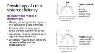
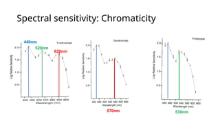
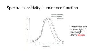
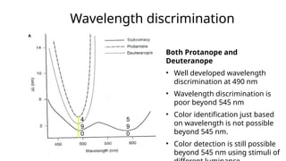
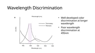
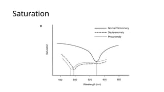
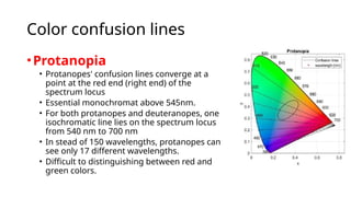
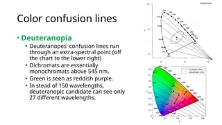
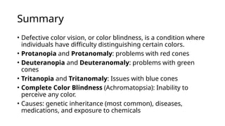
• Understand types of hereditary color vision deficiencies.
• Understand the physiology of color vision deficiencies (spectral
sensitivity, wavelength discrimination and saturation)
• Understand the replacement model of dichromacy.
• Understand color confusion lines, color discrimination, and labelling
associated with color vision deficiencies.
• Understand the differences between hereditary and acquired color
vision deficiencies.
• Understand the transmittance pattern of X-linked red-green defects.
• Understand the Kollner’s rule and its exception to color vision
deficiencies.
Pretest- MCQs
Q1. The most common type of
color vision defect is:
• A) Monochromacy
• B) Deuteranomaly
• C) Tritanopia
• D) Protanopia
Q2. Protanopia is a type of color
vision defect associated with:
• A) Red defect
• B) Blue defect
• C) Green defect
• D) Yellow defect
Pretest- MCQs
Q3 Deuteranomaly primarily
affects the perception of which
colors?
• A) Red and yellow
• B) Blue and green
• C) Red and green
• D) Yellow and blue
Q4. Tritanopia is a rare color
vision defect affecting the
perception of which colors?
• A) Red and green
• B) Blue and yellow
• C) Red and yellow
• D) Green and blue
Pretest- MCQs
Q5. Color vision defects are more
common in men than in women
because:
• A) Men have more rod cells than
cone cells
• B) The defect is carried on the Y
chromosome
• C) Men have only one X
chromosome
• D) Women have better overall vision
Q6. Which type of color vision
defect is characterized by the
inability to perceive any color,
seeing everything in shades of gray?
• A) Deuteranopia
• B) Protanopia
• C) Monochromacy
• D) Tritanopia
Pretest: true/false statement
1. Color vision defects are more common in females than in males.
(True/False)
2. People with deuteranopia cannot distinguish between red and green
colors. (True/False)
3. Tritanopia is a rare type of color vision defect affecting the perception
of blue and yellow colors. (True/False)
4. Color vision defects can be completely corrected with glasses or
contact lenses. (True/False)
Background
• Color vision deficiency (CVD) is the decreased ability to see
color or difference in color.
Types of color vision deficiency
• Monochromacy: Only one pigment type is present
• Dichromacy: Only two pigment types are present
• Anomalous trichromacy: All three pigment types are present
Monochromacy
• Monochromacy non-
progressive complete color
deficiency (blindness)
• Patients with monochromacy
perceives objects with shades
of gray and use other cues to
label colors.
• These patients match any
wavelength in the spectrum
by changing the intensity of
another wavelength.
Monochromacy
• Rod monochromats: the
most common type,
affecting 10 in 1 million
people
• No cones, extremely poor
visual acuity.
• Blue cone monochromats:
colorblindness in photopic
and scotopic conditions
• Narrow range of color
perception in mesopic
conditions
700
ROD
L
M
S
Rod
Monochromats
Normal spectral sensitivity of retinal
photoreceptors
Dichromacy: One of three cones is missing or not
functioning.
• Protanopia
• Missing or defective L-
cones; x-linked
• 1% males, 0.02%
females
• Individuals see short-
wavelengths as blue
• Neutral point occurs at
492 nm
• Above neutral point,
they see yellow.
Dichromacy
• Deuteranopia
• Missing or defective M –
cones; x-linked
• 1% males, 0.01% females
• Individuals see short-
wavelengths as blue
• Neutral point occurs at 498
nm
• their brightness vision is
more like that of color-
normal.
• Above neutral point, they see
yellow.
Dichromacy
• Tritanopia
• Missing or defective S – cones;
autosomal dominant
• 0.002% males, 0.001% females.
• Individual see short-
wavelengths as green
• Neutral point occurs at 570
nm.
• Above neutral point they see
red.
chrome-extension://gphandlahdpffmccakmbngmbjnjiiahp/http://courses.washington.edu/psy333/lecture_pdfs/Week7_Day2.pdf
Neutral Points
• The wavelength where the
two remaining pigments
crosses.
• Colors look very
desaturated and whitish at
that point.
• Patients cannot
distinguish colors at this
area of the spectrum.
• Colors at this wavelength
are perceived as gray.
570
Anomalous Trichromacy - three pigment types, reduced
spectral sensitivity of one cone type. There is no neutral point.
• Protanomaly –
• Shifted red pigments, red-weakness, red-pale
• Red, orange, yellow and yellow-green appear somewhat
shifted in hue towards green.
• Violet or lavender color appears bluish.
• In dazzling sunlight or rain or fog, blinking red traffic light
from blinking yellow may not be appreciable.
• Incidence 1% males.
Anomalous Trichromacy
• Deuteranomaly
• Shifted green pigments, green-weakness, green-pale
• Red, orange, yellow and yellow-green appear somewhat
shifted in hue towards red.
• No loss of brightness unlike in protanomaly.
• Incidence 5% males.
• Tritanomaly
• Shifted blue pigment
• Will be hard to tell the difference between blue and green
and between yellow and red.
Hereditary Color Vision Defects
DEFECT PREV. IN MALES TRANSMISSION
Deuteranopia 1% X-linked recessive
Protanopia 1% X-linked recessive
Deuteranomaly 5% (Most Common) X-linked recessive
Protonomaly 1% X-linked recessive
Tritanopia &
tritanomaly
.005% (Most Rare) Autosomal
Dominant
Physiology of color
vision deficiency
Replacement model of
dichromacy
• Missing photopigment is replaced
by a remaining photopigments
• Deuteranope: missing green
cones are replaced by red-cones
• Protanope: missing red cones are
replaced by green cones
• Exception: missing blue-cones are
not replaced by another
photopigment
Deuteranomal
y:
M-cone
displaced
toward long
wavelengths
Protanomaly:
L-cone displaced
toward short
wavelengths
Spectral sensitivity: Chromaticity
440nm
520nm
620nm
570nm 530nm
Spectral sensitivity: Luminance function
Protanopes can
not see light of
wavelength
above 660nm
Wavelength discrimination
Both Protanope and
Deuteranope
• Well developed wavelength
discrimination at 490 nm
• Wavelength discrimination is
poor beyond 545 nm
• Color identification just based
on wavelength is not possible
beyond 545 nm.
• Color detection is still possible
beyond 545 nm using stimuli of
4
9
0
5
9
0
Wavelength Discrimination
• Well developed color
discrimination at longer
wavelength
• Poor wavelength
discrimination at
495nm
Saturation
• Neutral point: desaturated
appearance of spectral
stimuli at specific
wavelength
570nm
Saturation
Color confusion lines
•Protanopia
• Protanopes' confusion lines converge at a
point at the red end (right end) of the
spectrum locus
• Essential monochromat above 545nm.
• For both protanopes and deuteranopes, one
isochromatic line lies on the spectrum locus
from 540 nm to 700 nm
• In stead of 150 wavelengths, protanopes can
see only 17 different wavelengths.
• Difficult to distinguishing between red and
green colors.
Color confusion lines
• Deuteranopia
• Deuteranopes' confusion lines run
through an extra-spectral point (off
the chart to the lower right)
• Dichromats are essentially
monochromats above 545 nm.
• Green is seen as reddish purple.
• In stead of 150 wavelengths,
deuteranopic candidate can see only
27 different wavelengths.
Color confusion lines
• Tritanopia
• Tritanopes confuse colors (of the
same brightness) that lie along
lines passing through a point near
the blue end of the spectrum locus
in the xy diagram
• Difficult to distinguishing between
blue and green colors and
between yellow and violet.
Practical implication
Color discrimination and labeling
• Color labeling is remarkably good in color vision deficiency.
• However, Industrial color labeling is difficult, such as
stripe/pattern analysis.
Color discrimination and labeling
Color Vision and Females
• Tend to be carriers
• Tend to have normal color vision
• 20 times less likely to have a color vision deficiency
Transmission of X linked, red-green defects
from parents to offspring
X X
X XX XX
Y XY XY
Father Normal (XY)
Mother Normal (XX)
X X
  X  X
Y XY XY
Father Defective ( Y)
Mother Normal (XX)
Daughters Carrier
Sons Normal
 
X  X  X
Y  Y  Y
Father Normal (XY)
Mother Defective ( )
Daughters Carrier
Sons Defective
Transmission of X linked, red-green defects
from parents to offspring
 X
    X
Y  Y XY
 X
X  X X X
Y  Y XY
Father Normal (XY)
Mother carrier ( X)
Daughter: 1 carrier, 1 normal
Sons: 1 defective, 1 normall
Father Defective ( Y)
Mother Carrier ( X)
Daughters: 1 carrier, 1 defective
Sons: 1defective, 1 Normal
Congenital vs. Acquired
CONGENITAL ACQUIRED
Affects both eyes equally One eye only OR asymmetric
Usually a R-G defect B-Y OR R-G
Other visual functions normal Other visual functions abnormal
Stable through lifetime Variable, dependent on test and diseases conditions
Learned to adapt-can label
objects
Cannot name color correctly
More prevalent in male Equally prevalent in male and female
Not associated diseases or
toxicity
Classification not straightforward with standard
clinical color test
Kollner’s Rule
Location Defect condition
Media B-Y Nuclear sclerosis
Outer retina B-Y AMD, DR
Inner retina R-G Optic atrophy, toxic amblyopia
Pathway R-G Lesions
• Outer retinal diseases and media changes result in blue-yellow color vision
defects
• Inner retina, optic nerve visual pathways and visual cortex diseases result in
red-green defects
Exception
to The
Kollner’s
Rule
Summary
• Defective color vision, or color blindness, is a condition where
individuals have difficulty distinguishing certain colors.
• Protanopia and Protanomaly: problems with red cones
• Deuteranopia and Deuteranomaly: problems with green
cones
• Tritanopia and Tritanomaly: Issues with blue cones
• Complete Color Blindness (Achromatopsia): Inability to
perceive any color.
• Causes: genetic inheritance (most common), diseases,
medications, and exposure to chemicals
Thank you

Understanding Color Vision Defects in Human.pptx

  • 1.
    Color Vision Defect DrGauri Sr Shrestha Lecturer, MMC, IOM, TU
  • 2.
    Learning objectives • Understandtypes of hereditary color vision deficiencies. • Understand the physiology of color vision deficiencies (spectral sensitivity, wavelength discrimination and saturation) • Understand the replacement model of dichromacy. • Understand color confusion lines, color discrimination, and labelling associated with color vision deficiencies. • Understand the differences between hereditary and acquired color vision deficiencies. • Understand the transmittance pattern of X-linked red-green defects. • Understand the Kollner’s rule and its exception to color vision deficiencies.
  • 3.
    Pretest- MCQs Q1. Themost common type of color vision defect is: • A) Monochromacy • B) Deuteranomaly • C) Tritanopia • D) Protanopia Q2. Protanopia is a type of color vision defect associated with: • A) Red defect • B) Blue defect • C) Green defect • D) Yellow defect
  • 4.
    Pretest- MCQs Q3 Deuteranomalyprimarily affects the perception of which colors? • A) Red and yellow • B) Blue and green • C) Red and green • D) Yellow and blue Q4. Tritanopia is a rare color vision defect affecting the perception of which colors? • A) Red and green • B) Blue and yellow • C) Red and yellow • D) Green and blue
  • 5.
    Pretest- MCQs Q5. Colorvision defects are more common in men than in women because: • A) Men have more rod cells than cone cells • B) The defect is carried on the Y chromosome • C) Men have only one X chromosome • D) Women have better overall vision Q6. Which type of color vision defect is characterized by the inability to perceive any color, seeing everything in shades of gray? • A) Deuteranopia • B) Protanopia • C) Monochromacy • D) Tritanopia
  • 6.
    Pretest: true/false statement 1.Color vision defects are more common in females than in males. (True/False) 2. People with deuteranopia cannot distinguish between red and green colors. (True/False) 3. Tritanopia is a rare type of color vision defect affecting the perception of blue and yellow colors. (True/False) 4. Color vision defects can be completely corrected with glasses or contact lenses. (True/False)
  • 7.
    Background • Color visiondeficiency (CVD) is the decreased ability to see color or difference in color. Types of color vision deficiency • Monochromacy: Only one pigment type is present • Dichromacy: Only two pigment types are present • Anomalous trichromacy: All three pigment types are present
  • 8.
    Monochromacy • Monochromacy non- progressivecomplete color deficiency (blindness) • Patients with monochromacy perceives objects with shades of gray and use other cues to label colors. • These patients match any wavelength in the spectrum by changing the intensity of another wavelength.
  • 9.
    Monochromacy • Rod monochromats:the most common type, affecting 10 in 1 million people • No cones, extremely poor visual acuity. • Blue cone monochromats: colorblindness in photopic and scotopic conditions • Narrow range of color perception in mesopic conditions 700 ROD L M S Rod Monochromats Normal spectral sensitivity of retinal photoreceptors
  • 10.
    Dichromacy: One ofthree cones is missing or not functioning. • Protanopia • Missing or defective L- cones; x-linked • 1% males, 0.02% females • Individuals see short- wavelengths as blue • Neutral point occurs at 492 nm • Above neutral point, they see yellow.
  • 11.
    Dichromacy • Deuteranopia • Missingor defective M – cones; x-linked • 1% males, 0.01% females • Individuals see short- wavelengths as blue • Neutral point occurs at 498 nm • their brightness vision is more like that of color- normal. • Above neutral point, they see yellow.
  • 12.
    Dichromacy • Tritanopia • Missingor defective S – cones; autosomal dominant • 0.002% males, 0.001% females. • Individual see short- wavelengths as green • Neutral point occurs at 570 nm. • Above neutral point they see red. chrome-extension://gphandlahdpffmccakmbngmbjnjiiahp/http://courses.washington.edu/psy333/lecture_pdfs/Week7_Day2.pdf
  • 13.
    Neutral Points • Thewavelength where the two remaining pigments crosses. • Colors look very desaturated and whitish at that point. • Patients cannot distinguish colors at this area of the spectrum. • Colors at this wavelength are perceived as gray. 570
  • 14.
    Anomalous Trichromacy -three pigment types, reduced spectral sensitivity of one cone type. There is no neutral point. • Protanomaly – • Shifted red pigments, red-weakness, red-pale • Red, orange, yellow and yellow-green appear somewhat shifted in hue towards green. • Violet or lavender color appears bluish. • In dazzling sunlight or rain or fog, blinking red traffic light from blinking yellow may not be appreciable. • Incidence 1% males.
  • 15.
    Anomalous Trichromacy • Deuteranomaly •Shifted green pigments, green-weakness, green-pale • Red, orange, yellow and yellow-green appear somewhat shifted in hue towards red. • No loss of brightness unlike in protanomaly. • Incidence 5% males. • Tritanomaly • Shifted blue pigment • Will be hard to tell the difference between blue and green and between yellow and red.
  • 16.
    Hereditary Color VisionDefects DEFECT PREV. IN MALES TRANSMISSION Deuteranopia 1% X-linked recessive Protanopia 1% X-linked recessive Deuteranomaly 5% (Most Common) X-linked recessive Protonomaly 1% X-linked recessive Tritanopia & tritanomaly .005% (Most Rare) Autosomal Dominant
  • 17.
    Physiology of color visiondeficiency Replacement model of dichromacy • Missing photopigment is replaced by a remaining photopigments • Deuteranope: missing green cones are replaced by red-cones • Protanope: missing red cones are replaced by green cones • Exception: missing blue-cones are not replaced by another photopigment Deuteranomal y: M-cone displaced toward long wavelengths Protanomaly: L-cone displaced toward short wavelengths
  • 18.
  • 19.
    Spectral sensitivity: Luminancefunction Protanopes can not see light of wavelength above 660nm
  • 20.
    Wavelength discrimination Both Protanopeand Deuteranope • Well developed wavelength discrimination at 490 nm • Wavelength discrimination is poor beyond 545 nm • Color identification just based on wavelength is not possible beyond 545 nm. • Color detection is still possible beyond 545 nm using stimuli of 4 9 0 5 9 0
  • 21.
    Wavelength Discrimination • Welldeveloped color discrimination at longer wavelength • Poor wavelength discrimination at 495nm
  • 22.
    Saturation • Neutral point:desaturated appearance of spectral stimuli at specific wavelength 570nm
  • 23.
  • 24.
    Color confusion lines •Protanopia •Protanopes' confusion lines converge at a point at the red end (right end) of the spectrum locus • Essential monochromat above 545nm. • For both protanopes and deuteranopes, one isochromatic line lies on the spectrum locus from 540 nm to 700 nm • In stead of 150 wavelengths, protanopes can see only 17 different wavelengths. • Difficult to distinguishing between red and green colors.
  • 25.
    Color confusion lines •Deuteranopia • Deuteranopes' confusion lines run through an extra-spectral point (off the chart to the lower right) • Dichromats are essentially monochromats above 545 nm. • Green is seen as reddish purple. • In stead of 150 wavelengths, deuteranopic candidate can see only 27 different wavelengths.
  • 26.
    Color confusion lines •Tritanopia • Tritanopes confuse colors (of the same brightness) that lie along lines passing through a point near the blue end of the spectrum locus in the xy diagram • Difficult to distinguishing between blue and green colors and between yellow and violet.
  • 27.
  • 28.
    Color discrimination andlabeling • Color labeling is remarkably good in color vision deficiency. • However, Industrial color labeling is difficult, such as stripe/pattern analysis.
  • 29.
  • 30.
    Color Vision andFemales • Tend to be carriers • Tend to have normal color vision • 20 times less likely to have a color vision deficiency
  • 31.
    Transmission of Xlinked, red-green defects from parents to offspring X X X XX XX Y XY XY Father Normal (XY) Mother Normal (XX) X X   X  X Y XY XY Father Defective ( Y) Mother Normal (XX) Daughters Carrier Sons Normal   X  X  X Y  Y  Y Father Normal (XY) Mother Defective ( ) Daughters Carrier Sons Defective
  • 32.
    Transmission of Xlinked, red-green defects from parents to offspring  X     X Y  Y XY  X X  X X X Y  Y XY Father Normal (XY) Mother carrier ( X) Daughter: 1 carrier, 1 normal Sons: 1 defective, 1 normall Father Defective ( Y) Mother Carrier ( X) Daughters: 1 carrier, 1 defective Sons: 1defective, 1 Normal
  • 33.
    Congenital vs. Acquired CONGENITALACQUIRED Affects both eyes equally One eye only OR asymmetric Usually a R-G defect B-Y OR R-G Other visual functions normal Other visual functions abnormal Stable through lifetime Variable, dependent on test and diseases conditions Learned to adapt-can label objects Cannot name color correctly More prevalent in male Equally prevalent in male and female Not associated diseases or toxicity Classification not straightforward with standard clinical color test
  • 34.
    Kollner’s Rule Location Defectcondition Media B-Y Nuclear sclerosis Outer retina B-Y AMD, DR Inner retina R-G Optic atrophy, toxic amblyopia Pathway R-G Lesions • Outer retinal diseases and media changes result in blue-yellow color vision defects • Inner retina, optic nerve visual pathways and visual cortex diseases result in red-green defects
  • 35.
  • 36.
    Summary • Defective colorvision, or color blindness, is a condition where individuals have difficulty distinguishing certain colors. • Protanopia and Protanomaly: problems with red cones • Deuteranopia and Deuteranomaly: problems with green cones • Tritanopia and Tritanomaly: Issues with blue cones • Complete Color Blindness (Achromatopsia): Inability to perceive any color. • Causes: genetic inheritance (most common), diseases, medications, and exposure to chemicals
  • 37.

Editor's Notes

  • #3 1B, 2A
  • #4 3C, 4B
  • #5 5C, 6C
  • #6 1F, 2T, 3T, 4F
  • #8 Cone Monochromats Rarest Type: Only 1 in 100 million people Acuity is fine during photopic conditions
  • #10 Is more common than monochromacy
  • #11 Is more common than monochromacy
  • #12 Is more common than monochromacy
  • #18 Presentation: Large stimuli (1°) and long duration (200msec) background moderately bright (1000 trolands)
  • #25 Deuteranopes' confusion lines run through an extra-spectral point (off the chart to the lower right) and their brightness vision is more like that of color normal. 
  • #27 https://colorusage.arc.nasa.gov/indiv_diffs.php#:~:text=Tritanopes%20confuse%20colors%20(of%20the,for%20a%20color%20normal%20observer.
  • #36 Chromatopsia These patients have no trouble distinguishing wavelengths, but perception of colors is altered Similar to wearing a filter over the eyes May occur in patients taking various medications Xanthopsia-slightly yellow tint (Digitalis) Cataract Formation Patients notice blue tint upon cataract removal