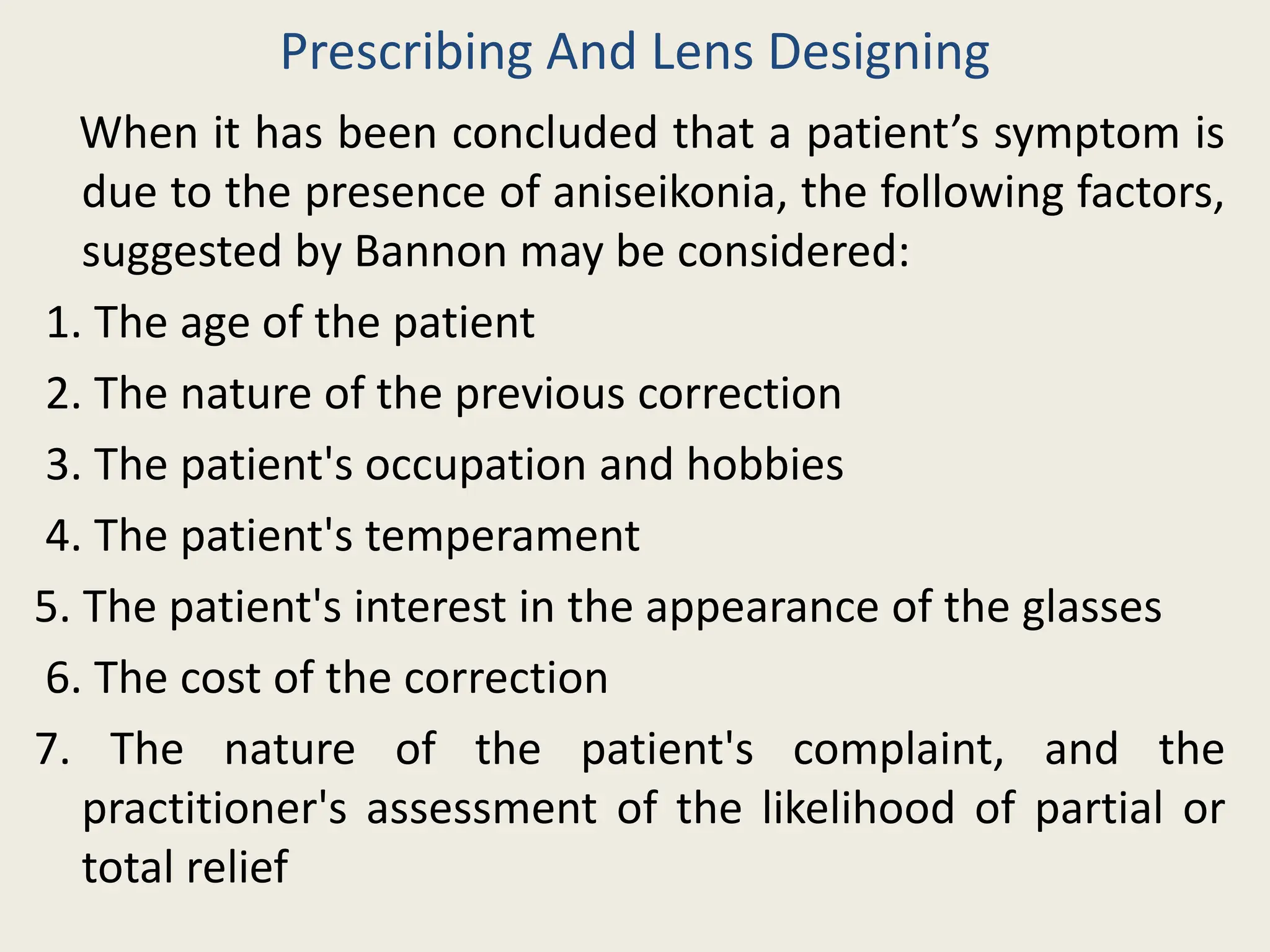
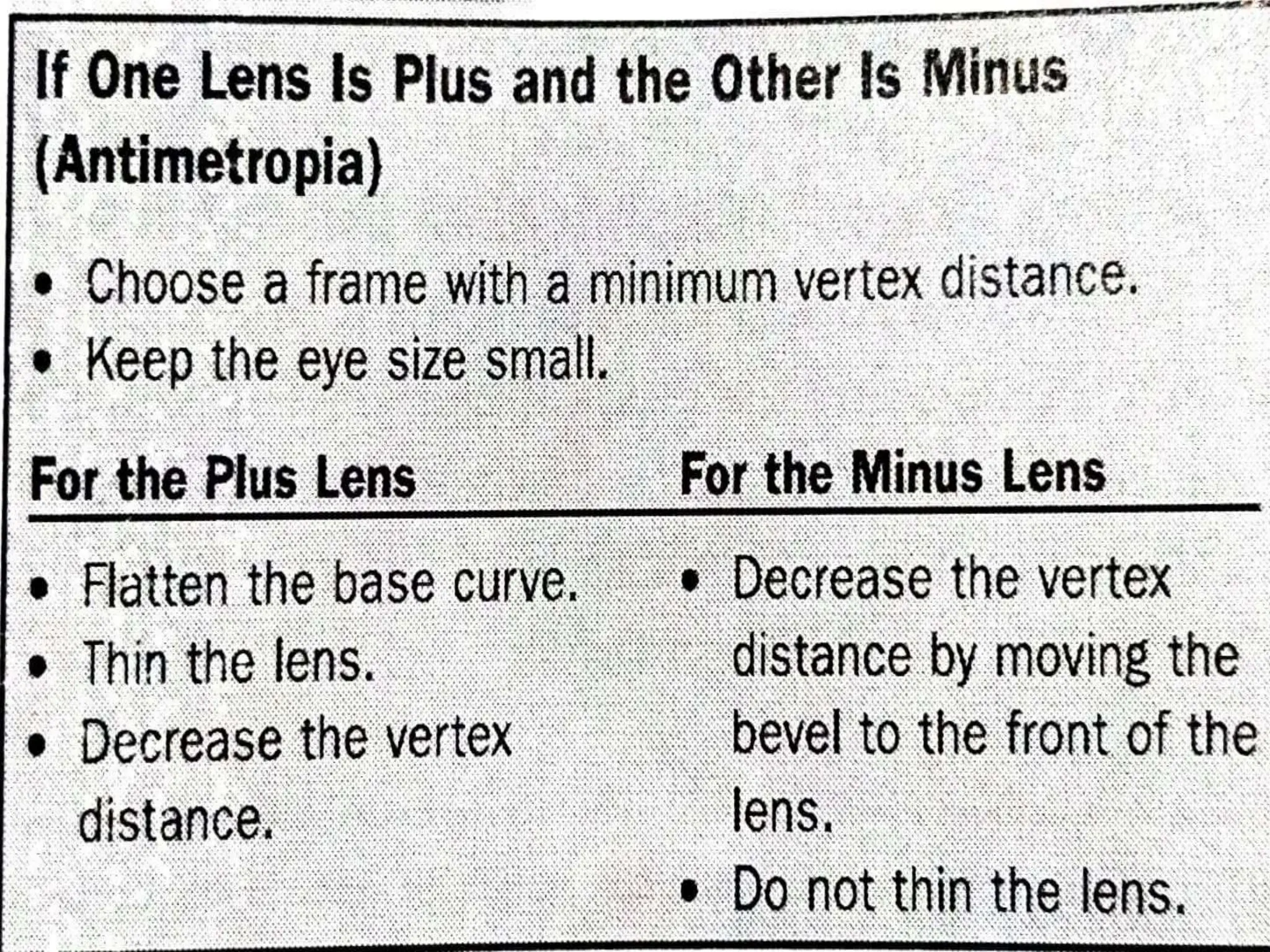
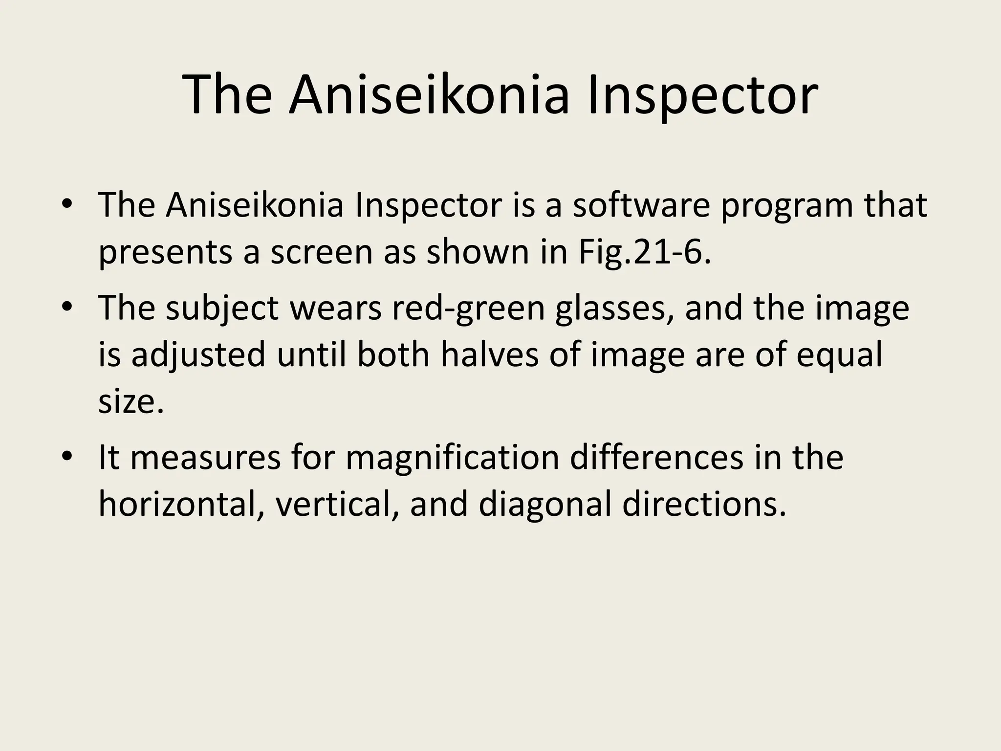
Ophthalmic Dispensing In
Anisometropia And Aniseikonia
Presented by:
Sujit Kumar Shah
B.Optometry, 2nd year
Introduction
•Anisometropia is when there is a difference in
refractive power between the left and right eyes.
•Anisometropia can works to an indivisual’s favour
in presbyopia.
•When one eye is emmetropic and needs no
correction and other is somewhat myopic, such a
person can avoid the need for reading glasses. One
eye is used to see for distance vision, and other for
near.
•On the whole, however, a significant amount of
anisometropia ends up creating problems.
Aniseikonia
• Aniseikonia is a relative difference in the size
and/or the shape of the images seen by the
right and the left eyes.
Types of Aniseikonia
 Physiologic Aniseikonia
• Aniseikonia occurs in limited but useful amount even
for indivisuals with eyes that are identical to one
another.
Suppose a person turns their eyes to the left to look
at an object. The right eye will be slightly farther
away from the object than the left eye. The image of
the object in rt. eye will be slightly smaller than the
image seen by the left eye. These differences give
clue that help in localizing the object in space. This
type of aniseikonia is expected and is referred to as
physiologic or natural aniseikonia.
Symmetrical Aniseikonia
• One eye may see an image that is symmetrically
larger than the other eye (i.e., it is equally larger in
every meridian). This is called symmetrical
aniseikonia
Asymmetrical Aniseikonia
• It is when there is progressive increase or
decrease across the visual field.
Detecting Clinically Significant Aniseikonia
• Although there are both obvious and not so obvious
signs and symptoms that may indicate clinically
significant aniseikonia, it is sometimes difficult to
recognize .
• Aniseikonia symptoms are often the same symptoms
as experienced with uncorrected refractive errors or
oculomotor imbalances.
• In addition to those, here are some indications of
clinically significant aniseikonia:
Continued….
1. High anisometropia or high astigmatism.
2. The presence of certain factors that physically alter the
eye, such as pseudophakia, scleral buckling, corneal
transplantation, refractive surgery, and optic atrophy.
3. Complaints about spatial distortion, such as slanting
floors, tilted walls, or ground too close or too far away.
4. Better optical comfort when only one eye is used.
It is helpful to notice if the symptoms occurred after a
prescription change or after the dispensing of new
glasses. Assuming the refraction is correct and the
lenses verify as they should, when anisometropia is
present, aniseikonia is likely.
Prescribing And Lens Designing
When it has been concluded that a patient’s symptom is
due to the presence of aniseikonia, the following factors,
suggested by Bannon may be considered:
1. The age of the patient
2. The nature of the previous correction
3. The patient's occupation and hobbies
4. The patient's temperament
5. The patient's interest in the appearance of the glasses
6. The cost of the correction
7. The nature of the patient's complaint, and the
practitioner's assessment of the likelihood of partial or
total relief
Frame selection
• Since these lenses are thicker and therefore heavier
than most other lenses, the eye size should be kept
small, both to reduce weight and to allow fitting the
lens at as short a vertex distance as possible.
• A plastic frame will conceal the thick edge better
than will a metal frame or a rimless mounting.
• An eikonic lens tends to have highly curved surfaces
and a correspondingly curved bevel, and therefore
may be difficult to mount with out warping the
frame: A symmetrical lens shape is therefore
recommended.
• The patient should be seen often during the first few
weeks after the correction is dispensed, in order to
check the adjustment and to provide reassurance
and additional instruction if needed.
Correcting Aniseikonia With Spectacle Lenses
• There are several ways to approach the problem of
aniseikonia:
1. If you are concerned that aniseikonia might be a
problem, but have no evidence, use a “ First Pass
Method.”
2. If you are fairly certain aniseikonia is present, want to
address it yourself, but have no way of measuring it,
then make “directionally correct magnification
chnages” to each lens indivisually.
3. Estimate percent magnification difference based on
the refractive prescription and change lens
parameters accordingly.
4. Measure the present magnification differences
between two eyes and change the lens paramters
accordingly
Using a “First Pass Method” to prevent Possible
Problems
• This can done before anything else and will not hurt
anything, even if aniseikonia is not a problem at all.
1. Use a frame with a short vertex distance and, if
nosepads are present, further reduce the vertex
distance.
2. Use a frame with a small size. This secondarily
reduces vertex distance.
3. Use an aspheric lens design. This usually flattens the
base curve.
4. Use a high index materials. This will thin plus lens
center thickness.
Making “Directionally correct” Magnification
Changes
• What is done with each lens will depend upon the
power of the right and left lenses compared with one
another.
Estimating Percent Magnification Differences
• Estimates of how much magnification changes
per diopter of power vary.
• Linksz and Bannon say we can expect 1.5% per
diopter of anisometropia.
Measuring Percent Magnification Differences
• The ideal way to correct for aniseikonia is to measure
it directly.
• Historically the classical method was to use a space
eikonometer.
• A space eikonometer is used to quatitatively
measure image size differences.
• It is no longer made.
The Aniseikonia Inspector
• The Aniseikonia Inspector is a software program that
presents a screen as shown in Fig.21-6.
• The subject wears red-green glasses, and the image
is adjusted until both halves of image are of equal
size.
• It measures for magnification differences in the
horizontal, vertical, and diagonal directions.
Magnification
‘+ lens’
Magnification
‘- lens’
Steepen base curve
Flatten base curve
Increase
Decrease
Decrease
Increase
Thickness increase
Decrease thickness
Increase
Decrease
Decrease
Increase
Vertex distance increase
Vertex distance decrease
Increase
Decrease
Increase
Decrease
How Helpful Is Correcting For Aniseikonia
• In a study done at Emory Eye centre, Achiron et al
compared corrections for 34 anisometropes.
• They found that modifying lens design to equalize
relative spectacle magnification both reduced
aniseikonia and improved subjective comfort and
performance.
• At the conclusion of the study, 93% of the study
subjects preferred the spectacles that had been
modified to correct for aniseikonia over traditional
spectacles.
References
• System For Ophthalmic Dispensing, Third Edition
• Fannin.TroyE, Clinical Optics.USA, Butterworth
Publisher
Ophthalmic dispensing in Anisometropia and Aniseikonia.pptx

Ophthalmic dispensing in Anisometropia and Aniseikonia.pptx

  • 1.
    Ophthalmic Dispensing In AnisometropiaAnd Aniseikonia Presented by: Sujit Kumar Shah B.Optometry, 2nd year
  • 2.
    Introduction •Anisometropia is whenthere is a difference in refractive power between the left and right eyes. •Anisometropia can works to an indivisual’s favour in presbyopia. •When one eye is emmetropic and needs no correction and other is somewhat myopic, such a person can avoid the need for reading glasses. One eye is used to see for distance vision, and other for near. •On the whole, however, a significant amount of anisometropia ends up creating problems.
  • 3.
    Aniseikonia • Aniseikonia isa relative difference in the size and/or the shape of the images seen by the right and the left eyes.
  • 4.
    Types of Aniseikonia Physiologic Aniseikonia • Aniseikonia occurs in limited but useful amount even for indivisuals with eyes that are identical to one another. Suppose a person turns their eyes to the left to look at an object. The right eye will be slightly farther away from the object than the left eye. The image of the object in rt. eye will be slightly smaller than the image seen by the left eye. These differences give clue that help in localizing the object in space. This type of aniseikonia is expected and is referred to as physiologic or natural aniseikonia.
  • 5.
    Symmetrical Aniseikonia • Oneeye may see an image that is symmetrically larger than the other eye (i.e., it is equally larger in every meridian). This is called symmetrical aniseikonia Asymmetrical Aniseikonia • It is when there is progressive increase or decrease across the visual field.
  • 7.
    Detecting Clinically SignificantAniseikonia • Although there are both obvious and not so obvious signs and symptoms that may indicate clinically significant aniseikonia, it is sometimes difficult to recognize . • Aniseikonia symptoms are often the same symptoms as experienced with uncorrected refractive errors or oculomotor imbalances. • In addition to those, here are some indications of clinically significant aniseikonia:
  • 8.
    Continued…. 1. High anisometropiaor high astigmatism. 2. The presence of certain factors that physically alter the eye, such as pseudophakia, scleral buckling, corneal transplantation, refractive surgery, and optic atrophy. 3. Complaints about spatial distortion, such as slanting floors, tilted walls, or ground too close or too far away. 4. Better optical comfort when only one eye is used. It is helpful to notice if the symptoms occurred after a prescription change or after the dispensing of new glasses. Assuming the refraction is correct and the lenses verify as they should, when anisometropia is present, aniseikonia is likely.
  • 9.
    Prescribing And LensDesigning When it has been concluded that a patient’s symptom is due to the presence of aniseikonia, the following factors, suggested by Bannon may be considered: 1. The age of the patient 2. The nature of the previous correction 3. The patient's occupation and hobbies 4. The patient's temperament 5. The patient's interest in the appearance of the glasses 6. The cost of the correction 7. The nature of the patient's complaint, and the practitioner's assessment of the likelihood of partial or total relief
  • 10.
    Frame selection • Sincethese lenses are thicker and therefore heavier than most other lenses, the eye size should be kept small, both to reduce weight and to allow fitting the lens at as short a vertex distance as possible. • A plastic frame will conceal the thick edge better than will a metal frame or a rimless mounting. • An eikonic lens tends to have highly curved surfaces and a correspondingly curved bevel, and therefore may be difficult to mount with out warping the frame: A symmetrical lens shape is therefore recommended.
  • 11.
    • The patientshould be seen often during the first few weeks after the correction is dispensed, in order to check the adjustment and to provide reassurance and additional instruction if needed.
  • 12.
    Correcting Aniseikonia WithSpectacle Lenses • There are several ways to approach the problem of aniseikonia: 1. If you are concerned that aniseikonia might be a problem, but have no evidence, use a “ First Pass Method.” 2. If you are fairly certain aniseikonia is present, want to address it yourself, but have no way of measuring it, then make “directionally correct magnification chnages” to each lens indivisually. 3. Estimate percent magnification difference based on the refractive prescription and change lens parameters accordingly. 4. Measure the present magnification differences between two eyes and change the lens paramters accordingly
  • 13.
    Using a “FirstPass Method” to prevent Possible Problems • This can done before anything else and will not hurt anything, even if aniseikonia is not a problem at all. 1. Use a frame with a short vertex distance and, if nosepads are present, further reduce the vertex distance. 2. Use a frame with a small size. This secondarily reduces vertex distance. 3. Use an aspheric lens design. This usually flattens the base curve. 4. Use a high index materials. This will thin plus lens center thickness.
  • 14.
    Making “Directionally correct”Magnification Changes • What is done with each lens will depend upon the power of the right and left lenses compared with one another.
  • 19.
    Estimating Percent MagnificationDifferences • Estimates of how much magnification changes per diopter of power vary. • Linksz and Bannon say we can expect 1.5% per diopter of anisometropia.
  • 20.
    Measuring Percent MagnificationDifferences • The ideal way to correct for aniseikonia is to measure it directly. • Historically the classical method was to use a space eikonometer. • A space eikonometer is used to quatitatively measure image size differences. • It is no longer made.
  • 21.
    The Aniseikonia Inspector •The Aniseikonia Inspector is a software program that presents a screen as shown in Fig.21-6. • The subject wears red-green glasses, and the image is adjusted until both halves of image are of equal size. • It measures for magnification differences in the horizontal, vertical, and diagonal directions.
  • 23.
    Magnification ‘+ lens’ Magnification ‘- lens’ Steepenbase curve Flatten base curve Increase Decrease Decrease Increase Thickness increase Decrease thickness Increase Decrease Decrease Increase Vertex distance increase Vertex distance decrease Increase Decrease Increase Decrease
  • 24.
    How Helpful IsCorrecting For Aniseikonia • In a study done at Emory Eye centre, Achiron et al compared corrections for 34 anisometropes. • They found that modifying lens design to equalize relative spectacle magnification both reduced aniseikonia and improved subjective comfort and performance. • At the conclusion of the study, 93% of the study subjects preferred the spectacles that had been modified to correct for aniseikonia over traditional spectacles.
  • 25.
    References • System ForOphthalmic Dispensing, Third Edition • Fannin.TroyE, Clinical Optics.USA, Butterworth Publisher