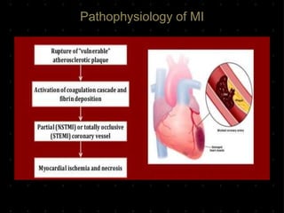
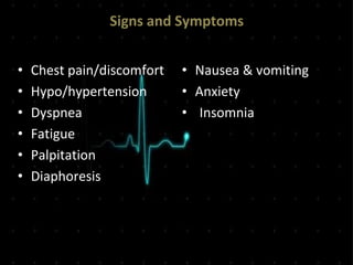
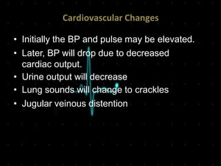
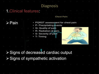
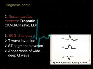
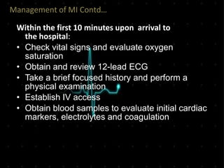
Acute Myocardial Infarction
RN MINTU THOMAS
Definition
• Myocardial infarction refers to a condition
when the blood flow decreases or stops to
a part of heart which leads to ischemia
and myocardial cell damage.
• Ischemia usually occurs due to blockage
of the coronary vessels.
Definition contd…
• This blockage is often the result of
thrombus that is superimposed on an
ulcerated or unstable atherosclerotic
plaque formation in the coronary artery
Coronary Artery Anatomy
Coronary Artery Events
• Ischemia – Outer most area, source of
arrhythmias, viable if no further infarction.
• Injury – Viable tissue found between
ischemic and infarcted areas.
• Infarction/necrosis – Center area, dead
not viable tissue that turn into scar.
MI Classifications
• MI’s can be subcategorized by anatomy
and clinical diagnostic information.
Anatomic
 Transmural – infarction that involves
the whole thickness of heart muscle.
 Subendocardial – infarction that
involves the innermost layer and
parts of myocardium but does not
extend to epicardium.
Diagnostic
 ST elevation MI (STEMI)
 Non ST elevation MI (NSTEMI).
Risk Factors
Non Modifiable
• Age
• Gender
• Family history
Modifiable
• Smoking
• Diabetes
• Hypertension
• Hyperlipidemia
• Obesity & Physical
Inactivity
Pathophysiology of MI
Signs and Symptoms
• Chest pain/discomfort
• Hypo/hypertension
• Dyspnea
• Fatigue
• Palpitation
• Diaphoresis
• Nausea & vomiting
• Anxiety
• Insomnia
Cardiovascular Changes
• Initially the BP and pulse may be elevated.
• Later, BP will drop due to decreased
cardiac output.
• Urine output will decrease
• Lung sounds will change to crackles
• Jugular veinous distention
Diagnosis
1.Clinical features:
Pain
Signs of decreased cardiac output
Signs of sympathetic activation
Diagnosis contd…
2. Serum cardiac
markers: Troponin ,
CKMB/CK ratio, LDH
3. ECG changes:
 T wave inversion
 ST segment elevation
 Appearance of wide
deep Q wave
Localization of MI on ECG
The changes of ischemia , injury or infarction are
seen in the leads overlying the area involved.
Diagnosis contd..
4. Echocardiogram
5.Chest Xray
6.Angiography
Complications of MI
Arrhythmias
Cardiogenic Shock
Congestive Heart Failure
Cardiac Tamponade
Pericarditis
Thromboembolism
Recurrent Infarction
Managementof MI
The immediate goal for any acute myocardial
infarction is to restore normal coronary blood
flow to vessels and salvage myocardium
Mnemonic for the management of MI
M – Morphine
O – Oxygen Therapy
N – Nitroglycerine
A – Aspirin
Management of MI Contd…
Within the first 10 minutes upon arrival to
the hospital:
• Check vital signs and evaluate oxygen
saturation
• Obtain and review 12-lead ECG
• Take a brief focused history and perform a
physical examination
• Establish IV access
• Obtain blood samples to evaluate initial cardiac
markers, electrolytes and coagulation
Management of MI contd…
Treatment options for MI:
Reperfusion as soon as possible(within
12hours of onset of symptoms)
Antiplatelet therapy(aspirin , clopidogrel +/-
Glycoprotein iib/iiia inhibitors)
Anticoagulants (unfractionated heparin or
LMWH)
Immediate PCI or Fibrinolytic therapy –
PCI has higher reperfusion rate and is better
if patient present >1hr but thrombolysis is
gold standard if patient arrives within 1hr
Management for MI contd…
 Subsequent management of MI:
 Aspirin and clopidogrel, statins, ACE inhibitors,
Betablockers
 Anticoagulation therapy to prevent
thromboembolism
 Nitrates as needed
 Cardiac rehabilitation
 Antiplatelet therapy post stent:
 Aspirin for life
 Clopidogrel for the recommended period
Long Term Care
• Lifestyle modifications.
• Healthy eating.
• Maintaining blood pressure ,blood sugar and
cholesterol under control.
• Smoking cessation
Myocardial infarction

Myocardial infarction

  • 1.
  • 2.
    Definition • Myocardial infarctionrefers to a condition when the blood flow decreases or stops to a part of heart which leads to ischemia and myocardial cell damage. • Ischemia usually occurs due to blockage of the coronary vessels.
  • 3.
    Definition contd… • Thisblockage is often the result of thrombus that is superimposed on an ulcerated or unstable atherosclerotic plaque formation in the coronary artery
  • 4.
  • 5.
  • 6.
    Coronary Artery Events •Ischemia – Outer most area, source of arrhythmias, viable if no further infarction. • Injury – Viable tissue found between ischemic and infarcted areas. • Infarction/necrosis – Center area, dead not viable tissue that turn into scar.
  • 7.
    MI Classifications • MI’scan be subcategorized by anatomy and clinical diagnostic information. Anatomic  Transmural – infarction that involves the whole thickness of heart muscle.  Subendocardial – infarction that involves the innermost layer and parts of myocardium but does not extend to epicardium. Diagnostic  ST elevation MI (STEMI)  Non ST elevation MI (NSTEMI).
  • 8.
    Risk Factors Non Modifiable •Age • Gender • Family history Modifiable • Smoking • Diabetes • Hypertension • Hyperlipidemia • Obesity & Physical Inactivity
  • 9.
  • 10.
    Signs and Symptoms •Chest pain/discomfort • Hypo/hypertension • Dyspnea • Fatigue • Palpitation • Diaphoresis • Nausea & vomiting • Anxiety • Insomnia
  • 11.
    Cardiovascular Changes • Initiallythe BP and pulse may be elevated. • Later, BP will drop due to decreased cardiac output. • Urine output will decrease • Lung sounds will change to crackles • Jugular veinous distention
  • 12.
    Diagnosis 1.Clinical features: Pain Signs ofdecreased cardiac output Signs of sympathetic activation
  • 13.
    Diagnosis contd… 2. Serumcardiac markers: Troponin , CKMB/CK ratio, LDH 3. ECG changes:  T wave inversion  ST segment elevation  Appearance of wide deep Q wave
  • 14.
    Localization of MIon ECG The changes of ischemia , injury or infarction are seen in the leads overlying the area involved.
  • 15.
  • 16.
    Complications of MI Arrhythmias CardiogenicShock Congestive Heart Failure Cardiac Tamponade Pericarditis Thromboembolism Recurrent Infarction
  • 17.
    Managementof MI The immediategoal for any acute myocardial infarction is to restore normal coronary blood flow to vessels and salvage myocardium Mnemonic for the management of MI M – Morphine O – Oxygen Therapy N – Nitroglycerine A – Aspirin
  • 18.
    Management of MIContd… Within the first 10 minutes upon arrival to the hospital: • Check vital signs and evaluate oxygen saturation • Obtain and review 12-lead ECG • Take a brief focused history and perform a physical examination • Establish IV access • Obtain blood samples to evaluate initial cardiac markers, electrolytes and coagulation
  • 19.
    Management of MIcontd… Treatment options for MI: Reperfusion as soon as possible(within 12hours of onset of symptoms) Antiplatelet therapy(aspirin , clopidogrel +/- Glycoprotein iib/iiia inhibitors) Anticoagulants (unfractionated heparin or LMWH) Immediate PCI or Fibrinolytic therapy – PCI has higher reperfusion rate and is better if patient present >1hr but thrombolysis is gold standard if patient arrives within 1hr
  • 20.
    Management for MIcontd…  Subsequent management of MI:  Aspirin and clopidogrel, statins, ACE inhibitors, Betablockers  Anticoagulation therapy to prevent thromboembolism  Nitrates as needed  Cardiac rehabilitation  Antiplatelet therapy post stent:  Aspirin for life  Clopidogrel for the recommended period
  • 21.
    Long Term Care •Lifestyle modifications. • Healthy eating. • Maintaining blood pressure ,blood sugar and cholesterol under control. • Smoking cessation