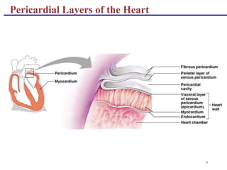
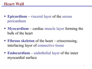
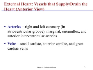
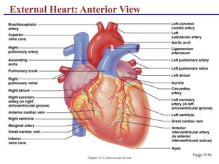
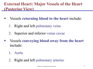
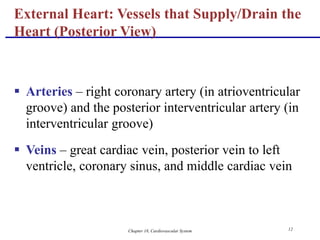
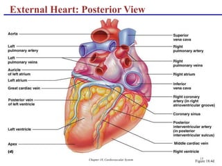
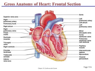
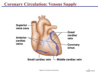
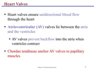
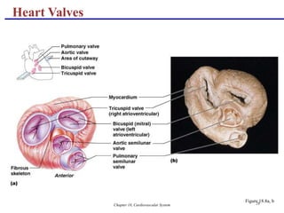
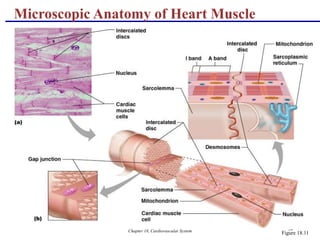
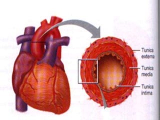
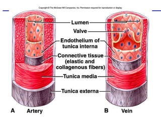
The document describes the anatomy and structure of the cardiovascular system, including the heart and blood vessels. It details the layers that make up the heart walls and pericardium. It explains the coronary circulation that supplies blood to the heart muscle and lists the major arteries and veins involved in systemic and pulmonary circulation. Key anatomical features like heart valves and chambers are defined along with common congenital defects. Microscopic views of heart muscle and blood vessels are provided.
A closed system of the heart and blood vessels
The heart pumps blood
Blood vessels allow blood to circulate to all parts of the body
The function of the cardiovascular system is to deliver oxygen and nutrients and to remove carbon dioxide and other waste products
A closed system of the heart and blood vessels
The heart pumps blood
Blood vessels allow blood to circulate to all parts of the body
The function of the cardiovascular system is to deliver oxygen and nutrients and to remove carbon dioxide and other waste products
anatomy of Left atrium and left ventricle of the human heartGeetanjaliKarle1
left atrium- interior of auricle is rough due to musculi pectinate, rest chamber is smooth. fossa lunate is present on septal wall. 4 pulmonary veins open on posterior wall.
left ventricle- inflowing part is rough due to mitral or bicuspid valve apparatus, trabeculae carneae.
outflowing part is smooth called infundibulum. ascending aorta starts from infundibulum. aortic valve guards opening of ascending aorta
HEART RATE
REGULATION OF HEART RATE
VASOMOTOR CENTER – CARDIAC CENTER
MOTOR (EFFERENT) NERVE FIBERS TO HEART
FACTORS AFFECTING VASOMOTOR CENTER
for all medical & health care students
Hey Guys
im happy you are enjoying my content. please subscribe to my channel on youtube as i will make more videos soon. https://bit.ly/2XXNyTT
thank you as you subscribe.
Heart is the center of your cardiovascular system. It is responsible for pumping blood through your body, carrying nutrients in and taking waste out. Having good cardiovascular health is an important factor of optimal wellness.
An adult has a mass of 250-350 grams.
Each day, the average heart beats 100,000 times, pumping about 2,000 gallons (7,571 liters) of blood.
By the end of a long life, a person's heart may have beat (expanded and contracted) more than 3.5 billion times.
Heart has four chambers and four valves present in the heart control the flow of blood in the heart.
Approximately the size of your fist
Location
Superior surface of diaphragm
Left of the midline
Anterior to the vertebral column, posterior to the sternum
Coverings of the Heart: Anatomy
Pericardium – a double-walled sac around the heart composed of:
A superficial fibrous pericardium
A deep two-layer serous pericardium
The parietal layer lines the internal surface of the fibrous pericardium
The visceral layer or epicardium lines the surface of the heart
They are separated by the fluid-filled pericardial cavity
A closed system of the heart and blood vessels
The heart pumps blood
Blood vessels allow blood to circulate to all parts of the body
The function of the cardiovascular system is to deliver oxygen and nutrients and to remove carbon dioxide and other waste products
A closed system of the heart and blood vessels
The heart pumps blood
Blood vessels allow blood to circulate to all parts of the body
The function of the cardiovascular system is to deliver oxygen and nutrients and to remove carbon dioxide and other waste products
anatomy of Left atrium and left ventricle of the human heartGeetanjaliKarle1
left atrium- interior of auricle is rough due to musculi pectinate, rest chamber is smooth. fossa lunate is present on septal wall. 4 pulmonary veins open on posterior wall.
left ventricle- inflowing part is rough due to mitral or bicuspid valve apparatus, trabeculae carneae.
outflowing part is smooth called infundibulum. ascending aorta starts from infundibulum. aortic valve guards opening of ascending aorta
HEART RATE
REGULATION OF HEART RATE
VASOMOTOR CENTER – CARDIAC CENTER
MOTOR (EFFERENT) NERVE FIBERS TO HEART
FACTORS AFFECTING VASOMOTOR CENTER
for all medical & health care students
Hey Guys
im happy you are enjoying my content. please subscribe to my channel on youtube as i will make more videos soon. https://bit.ly/2XXNyTT
thank you as you subscribe.
Heart is the center of your cardiovascular system. It is responsible for pumping blood through your body, carrying nutrients in and taking waste out. Having good cardiovascular health is an important factor of optimal wellness.
An adult has a mass of 250-350 grams.
Each day, the average heart beats 100,000 times, pumping about 2,000 gallons (7,571 liters) of blood.
By the end of a long life, a person's heart may have beat (expanded and contracted) more than 3.5 billion times.
Heart has four chambers and four valves present in the heart control the flow of blood in the heart.
Approximately the size of your fist
Location
Superior surface of diaphragm
Left of the midline
Anterior to the vertebral column, posterior to the sternum
Coverings of the Heart: Anatomy
Pericardium – a double-walled sac around the heart composed of:
A superficial fibrous pericardium
A deep two-layer serous pericardium
The parietal layer lines the internal surface of the fibrous pericardium
The visceral layer or epicardium lines the surface of the heart
They are separated by the fluid-filled pericardial cavity
Flu Vaccine Alert in Bangalore Karnatakaaddon Scans
As flu season approaches, health officials in Bangalore, Karnataka, are urging residents to get their flu vaccinations. The seasonal flu, while common, can lead to severe health complications, particularly for vulnerable populations such as young children, the elderly, and those with underlying health conditions.
Dr. Vidisha Kumari, a leading epidemiologist in Bangalore, emphasizes the importance of getting vaccinated. "The flu vaccine is our best defense against the influenza virus. It not only protects individuals but also helps prevent the spread of the virus in our communities," he says.
This year, the flu season is expected to coincide with a potential increase in other respiratory illnesses. The Karnataka Health Department has launched an awareness campaign highlighting the significance of flu vaccinations. They have set up multiple vaccination centers across Bangalore, making it convenient for residents to receive their shots.
To encourage widespread vaccination, the government is also collaborating with local schools, workplaces, and community centers to facilitate vaccination drives. Special attention is being given to ensuring that the vaccine is accessible to all, including marginalized communities who may have limited access to healthcare.
Residents are reminded that the flu vaccine is safe and effective. Common side effects are mild and may include soreness at the injection site, mild fever, or muscle aches. These side effects are generally short-lived and far less severe than the flu itself.
Healthcare providers are also stressing the importance of continuing COVID-19 precautions. Wearing masks, practicing good hand hygiene, and maintaining social distancing are still crucial, especially in crowded places.
Protect yourself and your loved ones by getting vaccinated. Together, we can help keep Bangalore healthy and safe this flu season. For more information on vaccination centers and schedules, residents can visit the Karnataka Health Department’s official website or follow their social media pages.
Stay informed, stay safe, and get your flu shot today!
Factory Supply Best Quality Pmk Oil CAS 28578–16–7 PMK Powder in Stockrebeccabio
Factory Supply Best Quality Pmk Oil CAS 28578–16–7 PMK Powder in Stock
Telegram: bmksupplier
signal: +85264872720
threema: TUD4A6YC
You can contact me on Telegram or Threema
Communicate promptly and reply
Free of customs clearance, Double Clearance 100% pass delivery to USA, Canada, Spain, Germany, Netherland, Poland, Italy, Sweden, UK, Czech Republic, Australia, Mexico, Russia, Ukraine, Kazakhstan.Door to door service
Hot Selling Organic intermediates
The prostate is an exocrine gland of the male mammalian reproductive system
It is a walnut-sized gland that forms part of the male reproductive system and is located in front of the rectum and just below the urinary bladder
Function is to store and secrete a clear, slightly alkaline fluid that constitutes 10-30% of the volume of the seminal fluid that along with the spermatozoa, constitutes semen
A healthy human prostate measures (4cm-vertical, by 3cm-horizontal, 2cm ant-post ).
It surrounds the urethra just below the urinary bladder. It has anterior, median, posterior and two lateral lobes
It’s work is regulated by androgens which are responsible for male sex characteristics
Generalised disease of the prostate due to hormonal derangement which leads to non malignant enlargement of the gland (increase in the number of epithelial cells and stromal tissue)to cause compression of the urethra leading to symptoms (LUTS
Title: Sense of Taste
Presenter: Dr. Faiza, Assistant Professor of Physiology
Qualifications:
MBBS (Best Graduate, AIMC Lahore)
FCPS Physiology
ICMT, CHPE, DHPE (STMU)
MPH (GC University, Faisalabad)
MBA (Virtual University of Pakistan)
Learning Objectives:
Describe the structure and function of taste buds.
Describe the relationship between the taste threshold and taste index of common substances.
Explain the chemical basis and signal transduction of taste perception for each type of primary taste sensation.
Recognize different abnormalities of taste perception and their causes.
Key Topics:
Significance of Taste Sensation:
Differentiation between pleasant and harmful food
Influence on behavior
Selection of food based on metabolic needs
Receptors of Taste:
Taste buds on the tongue
Influence of sense of smell, texture of food, and pain stimulation (e.g., by pepper)
Primary and Secondary Taste Sensations:
Primary taste sensations: Sweet, Sour, Salty, Bitter, Umami
Chemical basis and signal transduction mechanisms for each taste
Taste Threshold and Index:
Taste threshold values for Sweet (sucrose), Salty (NaCl), Sour (HCl), and Bitter (Quinine)
Taste index relationship: Inversely proportional to taste threshold
Taste Blindness:
Inability to taste certain substances, particularly thiourea compounds
Example: Phenylthiocarbamide
Structure and Function of Taste Buds:
Composition: Epithelial cells, Sustentacular/Supporting cells, Taste cells, Basal cells
Features: Taste pores, Taste hairs/microvilli, and Taste nerve fibers
Location of Taste Buds:
Found in papillae of the tongue (Fungiform, Circumvallate, Foliate)
Also present on the palate, tonsillar pillars, epiglottis, and proximal esophagus
Mechanism of Taste Stimulation:
Interaction of taste substances with receptors on microvilli
Signal transduction pathways for Umami, Sweet, Bitter, Sour, and Salty tastes
Taste Sensitivity and Adaptation:
Decrease in sensitivity with age
Rapid adaptation of taste sensation
Role of Saliva in Taste:
Dissolution of tastants to reach receptors
Washing away the stimulus
Taste Preferences and Aversions:
Mechanisms behind taste preference and aversion
Influence of receptors and neural pathways
Impact of Sensory Nerve Damage:
Degeneration of taste buds if the sensory nerve fiber is cut
Abnormalities of Taste Detection:
Conditions: Ageusia, Hypogeusia, Dysgeusia (parageusia)
Causes: Nerve damage, neurological disorders, infections, poor oral hygiene, adverse drug effects, deficiencies, aging, tobacco use, altered neurotransmitter levels
Neurotransmitters and Taste Threshold:
Effects of serotonin (5-HT) and norepinephrine (NE) on taste sensitivity
Supertasters:
25% of the population with heightened sensitivity to taste, especially bitterness
Increased number of fungiform papillae
HOT NEW PRODUCT! BIG SALES FAST SHIPPING NOW FROM CHINA!! EU KU DB BK substit...GL Anaacs
Contact us if you are interested:
Email / Skype : kefaya1771@gmail.com
Threema: PXHY5PDH
New BATCH Ku !!! MUCH IN DEMAND FAST SALE EVERY BATCH HAPPY GOOD EFFECT BIG BATCH !
Contact me on Threema or skype to start big business!!
Hot-sale products:
NEW HOT EUTYLONE WHITE CRYSTAL!!
5cl-adba precursor (semi finished )
5cl-adba raw materials
ADBB precursor (semi finished )
ADBB raw materials
APVP powder
5fadb/4f-adb
Jwh018 / Jwh210
Eutylone crystal
Protonitazene (hydrochloride) CAS: 119276-01-6
Flubrotizolam CAS: 57801-95-3
Metonitazene CAS: 14680-51-4
Payment terms: Western Union,MoneyGram,Bitcoin or USDT.
Deliver Time: Usually 7-15days
Shipping method: FedEx, TNT, DHL,UPS etc.Our deliveries are 100% safe, fast, reliable and discreet.
Samples will be sent for your evaluation!If you are interested in, please contact me, let's talk details.
We specializes in exporting high quality Research chemical, medical intermediate, Pharmaceutical chemicals and so on. Products are exported to USA, Canada, France, Korea, Japan,Russia, Southeast Asia and other countries.
Tom Selleck Health: A Comprehensive Look at the Iconic Actor’s Wellness Journeygreendigital
Tom Selleck, an enduring figure in Hollywood. has captivated audiences for decades with his rugged charm, iconic moustache. and memorable roles in television and film. From his breakout role as Thomas Magnum in Magnum P.I. to his current portrayal of Frank Reagan in Blue Bloods. Selleck's career has spanned over 50 years. But beyond his professional achievements. fans have often been curious about Tom Selleck Health. especially as he has aged in the public eye.
Follow us on: Pinterest
Introduction
Many have been interested in Tom Selleck health. not only because of his enduring presence on screen but also because of the challenges. and lifestyle choices he has faced and made over the years. This article delves into the various aspects of Tom Selleck health. exploring his fitness regimen, diet, mental health. and the challenges he has encountered as he ages. We'll look at how he maintains his well-being. the health issues he has faced, and his approach to ageing .
Early Life and Career
Childhood and Athletic Beginnings
Tom Selleck was born on January 29, 1945, in Detroit, Michigan, and grew up in Sherman Oaks, California. From an early age, he was involved in sports, particularly basketball. which played a significant role in his physical development. His athletic pursuits continued into college. where he attended the University of Southern California (USC) on a basketball scholarship. This early involvement in sports laid a strong foundation for his physical health and disciplined lifestyle.
Transition to Acting
Selleck's transition from an athlete to an actor came with its physical demands. His first significant role in "Magnum P.I." required him to perform various stunts and maintain a fit appearance. This role, which he played from 1980 to 1988. necessitated a rigorous fitness routine to meet the show's demands. setting the stage for his long-term commitment to health and wellness.
Fitness Regimen
Workout Routine
Tom Selleck health and fitness regimen has evolved. adapting to his changing roles and age. During his "Magnum, P.I." days. Selleck's workouts were intense and focused on building and maintaining muscle mass. His routine included weightlifting, cardiovascular exercises. and specific training for the stunts he performed on the show.
Selleck adjusted his fitness routine as he aged to suit his body's needs. Today, his workouts focus on maintaining flexibility, strength, and cardiovascular health. He incorporates low-impact exercises such as swimming, walking, and light weightlifting. This balanced approach helps him stay fit without putting undue strain on his joints and muscles.
Importance of Flexibility and Mobility
In recent years, Selleck has emphasized the importance of flexibility and mobility in his fitness regimen. Understanding the natural decline in muscle mass and joint flexibility with age. he includes stretching and yoga in his routine. These practices help prevent injuries, improve posture, and maintain mobilit
These simplified slides by Dr. Sidra Arshad present an overview of the non-respiratory functions of the respiratory tract.
Learning objectives:
1. Enlist the non-respiratory functions of the respiratory tract
2. Briefly explain how these functions are carried out
3. Discuss the significance of dead space
4. Differentiate between minute ventilation and alveolar ventilation
5. Describe the cough and sneeze reflexes
Study Resources:
1. Chapter 39, Guyton and Hall Textbook of Medical Physiology, 14th edition
2. Chapter 34, Ganong’s Review of Medical Physiology, 26th edition
3. Chapter 17, Human Physiology by Lauralee Sherwood, 9th edition
4. Non-respiratory functions of the lungs https://academic.oup.com/bjaed/article/13/3/98/278874
Report Back from SGO 2024: What’s the Latest in Cervical Cancer?bkling
Are you curious about what’s new in cervical cancer research or unsure what the findings mean? Join Dr. Emily Ko, a gynecologic oncologist at Penn Medicine, to learn about the latest updates from the Society of Gynecologic Oncology (SGO) 2024 Annual Meeting on Women’s Cancer. Dr. Ko will discuss what the research presented at the conference means for you and answer your questions about the new developments.
MANAGEMENT OF ATRIOVENTRICULAR CONDUCTION BLOCK.pdfJim Jacob Roy
Cardiac conduction defects can occur due to various causes.
Atrioventricular conduction blocks ( AV blocks ) are classified into 3 types.
This document describes the acute management of AV block.
2. 2
Heart Anatomy
Approximately the size of your fist
Location
Superior surface of diaphragm
Left of the midline
Anterior to the vertebral column, posterior to the
sternum
4. 4
Coverings of the Heart: Anatomy
Pericardium – a double-walled sac around the
heart composed of:
1. A superficial fibrous pericardium
2. A deep two-layer serous pericardium
a. The parietal layer lines the internal surface of
the fibrous pericardium
b. The visceral layer or epicardium lines the
surface of the heart
They are separated by the fluid-filled
pericardial cavity
5. Chapter 18, Cardiovascular System 5
Coverings of the Heart: Physiology
The Function of the Pericardium:
Protects and anchors the heart
Prevents overfilling of the heart with blood
Allows for the heart to work in a relatively friction-
free environment
7. 7
Heart Wall
Epicardium – visceral layer of the serous
pericardium
Myocardium – cardiac muscle layer forming the
bulk of the heart
Fibrous skeleton of the heart – crisscrossing,
interlacing layer of connective tissue
Endocardium – endothelial layer of the inner
myocardial surface
8. Chapter 18, Cardiovascular System 8
Vessels returning blood to the heart include:
1. Superior and inferior venae cavae
2. Right and left pulmonary veins
Vessels conveying blood away from the heart include:
1. Pulmonary trunk, which splits into right and left
pulmonary arteries
2. Ascending aorta (three branches) –
a. Brachiocephalic
b. Left common carotid
c. Subclavian arteries
External Heart: Major Vessels of the Heart
(Anterior View)
9. Chapter 18, Cardiovascular System 9
Arteries – right and left coronary (in
atrioventricular groove), marginal, circumflex, and
anterior interventricular arteries
Veins – small cardiac, anterior cardiac, and great
cardiac veins
External Heart: Vessels that Supply/Drain the
Heart (Anterior View)
11. Chapter 18, Cardiovascular System 11
Vessels returning blood to the heart include:
1. Right and left pulmonary veins
2. Superior and inferior venae cavae
Vessels conveying blood away from the heart
include:
1. Aorta
2. Right and left pulmonary arteries
External Heart: Major Vessels of the Heart
(Posterior View)
12. Chapter 18, Cardiovascular System 12
Arteries – right coronary artery (in atrioventricular
groove) and the posterior interventricular artery (in
interventricular groove)
Veins – great cardiac vein, posterior vein to left
ventricle, coronary sinus, and middle cardiac vein
External Heart: Vessels that Supply/Drain the
Heart (Posterior View)
15. Chapter 18, Cardiovascular System 15
Atria of the Heart
Atria are the receiving chambers of the heart
Each atrium has a protruding auricle
Pectinate muscles mark atrial walls
Blood enters right atria from superior and inferior
venae cavae and coronary sinus
Blood enters left atria from pulmonary veins
16. Chapter 18, Cardiovascular System 16
Ventricles of the Heart
Ventricles are the discharging chambers of the heart
Papillary muscles and trabeculae carneae muscles
mark ventricular walls
Right ventricle pumps blood into the pulmonary
trunk
Left ventricle pumps blood into the aorta
17. Chapter 18, Cardiovascular System 17
Myocardial Thickness and Function
Thickness of myocardium varies according to the function of the
chamber
Atria are thin walled, deliver blood to adjacent ventricles
Ventricle walls are much thicker and stronger
right ventricle supplies blood to the lungs (little flow resistance)
left ventricle wall is the thickest to supply systemic circulation
18. Chapter 18, Cardiovascular System 18
Thickness of Cardiac Walls
Myocardium of left ventricle is much thicker than the right.
23. Chapter 18, Cardiovascular System 23
Coronary Circulation
Coronary circulation is the functional blood supply
to the heart muscle itself
Collateral routes ensure blood delivery to heart
even if major vessels are occluded
26. Chapter 18, Cardiovascular System 26
Heart Valves
Heart valves ensure unidirectional blood flow
through the heart
Atrioventricular (AV) valves lie between the atria
and the ventricles
AV valves prevent backflow into the atria when
ventricles contract
Chordae tendineae anchor AV valves to papillary
muscles
27. Chapter 18, Cardiovascular System 27
Heart Valves
Semilunar valves prevent backflow of blood into the
ventricles
Aortic semilunar valve lies between the left
ventricle and the aorta
Pulmonary semilunar valve lies between the right
ventricle and pulmonary trunk
33. Chapter 18, Cardiovascular System 33
Microscopic Anatomy of Heart Muscle
Cardiac muscle is striated, short, fat, branched, and
interconnected
The connective tissue endomysium acts as both
tendon and insertion
Intercalated discs anchor cardiac cells together and
allow free passage of ions
Heart muscle behaves as a functional syncytium
InterActive Physiology®:
Cardiovascular System: Anatomy Review: The Heart
PLAY
35. Vascular System - the Blood Vessels
Arteries, veins, and capillaries comprise the vascular
system.
Arteries and veins run parallel throughout the body with
a web-like network of capillaries connecting them.
Arteries use vessel size, controlled by the sympathetic
nervous system, to move blood by pressure.
Veins use one-way valves controlled by muscle
contractions.
36.
37. Arteries
Arteries are strong, elastic vessels adapted for carrying
blood away from the heart at relatively high pumping
pressure. Arteries divide into progressively thinner
tubes and eventually become fine branches called
arterioles. Blood in arteries is oxygen-rich, with the
exception of the pulmonary artery, which carries blood
to the lungs to be oxygenated.
The aorta is the largest artery in the body, the main
artery for systemic circulation. The major branches of
the aorta (aortic arch, ascending aorta, descending aorta)
supply blood to the head, abdomen, and extremities. Of
special importance are the right and left coronary
arteries, that supply blood to the heart itself.
38. Arteries
The main arteries which branch off the aorta and take
blood to specific parts of the body are:
Carotid arteries, which take blood to the neck and head
Coronary arteries, which provide blood supply to the
heart itself
Hepatic artery, which takes blood to the liver with
branches going to the stomach
Mesenteric artery, which takes blood to the intestines
Renal arteries, which takes blood to the kidneys
Femoral arteries, which take blood to the legs
39. Capillaries
The arterioles branch into the microscopic capillaries, or
capillary beds, which lie bathed in interstitial fluid, or
lymph, produced by the lymphatic system. Capillaries
are the points of exchange between the blood and
surrounding tissues. Materials cross in and out of the
capillaries by passing through or between the cells that
line the capillary. The extensive network of capillaries is
estimated at between 50,000 and 60,000 miles long.1
Microscopic--one cell layer thick
Network
Bathed in extracellular matrix of areolar tissue
Entire goal of C-V system is to get blood into capillaries
where diffusion takes place
40. Veins
Blood leaving the capillary beds flows into a series of
progressively larger vessels, called venules, which in
turn unite to form veins. Veins are responsible for
returning blood to the heart after the blood and the
body cells exchange gases, nutrients, and wastes.
Pressure in veins is low, so veins depend on nearby
muscular contractions to move blood along. Veins have
valves that prevent back-flow of blood.
Blood in veins is oxygen-poor, with the exception of the
pulmonary veins, which carry oxygenated blood from
the lungs back to the heart. The major veins, like their
companion arteries, often take the name of the organ
served. The exceptions are the superior vena cava and
the inferior vena cava, which collect body from all parts
of the body (except from the lungs) and channel it back
to the heart.
41. Artery/Vein Tissues
Arteries and veins have the same three tissue layers, but
the proportions of these layers differ. The innermost is
the intima; next comes the media; and the outermost is
the adventitia. Arteries have thick media to absorb the
pressure waves created by the heart's pumping. The
smooth-muscle media walls expand when pressure
surges, then snap back to push the blood forward when
the heart rests. Valves in the arteries prevent back-
flow. As blood enters the capillaries, the pressure falls
off. By the time blood reaches the veins, there is little
pressure. Thus, a thick media is no longer needed.
Surrounding muscles act to squeeze the blood along
veins. As with arteries, valves are again used to ensure
flow in the right direction.
44. DIFFERENCES BTWN VEINS
AND ARTERIES
Arteries (aa.) Veins (vv.)
Direction
of flow
Blood Away from
Heart
Blood to Heart
Pressure Higher Lower
45. DIFFERENCES BTWN VEINS
AND ARTERIES
Walls THICKER:
Tunica media
thicker than
tunica externa
THINNER:
Tunica externa
thicker than
tunica media
Lumen Smaller Larger
Valves No valves Valves (see
next)
46.
47. REVIEW
Describe the location of Heart in Chest
What Does C-V System do?
Describe how the heart works.
Describe the layers of veins & arteries.
List five differences between arteries and veins.
Name the five great vessels
49. Chapter 18, Cardiovascular System 49
Cardiac Muscle Contraction
Heart muscle:
Is stimulated by nerves and is self-excitable
(automaticity)
Contracts as a unit
Has a long (250 ms) absolute refractory period
Cardiac muscle contraction is similar to skeletal
muscle contraction
50. Chapter 18, Cardiovascular System 50
Heart Physiology: Intrinsic Conduction
System
Autorhythmic cells:
Initiate action potentials
Have unstable resting potentials called pacemaker
potentials
Use calcium influx (rather than sodium) for rising
phase of the action potential
52. Chapter 18, Cardiovascular System 52
Heart Physiology: Sequence of Excitation
Sinoatrial (SA) node generates impulses about 75
times/minute
Atrioventricular (AV) node delays the impulse
approximately 0.1 second
53. Chapter 18, Cardiovascular System 53
Heart Physiology: Sequence of Excitation
Impulse passes from atria to ventricles via the
atrioventricular bundle (bundle of His)
AV bundle splits into two pathways in the
interventricular septum (bundle branches)
1. Bundle branches carry the impulse toward the
apex of the heart
2. Purkinje fibers carry the impulse to the heart
apex and ventricular walls
56. Chapter 18, Cardiovascular System 56
Extrinsic Innervation of the Heart
Heart is stimulated
by the sympathetic
cardioacceleratory
center
Heart is inhibited by
the parasympathetic
cardioinhibitory
center
Figure 18.15
57. Chapter 18, Cardiovascular System 57
Electrocardiography
Electrical activity is recorded by electrocardiogram
(ECG)
P wave corresponds to depolarization of SA node
QRS complex corresponds to ventricular
depolarization
T wave corresponds to ventricular repolarization
Atrial repolarization record is masked by the larger
QRS complex
InterActive Physiology®:
Cardiovascular System: Intrinsic Conduction System
PLAY
59. Chapter 18, Cardiovascular System 59
Heart Sounds
Heart sounds (lub-dup) are associated with closing
of heart valves
First sound occurs as AV valves close and signifies
beginning of systole (contraction)
Second sound occurs when SL valves close at the
beginning of ventricular diastole (relaxation)
60. Chapter 18, Cardiovascular System 60
Cardiac Cycle
Cardiac cycle refers to all events associated with
blood flow through the heart
Systole – contraction of heart muscle
Diastole – relaxation of heart muscle
61. Chapter 18, Cardiovascular System 61
Phases of the Cardiac Cycle
Ventricular filling – mid-to-late diastole
Heart blood pressure is low as blood enters atria
(passively) and flows into ventricles
AV valves are open, then atrial systole occurs
62. Chapter 18, Cardiovascular System 62
Phases of the Cardiac Cycle
Ventricular systole (contraction)
Atria relax
Rising ventricular pressure results in closing of AV
valves
Isovolumetric contraction phase
Ventricular ejection phase opens semilunar valves
63. Chapter 18, Cardiovascular System 63
Phases of the Cardiac Cycle
Isovolumetric relaxation – early diastole
Ventricles relax
Backflow of blood in aorta and pulmonary trunk
closes semilunar valves
Dicrotic notch – brief rise in aortic pressure caused
by backflow of blood rebounding off semilunar
valves
InterActive Physiology®:
Cardiovascular System: Cardiac Cycle
PLAY
65. Chapter 18, Cardiovascular System 65
Cardiac Output (CO) and Reserve
Cardiac Output is the amount of blood pumped by
each ventricle in one minute
CO is the product of heart rate (HR) and stroke
volume (SV)
HR is the number of heart beats per minute
SV is the amount of blood pumped out by a
ventricle with each beat
Cardiac reserve is the difference between resting
and maximal CO
66. Chapter 18, Cardiovascular System 66
Cardiac Output: Example
CO (ml/min) = HR (75 beats/min) x SV (70 ml/beat)
CO = 5250 ml/min (5.25 L/min)
67. Chapter 18, Cardiovascular System 67
Regulation of Stroke Volume
SV = end diastolic volume (EDV) minus end
systolic volume (ESV)
EDV = amount of blood collected in a ventricle
during diastole
ESV = amount of blood remaining in a ventricle
after contraction
68. Chapter 18, Cardiovascular System 68
Factors Affecting Stroke Volume
Preload – amount ventricles are stretched by
contained blood
Contractility – cardiac cell contractile force due to
factors other than EDV
Afterload – back pressure exerted by blood in the
large arteries leaving the heart
69. Chapter 18, Cardiovascular System 69
Frank-Starling Law of the Heart
Preload, or degree of stretch, of cardiac muscle cells
before they contract is the critical factor controlling
stroke volume
Slow heartbeat and exercise increase venous return
to the heart, increasing SV
Blood loss and extremely rapid heartbeat decrease
SV
71. Chapter 18, Cardiovascular System 71
Extrinsic Factors Influencing Stroke Volume
Contractility is the increase in contractile strength,
independent of stretch and EDV
Increase in contractility comes from:
Increased sympathetic stimuli
Certain hormones
Ca2+ and some drugs
73. Chapter 18, Cardiovascular System 73
Contractility and Norepinephrine
Sympathetic
stimulation
releases
norepinephrine
and initiates a
cyclic AMP
second-
messenger
system
Figure 18.22
75. Chapter 18, Cardiovascular System 75
Sympathetic nervous system (SNS) stimulation is activated
by stress, anxiety, excitement, or exercise
Parasympathetic nervous system (PNS) stimulation is
mediated by acetylcholine and opposes the SNS
PNS dominates the autonomic stimulation, slowing heart
rate and causing vagal tone
If the Vagus Nerve was cut, the heart would lose its tone.
Thus, increasing the heart rate by 25 beats per minute.
Regulation of Heart Rate: Autonomic Nervous
System
76. Chapter 18, Cardiovascular System 76
Atrial (Bainbridge) Reflex
Atrial (Bainbridge) reflex – a sympathetic reflex
initiated by increased blood in the atria
Causes stimulation of the SA node
Stimulates baroreceptors in the atria, causing
increased SNS stimulation
77. Chapter 18, Cardiovascular System 77
Chemical Regulation of the Heart
The hormones epinephrine and thyroxine increase
heart rate
Intra- and extracellular ion concentrations must be
maintained for normal heart function
InterActive Physiology®:
Cardiovascular System: Cardiac Output
PLAY
81. Chapter 18, Cardiovascular System 81
Developmental Aspects of the Heart
Fetal heart structures that bypass pulmonary
circulation
Foramen ovale connects the two atria
Ductus arteriosus connects pulmonary trunk and
the aorta
83. Chapter 18, Cardiovascular System 83
Age-Related Changes Affecting the Heart
Sclerosis and thickening of valve flaps
Decline in cardiac reserve
Fibrosis of cardiac muscle
Atherosclerosis
84. Chapter 18, Cardiovascular System 84
Congestive Heart Failure
Causes of CHF
coronary artery disease, hypertension, MI, valve disorders,
congenital defects
Left side heart failure
less effective pump so more blood remains in ventricle
heart is overstretched & even more blood remains
blood backs up into lungs as pulmonary edema
suffocation & lack of oxygen to the tissues
Right side failure
fluid builds up in tissues as peripheral edema
85. Chapter 18, Cardiovascular System 85
Coronary Artery Disease
Heart muscle receiving
insufficient blood supply
narrowing of vessels---
atherosclerosis, artery
spasm or clot
atherosclerosis--smooth
muscle & fatty deposits in
walls of arteries
Treatment
drugs, bypass graft,
angioplasty, stent
86. Chapter 18, Cardiovascular System 86
Clinical Problems
MI = myocardial infarction
death of area of heart muscle from lack of O2
replaced with scar tissue
results depend on size & location of damage
Blood clot
use clot dissolving drugs streptokinase or t-PA & heparin
balloon angioplasty
Angina pectoris
heart pain from ischemia (lack of blood flow and oxygen )
of cardiac muscle