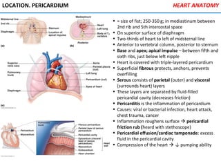
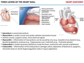
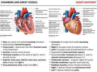
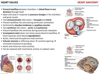
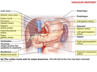
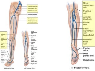
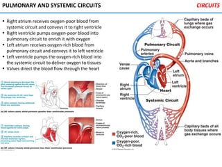
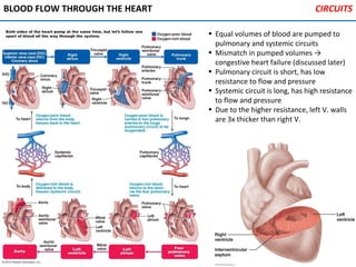
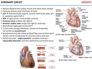
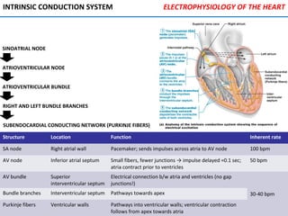
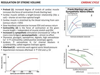
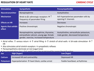
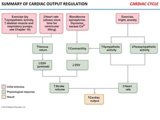
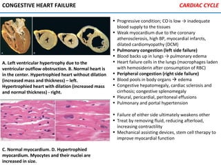
SlideShare a Scribd company logo
HEART ANATOMY
• ≈ size of fist; 250-350 g; in mediastinum between
2nd rib and 5th intercostal space
• On superior surface of diaphragm
• Two-thirds of heart to left of midsternal line
• Anterior to vertebral column, posterior to sternum
• Base and apex; apical impulse – between fifth and
sixth ribs, just below left nipple
• Heart is covered with triple-layered pericardium
• Superficial fibrous protects, anchors, prevents
overfilling
• Serous consists of parietal (outer) and visceral
(surrounds heart) layers
• These layers are separated by fluid-filled
pericardial cavity (decreases friction)
• Pericarditis is the inflammation of pericardium
• Causes: viral or bacterial infection, heart attack,
chest trauma, cancer
• Inflammation roughens surface  pericardial
friction rub (heard with stethoscope)
• Pericardial effusion/cardiac tamponade: excess
fluid in the pericardial cavity
• Compression of the heart  ↓ pumping ability
LOCATION. PERICARDIUM
THREE LAYERS OF THE HEART WALL
• Epicardium is visceral pericardium
• Myocardium is cardiac muscle and cardiac skeleton (connective tissue)
• Anchors muscle, supports valves, limits electrical signals
• Myocarditis – inflammation of myocardium; can be caused by virus (e.g., Coxackie virus), bacteria (e.g.,
Borellia burgdorferi), protozoa (e.g., Trypanosoma cruzi ), helminths (e.g., Trichinella spiralis)
• Endocardium is simple squamous epithelium inside the heart, continuous with vascular endothelium
• Endocarditis – inflammation of the endocardium; damages valves; deposition of bacterial (S. pyogenes,
rheumatic fever) or sterile (hypercoagulation state or lupus) vegetations
HEART ANATOMY
CHAMBERS AND GREAT VESSELS HEART ANATOMY
• Atria are small, thin-walled receiving chambers
• Separated by interatrial septum
• Fossa ovalis – depression left after foramen ovale
in the fetal heart
• Auricles increase atrial volume
• Pectinate muscles on the walls of right A. and in
the left auricle
• Superior vena cava, inferior vena cava, coronary
sinus empty into right A.
• Four pulmonary veins empty into left A.
• Ventricles are large thick-walled pumping
chambers
• Right V. occupies most of anterior surface
• Left V. occupies most of posteroinferior surface
• Separated by interventricular septum
• Associated sulci: coronary (between A. and V.),
anterior interventricular (between V.)
• Trabeculae carneae – irregular ridges of muscle
• Chordae tendineae regulate valve opening
• Papillary muscles anchor chordae tendineae
• Right V. delivers blood into pulmonary trunk
• Left V. delivers blood into aorta
HEART VALVES
• Prevent backflow between chambers → blood flows in one
direction through heart
• Open and close in response to pressure changes in the chambers
and great vessels
• Two atrioventricular (AV) valves – tricuspid and mitral
• Prevent backflow into atria during ventricular contraction
• Anchored by chordae tendineae to papillary muscles
• Two semilunar (SL) valves – aortic and pulmonary
• Prevent backflow into ventricles during ventricular relaxation
• Incompetent valve (does not close) causes blood to backflow 
heart repumps same blood (regurgitation)
• Aortic, mitral and tricuspid are most common
• Valvular stenosis is stiffening valves  constrict opening 
increased resistance to pumping
• Aortic and mitral are most common
• Can be replaced with mechanical, animal, or cadaver valve
HEART ANATOMY
VASCULAR ANATOMY
VASCULAR ANATOMY
VASCULAR ANATOMY
VASCULAR ANATOMY
VASCULAR ANATOMY
VASCULAR ANATOMY
VASCULAR ANATOMY
VASCULAR ANATOMY
VASCULAR ANATOMY
VASCULAR ANATOMY
PULMONARY AND SYSTEMIC CIRCUITS
• Right atrium receives oxygen-poor blood from
systemic circuit and conveys it to right ventricle
• Right ventricle pumps oxygen-poor blood into
pulmonary circuit to enrich it with oxygen
• Left atrium receives oxygen-rich blood from
pulmonary circuit and conveys it to left ventricle
• Left ventricle pumps the oxygen-rich blood into
the systemic circuit to deliver oxygen to tissues
• Valves direct the blood flow through the heart
CIRCUITS
BLOOD FLOW THROUGH THE HEART
• Equal volumes of blood are pumped to
pulmonary and systemic circuits
• Mismatch in pumped volumes →
congestive heart failure (discussed later)
• Pulmonary circuit is short, has low
resistance to flow and pressure
• Systemic circuit is long, has high resistance
to flow and pressure
• Due to the higher resistance, left V. walls
are 3x thicker than right V.
CIRCUITS
CORONARY CIRCUIT
• Delivers blood to the cardiac muscle only when heart relaxed
• Coronary arteries arise from base of aorta
• LCA  interventricular septum, anterior ventricular walls, left
A., posterior wall of left V.
• RCA  right atrium, most of right ventricle
• Coronary sinus empties into right A.
• Anterior cardiac veins empty into right A. anteriorly
• Most blood supply goes to the left V.
• Circuit varies among individuals; vessels are frequently
connected via anastomoses
• Anastomoses provide collateral blood flow around obstructed
vessel; cannot compensate for coronary artery occlusion
• Partial occlusion – angina pectoris; complete occlusion –
myocardial infarction; cells die; replaced by connective tissue
CIRCUITS
• Contractions are involuntary; do not require nervous
stimulation (pacemaker cells)
• Cells are striated, short, branched, 1-2 nuclei connected by
gap junctions; many mitochondria; wide T tubules
• Sarcoplasmic reticulum is simpler than in skeletal muscle
• Endomysium connects to cardiac skeleton; many capillaries
• Intercalated discs between cells contain desmosomes and
gap junctions
• Desmosomes hold cardiomyocytes together
• Gap junctions electrically couple adjacent cells → functional
syncytium
• Mechanism of contraction is similar to the skeletal muscle
ANATOMY OF A CARDIAC MUSCLE CARDIAC MUSCLE
ROLE OF CA2+
. SLIDING FILAMENTS
• Ca2+
low: tropomyosin (TPM) blocks myosin (M.) -binding sites on actin (A.) → M. heads can’t attach →
muscle fiber relaxed
• Ca2+
high: Ca2+
binds troponin (TN) → TN shifts TPM away from M.-binding sites on A. → M. heads attach to
A. → contraction
• Ca2+
is pumped back into SR → contraction stops
CARDIAC MUSCLE
Step Ion flow Voltage-gated Na+
channels
Voltage-gated K+
channels
Voltage-gated Ca+
channels
Depolarization Na+
in Open Closed Closed
Plateau Ca2+
in, K+
out Closed Open Open
Repolarization K+
out Inactivated Open Closed
PHYSIOLOGY OF CONTRACTION
• Rapid influx of Na+
→ depolarization of cardiomyocytes (1)
• Depolarization closes Na+
voltage-gated channels (VGC),
opens Ca2+
VGC
• Influx of Ca2+
maintains depolarization (plateau on the pic, 2)
• Plateau allows for extended contraction of the muscle
• Ca2+
VGC close, K+
VGC open; efflux of K+
→ repolarization
• When Na+
VGC close at the peak, they are inactivated –
nothing can open them → absolute refractory period
• Prevents possible tetanic contractions
• Metabolism is aerobic; little anaerobic ability
• Can use multiple fuel sources (i.e., lactic acid)
• Anaerobic respiration may cause damage to the cells
CARDIAC MUSCLE
• Ischemia anaerobic respiration  lactic acid  pH↓  Ca2+
↑  mitochondrial damage  decreased
ATP production  gap junctions close  lack of connection  fatal arrhythmias
PACEMAKER CELLS
• Heart can contract without nervous stimulation
• Heartbeat is coordinated by gap junctions and noncontractile autorhythmic (pacemaker) cells
• Pacemaker cells have unstable resting membrane potential
ELECTROPHYSIOLOGY OF THE HEART
Step Ion flow Na+
VGC K+
VGC Ca+
VGC
Pacemaker potential Na+
in Open Closed Closed
Depolarization Ca2+
in Open Closed Open
Repolarization K+
out Closed Open Inactivated
Structure Location Function Inherent rate
SA node Right atrial wall Pacemaker; sends impulses across atria to AV node 100 bpm
AV node Inferior atrial septum Small fibers, fewer junctions → impulse delayed ≈0.1 sec;
atria contract prior to ventricles
50 bpm
AV bundle Superior
interventricular septum
Electrical connection b/w atria and ventricles (no gap
junctions!)
30-40 bpmBundle branches Interventricular septum Pathways towards apex
Purkinje fibers Ventricular walls Pathways into ventricular walls; ventricular contraction
follows from apex towards atria
INTRINSIC CONDUCTION SYSTEM ELECTROPHYSIOLOGY OF THE HEART
SINOATRIAL NODE
ATRIOVENTRICULAR NODE
ATRIOVENTRICULAR BUNDLE
SUBENDOCARDIAL CONDUCTING NETWORK (PURKINJE FIBERS)
RIGHT AND LEFT BUNDLE BRANCHES
• All action potentials generated by nodal and contractile cells at given time
• P wave – depolarization of the SA node  atrial depolarization
• QRS complex – ventricular depolarization and atrial repolarization
• T wave – ventricular repolarization
• P-R interval: start of atrial depolarization to the start of ventricular depolarization
• S-T segment: entire ventricular myocardium depolarized
• Q-T interval: start of ventricular depolarization to the end of ventricular repolarization
NORMAL ELECTROCARDIOGRAM ELECTROPHYSIOLOGY OF THE HEART
• Cardioinhibitory (parasympathetic) center in medulla oblongata (MO)
• Normally inhibits SA, AV nodes via vagus nerve → normal heart rate (HR) @ 70 bpm
• Cardioacceleratory (sympathetic) center in MO stimulates SA, AV nodes via sympathetic nerves →↑ HR
REGULATION OF HEART RATE ELECTROPHYSIOLOGY OF THE HEART
Tachycardia
Fast heart rate (>100 bpm)
EKG normal otherwise; causes:
•fever (10 bpm per 1°C ↑)
•sympathetic stimulation
•toxins
•blood loss
Bradycardia
Slow heart rate (<60 bpm)
EKG normal otherwise; causes:
•athletic training
•parasympathetic stimulation (carotid
sinus syndrome – extremely sensitive
baroreceptors in carotid sinuses)
BLOCK OF INTRACARDIAL PATHWAYS ELECTROPHYSIOLOGY OF THE HEART
SA block
Signal is blocked at SA node before it
enters atrial muscle; no P waves;
ventricles set the rhythm (slower, 40-
60 bpm – junctional rhythm)
AV block
Decreased rate of impulse conduction
or complete blockage via AV bundle
•1st
degree – extended P-R interval
(delay of conduction)
•2nd
degree – dropped beat (AP
sometimes can’t get through)
•3rd
degree – complete block; rhythm
originates in AV node or AV bundle
•Too slow (30-40 bpm), pacemaker
needed
ECTOPIC FOCI. FIBRILLATION ELECTROPHYSIOLOGY OF THE HEART
Premature
atrial
contraction
Ectopic focus is in the atria; extended interval
between contractions is compensatory pause;
can be observed in healthy people; caffeine,
nicotine, lack of sleep can be a cause
AV nodal
/bundle
premature
contraction
Impulse travels to ventricles and backwards to
atria; P wave is superimposed on the QRS-T
complex; same importance and causes as
premature atrial contractions
Ventricular
premature
contraction
QRS complex is prolonged, with higher
voltage; T wave has inverted electrical
polarity; can have same causes as previous
two; sign for higher risk for ventricular
fibrillation
Atrial
fibrillation
Uncoordinated contractions of atrial muscle;
↓pumping for 20-30%; may be caused by
atrial enlargement due to the defective valves
Ventricular
fibrillation
Uncoordinated contractions of ventricular
muscle resulting in no pumping of blood;
caused by electric shock or ischemia
• Ectopic focus – pacemaker out of SA node; may be due local ischemia, calcifications, or chemicals
(caffeine, nicotine, drugs)
THE CARDIAC CYCLE
• Cardiac cycle describes blood flow through the heart during one complete heartbeat with all
accompanying electrical events, pressure changes, opening and closing of valves
• Systole is the contraction, and diastole is relaxation (of atria or ventricles)
• Mid-to-late diastole; AV valves open; SL
valves closed; Pa > Pv < Pvessel
• 80% of blood passively flows into ventricles
• Atrial systole delivers remaining 20%
• End diastolic volume (EDV): volume of
blood in the ventricle at the end of diastole
• Atria relax; ventricles contract
• Isovolumetric contraction : all valves
closed; Pa < Pv < Pvessel
• Ejection : AV valves closed, SL valves
open; Pa < Pv > Pvessel
• End systolic volume (ESV): volume of
blood in the ventricle after systole
• Ventricles relax
• Isovolumetric relaxation: all
valves closed; Pa < Pv < Pvessel
• Backflow of blood in aorta →
dicrotic notch (spike in BP)
• Goes back to ventricular filling
CARDIAC CYCLE
• Valves close, produce two
sounds (lub-dup)
• First as AV valves close;
beginning of ventricular
systole
• Second as SL valves close;
beginning of ventricular
diastole
• Pause – heart relaxation
• Heart murmurs - abnormal
heart sounds; usually
indicate incompetent or
stenotic valves
CARDIAC OUTPUT
• Volume of blood pumped by each ventricle in one minute: CO = heart rate (HR) × stroke volume (SV)
• HR = number of beats per minute; SV = volume of blood pumped out by one ventricle with each beat
CARDIAC OUTPUT ↑CARDIAC OUTPUT ↑
ARTERIAL PRESSURE ↓ARTERIAL PRESSURE ↓ FORCE OF CONTRACTION ↑FORCE OF CONTRACTION ↑
LENGTH OF DIASTOLELENGTH OF DIASTOLE VENOUS RETURN/PRESSUREVENOUS RETURN/PRESSURE
SYMPATHETIC STIMULATION ↑SYMPATHETIC STIMULATION ↑
HEART RATE ↑HEART RATE ↑ STROKE VOLUME ↑STROKE VOLUME ↑
EDV ↑EDV ↑ ESV ↓ESV ↓
CARDIAC CYCLE
CO HR SV EDV ESV
Increased if… Increased Increased Increased Decreased
Decreased if… Decreased Decreased Decreased Increased
• SV = EDV – ESV
• CO = HR × (EDV –
ESV)
• CO at rest (ml/min) = HR (75 beats/min) × SV (70 ml/beat) = 5.25 L/min
• COmax = 4–5x COrest in nonathletic people; 7x in trained athletes; cardiac reserve
(CR) = COmax - COrest
Volume Increased by Decreased by
EDV Long diastole, ↑ venous pressure Short diastole, ↓ venous return/pressure
ESV ↑ blood pressure, ↓ contractility ↓ BP, ↑ contractility
REGULATION OF STROKE VOLUME
• Preload (1): increased degree of stretch of cardiac muscle
increases the force of contraction (Frank-Starling law)
• Cardiac muscle exhibits a length-tension relationship (like a
coil) – shorter at rest than optimal length
• Cardiac muscle is stretched by the blood returning from vein
(venous return = EDV)
• Slow heartbeat and exercise increase EDV and venous return
• Contractility (2) is the force of contraction at the given
muscle length – independent of muscle stretch and EDV
• Increased by sympathetic stimulation (increased Ca2+
influx 
more cross bridges); parasympathetic – almost no effect
• Thyroxine, glucagon, epinephrine, digitalis, high extracellular
Ca2+
→ ↑ contractility; called positive ionotropic agents
• Acidosis, ↑ extracellular K+
, Ca2+
channel blockers →
↓contractility; called negative inotropic agents
• Afterload (3) – ventricles work against aortic blood pressure
• Hypertension increases afterload → ↑ ESV and ↓ SV
1
2
3
CARDIAC CYCLE
• Atrial reflex: ↑ venous return → ↑ atrial filling → ↑ stretch of atrial walls → SA node stimulation  ↑
HR
• Also stimulates atrial stretch receptors → sympathetic reflexes
• Parasympathetics dominate at rest (vagal tone)
• Ion concentrations (e.g., Ca2+
and K+
) must be maintained for normal heart function
Stimulation Sympathetic Parasympathethic
Neurotransmitter Norepinephrine Acetylcholine
Mechanism Binds to β1-adrenergic receptors  ↑
frequency of pacemaker firing
Ach hyperpolarizes pacemaker cells by
opening K+
channels
Effects on HR Increases Decreases
Agents that exhibit
effect
Positive chronotropic Negative chronotropic
Norepinephrine, epinephrine, thyroxine,
extracellular calcium, young age, female
gender, exercise, increased temperature
Acetylcholine, extracellular potassium,
male gender, decreased temperature
REGULATION OF HEART RATE CARDIAC CYCLE
Ion High (hyper) Low (hypo)
Calcium Increased HR and contractility Depressed HR
Potassium Hyperpolarization  heart block, cardiac arrest Feeble heartbeat; arrhythmias
SUMMARY OF CARDIAC OUTPUT REGULATION CARDIAC CYCLE
CONGESTIVE HEART FAILURE
• Progressive condition; CO is low → inadequate
blood supply to the tissues
• Weak myocardium due to the coronary
atherosclerosis, high BP, myocardial infarcts,
dilated cardiomyopathy (DCM)
• Pulmonary congestion (left side failure)
• Blood backs up in lungs → pulmonary edema
• Heart failure cells in the lungs (macrophages laden
with hemosiderin after consumption of RBC)
• Peripheral congestion (right side failure)
• Blood pools in body organs  edema
• Congestive hepatomegaly, cardiac sclerosis and
cirrhosis; congestive splenomegaly
• Pleural, pericardial, peritoneal effusions
• Pulmonary and portal hypertension
• Failure of either side ultimately weakens other
• Treat by removing fluid, reducing afterload,
increasing contractility
• Mechanical assisting devices, stem cell therapy to
improve myocardial function
A. Left ventricular hypertrophy due to the
ventricular outflow obstruction. B. Normal heart is
in the center. Hypertrophied heart without dilation
(increased mass and thickness) – left.
Hypertrophied heart with dilation (increased mass
and normal thickness) - right.
C. Normal myocardium. D. Hypertrophied
myocardium. Myocytes and their nuclei are
increased in size.
CARDIAC CYCLE
DDEVELOPING HEART. LEFT-TO-RIGHT SHUNTS
• Embryonic heart chambers: sinus venosus (1), atrium (2), ventricle (3), bulbus cordis (4)
• Fetal heart structures bypass pulmonary circulation; foramen ovale connects atria (fossa ovalis in adults)
• Ductus arteriosus connects pulmonary trunk to aorta (ligamentum arteriosum in adults); close after birth
CONGENITAL HEART DISEASE
Left-to-right shunts
Atrial septal defect (ASD)
Ventricular septal defect (VSD)
Patent ductus arteriosus (PDA)
Pulmonary hypertension, cyanosis, heart
failure, embolization, altered hemodynamics
with dilation or hypertrophy
LEFT-TO-RIGHT SHUNTS. OBSTRUCTIONS
Right-to-left
shunts
Tetralogy of Fallot (VSD, subpulmonary
stenosis, aorta overrides VSD, RV hypertrophy)
Transposition of great arteries (simultaneous
shunts are frequent)
Cyanosis
Complete separation of circuits (incompatible with life),
TV hypertrophy, LV hypotrophy
Obstructions Coarctation of aorta
Aortic stenosis or atresia
(blockade)
Cyanosis, high BP in upper
extremities and low BP in
lower extremities, arterial
insufficiency
LV hypoplasia (atresia) or
hypertrophy (stenosis)
CONGENITAL HEART DISEASE
AGING HEART CARDIAC DISEASE
WALL LAYERS
• Arteries carry blood away from heart, veins carry blood
toward heart; capillaries is the site of exchange in the tissues
Vessels Tunics Consist of…
Arteries
and veins
intima Endothelium (reduces friction, regulates vasomotor responses), basal lamina
media Smooth muscle (vasodialtion/constriction), elastic fibers
externa Collagen fibers (protection and structure), lymphatics, nerve fibers, vasa vasorum (blood supply
for the walls of large vessels)
Capillaries Endothelium and basal lamina (rapid exchange between blood and tissues)
BLOOD VESSELS
ARTERIES
Elastic Largest (aorta and branches); elastin in all tunics; large lumen = low
resistance; no active vasomotor responses; pressure reservoir (expand
and recoil)
Muscular Distal to elastic; deliver blood to organs; thick t. media; active
vasomotor responses
Arterioles Smallest; deliver blood to tissues; active in vasoconstriction
BLOOD VESSELS
VEINS
• Large lumens → little resistance; venous valves prevent backflow
• Blood goes back to the heart easy despite the low pressure
Venules Post-capillary (smallest, porous, fluid and WBC transport, no SMC); SMC
appear as size increases (venules converge)
Veins Larger lumens, thinner walls, and lower BP compared to arteries; thin t.
media (weak vasomotor responses); capacitance vessels (60% of blood is
in venules any given time)
Venous
sinuses
Flattened veins with extremely thin walls (e.g., coronary sinus of the
heart and dural sinuses of the brain)
BLOOD VESSELS
CAPILLARIES
• Smallest blood vessels
• Endothelium and basal lamina
• Pericytes control stability and permeability
• Diameter of a single cell
• Walls of thin tunica intima
• Not found in cartilage, epithelia, cornea, eye lens
• Direct access to almost every cell
• Exchange of gases, nutrients, wastes, hormones,
etc., between blood and interstitial fluid
BLOOD VESSELS
TYPES OF CAPILLARIES
Type Location Features
Continuous Skin, muscles, brain Tight junctions b/w endothelial
cells; intercellular clefts
(passage of fluids and small
molecules); blood-brain barrier
in the brain
Fenestrated Small intestine,
endocrine glands,
kidneys
More permeable; fenestrations;
absorption or filtration
Sinusoid Liver, bone marrow,
spleen, adrenal
medulla
Few tight junctions;
fenestrations; large clefts and
lumens; slow blood flow;
effective exchange;
macrophages in the lining
BLOOD VESSELS
BLOOD FLOW THROUGH CAPILLARY
BEDS
• Microcirculation: ≈ 95% vessels are microvessels
(Ø <100 µm)
• Networks of capillaries between arterioles and
venules – capillary bed; different properties in
different organs
• First signs of disease appear in microvessels before
signs are apparent in large vasculature
• Terminal arteriole → metarteriole → capillaries
and thoroughfare channel → postcapillary venule
• Thoroughfare channel is vascular shunt
(anastomose)
• Can be arterial (e.g., coronary circulation), venular
(frequent), or arteriovenular (thoroughfare
channel)
• 10 to 100 exchange vessels per capillary bed
• Branch from metarteriole and return to
thoroughfare channel
• Precapillary sphincters regulate capillary blood
flow
• Blood flow through the capillary bed is regulated
by local chemical cues, hormones, and nerves
PHYSIOLOGY OF CIRCULATION
Blood flow (BF) Volume of blood flowing through
vessel, organ, or entire circulation
in given period [ml/min]
Equivalent to cardiac output; constant at rest; local BF varies
between organs
Blood pressure
(BP)
Force per unit area exerted on wall
of blood vessel by blood [mm Hg]
Measured as systemic BP in the large arteries; proportional to
blood volume (BV); blood follows pressure gradient (∆BP)
Peripheral
resistance (PR)
Opposition to flow Amount of friction blood encounters with vessel walls, generally
in peripheral (systemic) circulation
FLOW, PRESSURE, AND RESISTANCE
BLOOD FLOW IN THE SYSTEMIC CIRCUIT
• Heart pumps blood → BF is generated
• BF is opposed by PR →BP is generated
• Systemic pressure is highest in aorta, declines
through the circuit, lowest in the right atrium
• Steepest BP drop in arterioles (resistance vessels)
• Arterial pressure depends on the elasticity of and
blood volume in the large arteries (aorta etc.)
• BP near heart is pulsatile
• Systolic (≈120 mm Hg), diastolic (≈70-80 mm Hg)
• Pulse pressure (PP) = SBP – DBP (this is pulse)
• Mean arterial pressure (MAP) propels blood to
tissues; MAP = DBP + 1/3 PP
• Ex. BP = 120/80; MAP = 93 mm Hg
• PP and MAP decrease away from heart
• Capillary from 17 to 35 mm Hg; lower is better
• Otherwise may cause damage and edema
• Venous is almost constant (≈15 mm Hg); low due to the low PR
• Venous return is amount of blood coming back to the heart from veins
• Muscular pump: contraction of skeletal muscles "milks" veins
• Respiratory pump: pressure changes during breathing
• Squeeze abdominal veins, expand thoracic veins
• Valves prevent backflow; venoconstriction is possible
PHYSIOLOGY OF CIRCULATION
REGULATION OF BLOOD PRESSURE
• Depends on and regulated by the changes in CO (heart), PR (blood vessels), BV (kidneys)
• Neural, chemical, and hormonal short-term mechanisms alter CO and PR; renal long-term responses – BV
PHYSIOLOGY OF CIRCULATION
NERVOUS, CHEMICAL, HORMONAL, HYPOTHALAMUS
• Baroreceptors (mechanoreceptors) sense ↑or ↓ stretch of the vessel wall and then respectively
• Signals to cardioinhibitory or cardioacceleratory centers in the medulla → ↓ or ↑ CO → ↓ or ↑ BP
• Cardiac inhibition is parasympathetic, acceleration is sympathetic
• Signals to vasomotor center in the medulla → vasodilation or vasoconstriction (entirely sympathetic)
• Chemoreceptors in aortic arch and large arteries
of neck detect ↑CO2, or ↓ pH or O2
• To cardioacceleratory center  ↑ CO → BP ↑
• To vasomotor center  ↑ vasoconstriction
• Altogether BF ↑, chemistry of the blood restored
• Hypothalamus can increase BP during stress
(involves cortical regulation) and during exercise
• Relaying signals through medulla oblongata
• (Nor)EPI ↑ BP via ↑ CO, vasoconstriction
• Angiotensin II, ADH ↑ BP via ↑ vasoconstriction
• ANP ↓ BP via ↓ BV (aldosterone antagonist)
SHORT-TERM CONTROLS
RENAL
• Baroreceptors quickly adapt to chronic BP changes
• BP controlled by altering BV via kidneys
• DIRECT MECHANISM (hormone-independent):
• ↑ BP or blood volume → ↑ urination → ↓ BP
• ↓ BP or blood volume → ↓ urination → ↑ BP
• INDIRECT MECHANISM (renin-angiotensin-
aldosterone system, RAAS)
∀ ↓ BP → ↑ renin
• Renin converts angiotensinogen (liver) to
angiotensin I
• Angiotensin converting enzyme (ACE) (lungs)
conversts angiotensin I to angiotensin II
• Angiotensin II → ↑ aldosterone and ADH
• Result: ↑ BV, thirst, vasoconstriction → ↑ BP
LONG-TERM CONTROLS
• ACE inhibitors are used to treat hypertension and heart failure
• First predecessor – from the venom of the South American pit viper
• Cushman and Ondetti studied structure of the first inhibitor
• Eventually developed captopril – the first ACE inhibitor drug
• Lasker Award 1999
SUMMARY OF BLOOD PRESSURE CONTROL PHYSIOLOGY OF CIRCULATION
MEASURING OF CIRCULATORY PARAMETERS AND BP
• Vital signs: pulse, blood pressure, respiratory rate, body temperature
• Pulse: pressure wave caused by expansion and recoil of arteries
• Pressure points where arteries are close to body surface; usually at the
wrist (radial artery)
• Systemic BP measured by auscultation (via sphygmomanometer)
• Cuff pressure > systolic pressure in brachial artery
• As pressure decreases, Korotkoff sounds examined by stethoscope
• Systolic BP: blood spurts through artery, sound appears
• Diastolic BP: blood flows freely, sound disappears
BLOOD PRESSURE
ALTERATIONS IN BP
• Transient changes: changes in posture, exercise,
stress, fever
• Age, sex, weight, race, mood → variations in BP
• Hypertension: sustained BP > 140/90 mm Hg
• Prehypertension: elevated BP
• Often persistent in obesity, diabetes
• Hypertension → heart failure, vasculopathy, renal
failure, atherosclerosis, cardiomyopathy, stroke
• Primary hypertension: 90% of HT
• Risk factors: genetics, diet, obesity, age, diabetes
mellitus, stress, and smoking
• No cure, can be controlled: ↓ salt, ↑ exercise, ↓
weight, XXX smoking, antihypertensive drugs
• Secondary hypertension is less common
• Causes; obstructed renal arteries, kidney disease,
endocrine disorders (hyperthyroidism, Cushing's)
• Treatment focuses on correcting underlying cause
• Hypotension: BP < 90/60 mm Hg; usually not a
concern; only if BF to tissues become inadequate
• Orthostatic hypotension: temporary low BP and
dizziness when standing up
• Chronic hypotension: hint of poor nutrition,
Addison's disease, hypothyroidism
• Acute hypotension: circulatory shock
BLOOD PRESSURE
Circulatory Shock
• Inadequate tissue perfusion and
circulation
• Hypovolemic shock: extreme
blood loss
CIRCULATORY SHOCK
• Vascular shock: vasodilation and ↓ PR
• Cardiogenic shock: cardiac insufficiency
→ inadequate circulation
TISSUE PERFUSION
• Delivery of O2 and nutrients, removal of CO2 and wastes
• Gas exchange (lungs), nutrient absorption of nutrients
(digestive tract), urine formation (kidneys)
• Flow matches tissue requirements
• Different in different tissues
• Inversely related to total cross-sectional area
• Fast in arteries; slow in capillaries (exchange); faster in veins
BLOOD FLOW THROUGH THE TISSUES
REGULATION OF TISSUE PERFUSION
• Tissue perfusion (TP) adjusts to tissue needs
• Organs control TP by altering arteriolar diameter
• Local control – independent of systemic BP
regulation (should be at the constant level)
• Regulates via metabolic AND myogenic controls
• METABOLIC CONTROLS
• Metabolically active tissues: ↑ CO2, ↓ pH, ↓ O2
• ↑ K+
, adenosine, prostaglandins
• Arterioles dilate, precapillary sphincters relax
• Endothelial cells release NO (vasodilator)
• NO acts on SMC, overcoming endothelin-mediated
vasoconstriction
• Inflammatory mediators also cause vasodilation
(e.g., kinins, prostaglandins, adenosine)
• MYOGENIC CONTROLS
• Balance changes in the systemic BP
• SMC respond to stretch
• ↑ stretch (↑ BP) → vasoconstriction → ↓ BF
• ↓ stretch → vasodilation → ↑ BF
• LONG-TERM REGULATION
• Angiogenesis: BF does not match tissue
needs in the long-term perspective
• Number of vessels ↑, existing vessels
enlarge
• Heart (coronary circuit), adaptation to
high altitude, cancer
BLOOD FLOW THROUGH THE TISSUES
MUSCLES. BRAIN. SKIN
Muscles • At rest, myogenic and neural mechanisms predominate (~ 1L /minute)
• Exercise hyperemia – blood flow increases in direct proportion to metabolic activity
• Metabolic controls override sympathetic vasoconstriction; can increase 10×
Brain • BF constant (neurons cannot survive without O2), ≈750 ml/min
• Metabolic controls: ↓ pH, ↑ CO2 → vasodilation
• Myogenic controls: ↓ BP → vasodilation → ↑ BF; ↑ BP) → vasoconstriction → ↓ BF (see pic)
• BP < 60 mm Hg → syncope (fainting); BP > 160 mm Hg → cerebral edema
Skin • BF to subcutaneous venous plexuses regulates body
temperature
• Varies from 50 ml/min to 2500 ml/min
• Controlled by sympathetic reflexes (initiated by
thermoreceptors)
• Hypothalamus → ↓ vasomotor center
• Vasodilation, heat dissipates
• Sweat contains bradykinin → vasodilation
Lungs • Arterial resistance and pressure are low (24/10 mm Hg)
• Metabolic regulation is opposite: ↓ O2 → vasoconstriction; ↑ O2 → vasodilation
• Blood flows to O2-rich areas of lung (ventilation-perfusion coupling)
Heart • Systole: coronary vessels are compressed, myocardial BF ↓, O2 from myoglobin
• Diastole: pressure from aorta forces blood through coronary circulation
• At rest ~ 250 ml/min; myogenic control
• Strenuous exercise: coronary vessels dilate (chemical regulation), BF ↑ 3x
BLOOD FLOW THROUGH THE TISSUES
CAPILLARY FLOW
• Capillary flow is slow and intermittent
• Diffusion of O2, CO2, nutrients, and wastes (b/w tissue and blood)
• Lipid-soluble – simple diffusion; water-soluble – clefts, fenestrations
(smaller molecules), pinocytosis (e.g., proteins)
• Fluid leaves capillaries at arterial
end, comes back at venular end
• Creates permanent fluid flow
through the tissues
BLOOD FLOW THROUGH THE TISSUES
CAPILLARY PRESSURES
Pressure Mechanism and
direction
Numbers
Capillary
hydrostatic
pressure (HPc)
Forces fluid
through the
capillary walls into
the tissue
Arterial end: 35
mm Hg
Venular end: 17
mm Hg
Interstitial fluid
hydrostatic
pressure (HPif)
Forces fluid
through the
capillary walls into
the blood
Assumed to be 0
due to the
lymphatic
drainage
Capillary colloid
osmotic pressure
(OPc)
Fluid goes
towards plasma
proteins; pulled
towards the blood
26 mmHg
Interstitial fluid
osmotic pressure
(OPif)
Fluid goes
towards (low)
proteins in the IF;
pulled into the
tissues
1 mmHg
BLOOD FLOW THROUGH THE TISSUES
NET FILTRATION PRESSURE: ARTERIAL END BLOOD FLOW THROUGH THE TISSUES
NET FILTRATION PRESSURE: VENULAR END BLOOD FLOW THROUGH THE TISSUES

More Related Content

What's hot

Anatomy and physiology of urinary system
Anatomy and physiology of urinary systemAnatomy and physiology of urinary system
Anatomy and physiology of urinary system
Pallavi Lokhande
 
The valves of the heart
The valves of the heartThe valves of the heart
The valves of the heart
Idris Siddiqui
 
Anatomy And Physiology of Human Heart
Anatomy And Physiology of Human Heart Anatomy And Physiology of Human Heart
Anatomy And Physiology of Human Heart
C L GUPTA EYE INSTITUTE MORADABAD UTTER PRADESH
 
Anatomy of heart
Anatomy of heartAnatomy of heart
Anatomy of heart
nmonty02
 
Blood vessels
Blood vesselsBlood vessels
Blood vessels
Vaibhav Srivastav
 
Chapter 15 - The Cardiovascular System - Part 1
Chapter 15 -  The Cardiovascular System - Part 1Chapter 15 -  The Cardiovascular System - Part 1
Chapter 15 - The Cardiovascular System - Part 1biol2074
 
Mediastinum
MediastinumMediastinum
Mediastinum
Dr Laxman Khanal
 
Cardiac Cycle.pptx
Cardiac Cycle.pptxCardiac Cycle.pptx
Cardiac Cycle.pptx
Vipin Chandran
 
1. pericardium
1. pericardium1. pericardium
1. pericardium
Dr. Mohammad Mahmoud
 
Cardio vascular system Anatomy and physiology
Cardio vascular system Anatomy and physiology Cardio vascular system Anatomy and physiology
Cardio vascular system Anatomy and physiology
Shafici Almis
 
Human heart
Human heartHuman heart
Human heart
Vijay Salvekar
 
Anatomy of Cardiac System
Anatomy of Cardiac SystemAnatomy of Cardiac System
Anatomy of Cardiac System
DR .PALLAVI PATHANIA
 
Heart Anatomy and Physiology:Review
Heart Anatomy and Physiology:ReviewHeart Anatomy and Physiology:Review
Heart Anatomy and Physiology:ReviewTosca Torres
 
Heart
HeartHeart
The coronary circulation
The coronary circulationThe coronary circulation
The coronary circulation
Idris Siddiqui
 
cardiovascular physiology
 cardiovascular physiology cardiovascular physiology
cardiovascular physiology
aliagr
 
ANATOMY AND PHYSIOLOGY OF THE HEART
ANATOMY AND PHYSIOLOGY OF THE HEARTANATOMY AND PHYSIOLOGY OF THE HEART
ANATOMY AND PHYSIOLOGY OF THE HEART
Praveen Nagula
 
Descripition of heart and purkinje fibres
Descripition of heart and purkinje fibres Descripition of heart and purkinje fibres
Descripition of heart and purkinje fibres
Ali Muntazir
 

What's hot (20)

Anatomy and physiology of urinary system
Anatomy and physiology of urinary systemAnatomy and physiology of urinary system
Anatomy and physiology of urinary system
 
The valves of the heart
The valves of the heartThe valves of the heart
The valves of the heart
 
Anatomy And Physiology of Human Heart
Anatomy And Physiology of Human Heart Anatomy And Physiology of Human Heart
Anatomy And Physiology of Human Heart
 
Anatomy of heart
Anatomy of heartAnatomy of heart
Anatomy of heart
 
Blood vessels
Blood vesselsBlood vessels
Blood vessels
 
Chapter 15 - The Cardiovascular System - Part 1
Chapter 15 -  The Cardiovascular System - Part 1Chapter 15 -  The Cardiovascular System - Part 1
Chapter 15 - The Cardiovascular System - Part 1
 
Mediastinum
MediastinumMediastinum
Mediastinum
 
Anatomy and function of the coronary arteries
Anatomy and function of the coronary arteriesAnatomy and function of the coronary arteries
Anatomy and function of the coronary arteries
 
Cardiac Cycle.pptx
Cardiac Cycle.pptxCardiac Cycle.pptx
Cardiac Cycle.pptx
 
1. pericardium
1. pericardium1. pericardium
1. pericardium
 
Cardio vascular system Anatomy and physiology
Cardio vascular system Anatomy and physiology Cardio vascular system Anatomy and physiology
Cardio vascular system Anatomy and physiology
 
Human heart
Human heartHuman heart
Human heart
 
Anatomy of Cardiac System
Anatomy of Cardiac SystemAnatomy of Cardiac System
Anatomy of Cardiac System
 
blood supply of heart
blood supply of heartblood supply of heart
blood supply of heart
 
Heart Anatomy and Physiology:Review
Heart Anatomy and Physiology:ReviewHeart Anatomy and Physiology:Review
Heart Anatomy and Physiology:Review
 
Heart
HeartHeart
Heart
 
The coronary circulation
The coronary circulationThe coronary circulation
The coronary circulation
 
cardiovascular physiology
 cardiovascular physiology cardiovascular physiology
cardiovascular physiology
 
ANATOMY AND PHYSIOLOGY OF THE HEART
ANATOMY AND PHYSIOLOGY OF THE HEARTANATOMY AND PHYSIOLOGY OF THE HEART
ANATOMY AND PHYSIOLOGY OF THE HEART
 
Descripition of heart and purkinje fibres
Descripition of heart and purkinje fibres Descripition of heart and purkinje fibres
Descripition of heart and purkinje fibres
 

Viewers also liked

ppt of anatomy of heart & ML imp
ppt of anatomy of heart & ML impppt of anatomy of heart & ML imp
ppt of anatomy of heart & ML impGopal Hargi
 
Heart Anatomy
Heart AnatomyHeart Anatomy
Heart Anatomy
Chy Yong
 
Heart anatomy
Heart anatomyHeart anatomy
Heart anatomy
Dr. Waqas Nawaz
 
Anatomy of the Heart
Anatomy of the HeartAnatomy of the Heart
Anatomy of the Heart
Kenechukwu Nnamani
 
Anatomy of the heart
Anatomy of the heartAnatomy of the heart
Anatomy of the heartKhalid Musa
 
Anatomy of the heart
Anatomy of the heartAnatomy of the heart
Anatomy of the heart
Zeyad Magdy
 
Anatomy of heart
Anatomy of heart Anatomy of heart
Anatomy of heart
Aditya Reza
 
Anatomy of Heart
Anatomy of HeartAnatomy of Heart
Anatomy of Heart
Arzoo Baltistani
 
Anatomy Unit 2: Heart Anatomy & Function Notes
Anatomy Unit 2: Heart Anatomy & Function NotesAnatomy Unit 2: Heart Anatomy & Function Notes
Anatomy Unit 2: Heart Anatomy & Function Notes
rozeka01
 
Ap2 chap18heartclass
Ap2 chap18heartclassAp2 chap18heartclass
Ap2 chap18heartclass
MissReith
 
MDCT Anatomy of Heart Dr. Muhammad Bin Zulfiqar
MDCT Anatomy of Heart Dr. Muhammad Bin Zulfiqar MDCT Anatomy of Heart Dr. Muhammad Bin Zulfiqar
MDCT Anatomy of Heart Dr. Muhammad Bin Zulfiqar
Dr. Muhammad Bin Zulfiqar
 
Peptic ulcer disease
Peptic ulcer diseasePeptic ulcer disease
Peptic ulcer disease
Nomin-Erdene Dorjsambuu
 
External features of heart
External features of heartExternal features of heart
External features of heart
mgmcri1234
 
Section 1, chapter 15: anatomy of the heart
Section 1, chapter 15: anatomy of the heartSection 1, chapter 15: anatomy of the heart
Section 1, chapter 15: anatomy of the heart
Michael Walls
 
The Heart (Anatomy of the Thorax)
The Heart (Anatomy of the Thorax)The Heart (Anatomy of the Thorax)
The Heart (Anatomy of the Thorax)
Dr. Sherif Fahmy
 
The Respiratory System
The Respiratory SystemThe Respiratory System
The Respiratory System
raj kumar
 

Viewers also liked (20)

ppt of anatomy of heart & ML imp
ppt of anatomy of heart & ML impppt of anatomy of heart & ML imp
ppt of anatomy of heart & ML imp
 
Anatomy of heart
Anatomy of heartAnatomy of heart
Anatomy of heart
 
Heart Anatomy
Heart AnatomyHeart Anatomy
Heart Anatomy
 
Heart Anatomy
Heart AnatomyHeart Anatomy
Heart Anatomy
 
Heart anatomy
Heart anatomyHeart anatomy
Heart anatomy
 
Anatomy of the Heart
Anatomy of the HeartAnatomy of the Heart
Anatomy of the Heart
 
Heart
HeartHeart
Heart
 
Anatomy of the heart
Anatomy of the heartAnatomy of the heart
Anatomy of the heart
 
Anatomy of the heart
Anatomy of the heartAnatomy of the heart
Anatomy of the heart
 
Anatomy of heart
Anatomy of heart Anatomy of heart
Anatomy of heart
 
Anatomy of Heart
Anatomy of HeartAnatomy of Heart
Anatomy of Heart
 
Anatomy Unit 2: Heart Anatomy & Function Notes
Anatomy Unit 2: Heart Anatomy & Function NotesAnatomy Unit 2: Heart Anatomy & Function Notes
Anatomy Unit 2: Heart Anatomy & Function Notes
 
The heart
The heartThe heart
The heart
 
Ap2 chap18heartclass
Ap2 chap18heartclassAp2 chap18heartclass
Ap2 chap18heartclass
 
MDCT Anatomy of Heart Dr. Muhammad Bin Zulfiqar
MDCT Anatomy of Heart Dr. Muhammad Bin Zulfiqar MDCT Anatomy of Heart Dr. Muhammad Bin Zulfiqar
MDCT Anatomy of Heart Dr. Muhammad Bin Zulfiqar
 
Peptic ulcer disease
Peptic ulcer diseasePeptic ulcer disease
Peptic ulcer disease
 
External features of heart
External features of heartExternal features of heart
External features of heart
 
Section 1, chapter 15: anatomy of the heart
Section 1, chapter 15: anatomy of the heartSection 1, chapter 15: anatomy of the heart
Section 1, chapter 15: anatomy of the heart
 
The Heart (Anatomy of the Thorax)
The Heart (Anatomy of the Thorax)The Heart (Anatomy of the Thorax)
The Heart (Anatomy of the Thorax)
 
The Respiratory System
The Respiratory SystemThe Respiratory System
The Respiratory System
 

Similar to Cardiovascular system

Clinical Anatomy of the Heart, Pericardium and.pptx
Clinical Anatomy of the Heart, Pericardium and.pptxClinical Anatomy of the Heart, Pericardium and.pptx
Clinical Anatomy of the Heart, Pericardium and.pptx
HafizMohd21
 
Anatomy & physiology for the EP professional part I 8.4.14
Anatomy & physiology for the EP professional part I 8.4.14Anatomy & physiology for the EP professional part I 8.4.14
Anatomy & physiology for the EP professional part I 8.4.14
lpesbens
 
Chapter 20
Chapter 20Chapter 20
Chapter 20
Yukti Sharma
 
Cardiac anatomy and physiology
Cardiac anatomy and physiologyCardiac anatomy and physiology
Cardiac anatomy and physiology
MitchellChapman5
 
Cardiac anatomy and physiology
Cardiac anatomy and physiologyCardiac anatomy and physiology
Cardiac anatomy and physiology
kamalrajkumar1
 
CVS 2 IMPULSE GENERATION.ppt
CVS 2 IMPULSE GENERATION.pptCVS 2 IMPULSE GENERATION.ppt
CVS 2 IMPULSE GENERATION.ppt
kimkosh279
 
Anatomy and embryology of heart
Anatomy and embryology of heartAnatomy and embryology of heart
Anatomy and embryology of heart
pankaj bhosale
 
circulatory system prakruthi.pptx
circulatory system prakruthi.pptxcirculatory system prakruthi.pptx
circulatory system prakruthi.pptx
prakruthi bargur
 
Anatomy of heart dr nikunj shekhada (mbbs,ms gen surg ,dnb cts SR) 11 6-18
Anatomy of heart dr nikunj shekhada (mbbs,ms gen surg ,dnb cts SR) 11 6-18Anatomy of heart dr nikunj shekhada (mbbs,ms gen surg ,dnb cts SR) 11 6-18
Anatomy of heart dr nikunj shekhada (mbbs,ms gen surg ,dnb cts SR) 11 6-18
DR NIKUNJ SHEKHADA
 
Physiology of cardiovascular system
Physiology of cardiovascular systemPhysiology of cardiovascular system
Physiology of cardiovascular system
hailelias enawgaw
 
heart.pptx
heart.pptxheart.pptx
heart.pptx
Tsionfirzgi
 
Introduction to CVS ZMCh.pptx
Introduction to CVS ZMCh.pptxIntroduction to CVS ZMCh.pptx
Introduction to CVS ZMCh.pptx
Pandian M
 
The anatomy of the cardiac valves and their abnormalities
The anatomy of the cardiac valves and their abnormalitiesThe anatomy of the cardiac valves and their abnormalities
The anatomy of the cardiac valves and their abnormalities
Ibrahim Tawfeeq
 
CARDIO VASCULAR SYSTEM THE HEART
CARDIO VASCULAR SYSTEM THE HEARTCARDIO VASCULAR SYSTEM THE HEART
CARDIO VASCULAR SYSTEM THE HEART
PARUL UNIVERSITY
 
the normal heart
the normal heartthe normal heart
the normal heart
Reni Indrastuti
 
Anatomy of the heart lecture.pptx
Anatomy of the heart lecture.pptxAnatomy of the heart lecture.pptx
Anatomy of the heart lecture.pptx
SayaliPatil790915
 
Physiology presntation Cardaic cycle
Physiology presntation  Cardaic cyclePhysiology presntation  Cardaic cycle
Physiology presntation Cardaic cycle
Qurba Kiran
 
Cardiovascular Physiology
Cardiovascular PhysiologyCardiovascular Physiology
Cardiovascular Physiology
ZahidSubhani3
 
CARDIO VASCULAR SYSTEM.pdf for bsc nursing students
CARDIO VASCULAR SYSTEM.pdf for bsc nursing studentsCARDIO VASCULAR SYSTEM.pdf for bsc nursing students
CARDIO VASCULAR SYSTEM.pdf for bsc nursing students
shanmukhadevi
 
Cardiovascular System.pptx
Cardiovascular System.pptxCardiovascular System.pptx
Cardiovascular System.pptx
NkosinathiManana2
 

Similar to Cardiovascular system (20)

Clinical Anatomy of the Heart, Pericardium and.pptx
Clinical Anatomy of the Heart, Pericardium and.pptxClinical Anatomy of the Heart, Pericardium and.pptx
Clinical Anatomy of the Heart, Pericardium and.pptx
 
Anatomy & physiology for the EP professional part I 8.4.14
Anatomy & physiology for the EP professional part I 8.4.14Anatomy & physiology for the EP professional part I 8.4.14
Anatomy & physiology for the EP professional part I 8.4.14
 
Chapter 20
Chapter 20Chapter 20
Chapter 20
 
Cardiac anatomy and physiology
Cardiac anatomy and physiologyCardiac anatomy and physiology
Cardiac anatomy and physiology
 
Cardiac anatomy and physiology
Cardiac anatomy and physiologyCardiac anatomy and physiology
Cardiac anatomy and physiology
 
CVS 2 IMPULSE GENERATION.ppt
CVS 2 IMPULSE GENERATION.pptCVS 2 IMPULSE GENERATION.ppt
CVS 2 IMPULSE GENERATION.ppt
 
Anatomy and embryology of heart
Anatomy and embryology of heartAnatomy and embryology of heart
Anatomy and embryology of heart
 
circulatory system prakruthi.pptx
circulatory system prakruthi.pptxcirculatory system prakruthi.pptx
circulatory system prakruthi.pptx
 
Anatomy of heart dr nikunj shekhada (mbbs,ms gen surg ,dnb cts SR) 11 6-18
Anatomy of heart dr nikunj shekhada (mbbs,ms gen surg ,dnb cts SR) 11 6-18Anatomy of heart dr nikunj shekhada (mbbs,ms gen surg ,dnb cts SR) 11 6-18
Anatomy of heart dr nikunj shekhada (mbbs,ms gen surg ,dnb cts SR) 11 6-18
 
Physiology of cardiovascular system
Physiology of cardiovascular systemPhysiology of cardiovascular system
Physiology of cardiovascular system
 
heart.pptx
heart.pptxheart.pptx
heart.pptx
 
Introduction to CVS ZMCh.pptx
Introduction to CVS ZMCh.pptxIntroduction to CVS ZMCh.pptx
Introduction to CVS ZMCh.pptx
 
The anatomy of the cardiac valves and their abnormalities
The anatomy of the cardiac valves and their abnormalitiesThe anatomy of the cardiac valves and their abnormalities
The anatomy of the cardiac valves and their abnormalities
 
CARDIO VASCULAR SYSTEM THE HEART
CARDIO VASCULAR SYSTEM THE HEARTCARDIO VASCULAR SYSTEM THE HEART
CARDIO VASCULAR SYSTEM THE HEART
 
the normal heart
the normal heartthe normal heart
the normal heart
 
Anatomy of the heart lecture.pptx
Anatomy of the heart lecture.pptxAnatomy of the heart lecture.pptx
Anatomy of the heart lecture.pptx
 
Physiology presntation Cardaic cycle
Physiology presntation  Cardaic cyclePhysiology presntation  Cardaic cycle
Physiology presntation Cardaic cycle
 
Cardiovascular Physiology
Cardiovascular PhysiologyCardiovascular Physiology
Cardiovascular Physiology
 
CARDIO VASCULAR SYSTEM.pdf for bsc nursing students
CARDIO VASCULAR SYSTEM.pdf for bsc nursing studentsCARDIO VASCULAR SYSTEM.pdf for bsc nursing students
CARDIO VASCULAR SYSTEM.pdf for bsc nursing students
 
Cardiovascular System.pptx
Cardiovascular System.pptxCardiovascular System.pptx
Cardiovascular System.pptx
 

Recently uploaded

June 3, 2024 Anti-Semitism Letter Sent to MIT President Kornbluth and MIT Cor...
June 3, 2024 Anti-Semitism Letter Sent to MIT President Kornbluth and MIT Cor...June 3, 2024 Anti-Semitism Letter Sent to MIT President Kornbluth and MIT Cor...
June 3, 2024 Anti-Semitism Letter Sent to MIT President Kornbluth and MIT Cor...
Levi Shapiro
 
Unit 8 - Information and Communication Technology (Paper I).pdf
Unit 8 - Information and Communication Technology (Paper I).pdfUnit 8 - Information and Communication Technology (Paper I).pdf
Unit 8 - Information and Communication Technology (Paper I).pdf
Thiyagu K
 
The Accursed House by Émile Gaboriau.pptx
The Accursed House by Émile Gaboriau.pptxThe Accursed House by Émile Gaboriau.pptx
The Accursed House by Émile Gaboriau.pptx
DhatriParmar
 
Honest Reviews of Tim Han LMA Course Program.pptx
Honest Reviews of Tim Han LMA Course Program.pptxHonest Reviews of Tim Han LMA Course Program.pptx
Honest Reviews of Tim Han LMA Course Program.pptx
timhan337
 
Home assignment II on Spectroscopy 2024 Answers.pdf
Home assignment II on Spectroscopy 2024 Answers.pdfHome assignment II on Spectroscopy 2024 Answers.pdf
Home assignment II on Spectroscopy 2024 Answers.pdf
Tamralipta Mahavidyalaya
 
special B.ed 2nd year old paper_20240531.pdf
special B.ed 2nd year old paper_20240531.pdfspecial B.ed 2nd year old paper_20240531.pdf
special B.ed 2nd year old paper_20240531.pdf
Special education needs
 
2024.06.01 Introducing a competency framework for languag learning materials ...
2024.06.01 Introducing a competency framework for languag learning materials ...2024.06.01 Introducing a competency framework for languag learning materials ...
2024.06.01 Introducing a competency framework for languag learning materials ...
Sandy Millin
 
Model Attribute Check Company Auto Property
Model Attribute  Check Company Auto PropertyModel Attribute  Check Company Auto Property
Model Attribute Check Company Auto Property
Celine George
 
Acetabularia Information For Class 9 .docx
Acetabularia Information For Class 9  .docxAcetabularia Information For Class 9  .docx
Acetabularia Information For Class 9 .docx
vaibhavrinwa19
 
678020731-Sumas-y-Restas-Para-Colorear.pdf
678020731-Sumas-y-Restas-Para-Colorear.pdf678020731-Sumas-y-Restas-Para-Colorear.pdf
678020731-Sumas-y-Restas-Para-Colorear.pdf
CarlosHernanMontoyab2
 
Phrasal Verbs.XXXXXXXXXXXXXXXXXXXXXXXXXX
Phrasal Verbs.XXXXXXXXXXXXXXXXXXXXXXXXXXPhrasal Verbs.XXXXXXXXXXXXXXXXXXXXXXXXXX
Phrasal Verbs.XXXXXXXXXXXXXXXXXXXXXXXXXX
MIRIAMSALINAS13
 
How to Make a Field invisible in Odoo 17
How to Make a Field invisible in Odoo 17How to Make a Field invisible in Odoo 17
How to Make a Field invisible in Odoo 17
Celine George
 
Guidance_and_Counselling.pdf B.Ed. 4th Semester
Guidance_and_Counselling.pdf B.Ed. 4th SemesterGuidance_and_Counselling.pdf B.Ed. 4th Semester
Guidance_and_Counselling.pdf B.Ed. 4th Semester
Atul Kumar Singh
 
Overview on Edible Vaccine: Pros & Cons with Mechanism
Overview on Edible Vaccine: Pros & Cons with MechanismOverview on Edible Vaccine: Pros & Cons with Mechanism
Overview on Edible Vaccine: Pros & Cons with Mechanism
DeeptiGupta154
 
Lapbook sobre os Regimes Totalitários.pdf
Lapbook sobre os Regimes Totalitários.pdfLapbook sobre os Regimes Totalitários.pdf
Lapbook sobre os Regimes Totalitários.pdf
Jean Carlos Nunes Paixão
 
1.4 modern child centered education - mahatma gandhi-2.pptx
1.4 modern child centered education - mahatma gandhi-2.pptx1.4 modern child centered education - mahatma gandhi-2.pptx
1.4 modern child centered education - mahatma gandhi-2.pptx
JosvitaDsouza2
 
The French Revolution Class 9 Study Material pdf free download
The French Revolution Class 9 Study Material pdf free downloadThe French Revolution Class 9 Study Material pdf free download
The French Revolution Class 9 Study Material pdf free download
Vivekanand Anglo Vedic Academy
 
The Roman Empire A Historical Colossus.pdf
The Roman Empire A Historical Colossus.pdfThe Roman Empire A Historical Colossus.pdf
The Roman Empire A Historical Colossus.pdf
kaushalkr1407
 
aaaaaaaaaaaaaaaaaaaaaaaaaaaaaaaaaaaaaaaaa
aaaaaaaaaaaaaaaaaaaaaaaaaaaaaaaaaaaaaaaaaaaaaaaaaaaaaaaaaaaaaaaaaaaaaaaaaaaaaaaaaa
aaaaaaaaaaaaaaaaaaaaaaaaaaaaaaaaaaaaaaaaa
siemaillard
 
Additional Benefits for Employee Website.pdf
Additional Benefits for Employee Website.pdfAdditional Benefits for Employee Website.pdf
Additional Benefits for Employee Website.pdf
joachimlavalley1
 

Recently uploaded (20)

June 3, 2024 Anti-Semitism Letter Sent to MIT President Kornbluth and MIT Cor...
June 3, 2024 Anti-Semitism Letter Sent to MIT President Kornbluth and MIT Cor...June 3, 2024 Anti-Semitism Letter Sent to MIT President Kornbluth and MIT Cor...
June 3, 2024 Anti-Semitism Letter Sent to MIT President Kornbluth and MIT Cor...
 
Unit 8 - Information and Communication Technology (Paper I).pdf
Unit 8 - Information and Communication Technology (Paper I).pdfUnit 8 - Information and Communication Technology (Paper I).pdf
Unit 8 - Information and Communication Technology (Paper I).pdf
 
The Accursed House by Émile Gaboriau.pptx
The Accursed House by Émile Gaboriau.pptxThe Accursed House by Émile Gaboriau.pptx
The Accursed House by Émile Gaboriau.pptx
 
Honest Reviews of Tim Han LMA Course Program.pptx
Honest Reviews of Tim Han LMA Course Program.pptxHonest Reviews of Tim Han LMA Course Program.pptx
Honest Reviews of Tim Han LMA Course Program.pptx
 
Home assignment II on Spectroscopy 2024 Answers.pdf
Home assignment II on Spectroscopy 2024 Answers.pdfHome assignment II on Spectroscopy 2024 Answers.pdf
Home assignment II on Spectroscopy 2024 Answers.pdf
 
special B.ed 2nd year old paper_20240531.pdf
special B.ed 2nd year old paper_20240531.pdfspecial B.ed 2nd year old paper_20240531.pdf
special B.ed 2nd year old paper_20240531.pdf
 
2024.06.01 Introducing a competency framework for languag learning materials ...
2024.06.01 Introducing a competency framework for languag learning materials ...2024.06.01 Introducing a competency framework for languag learning materials ...
2024.06.01 Introducing a competency framework for languag learning materials ...
 
Model Attribute Check Company Auto Property
Model Attribute  Check Company Auto PropertyModel Attribute  Check Company Auto Property
Model Attribute Check Company Auto Property
 
Acetabularia Information For Class 9 .docx
Acetabularia Information For Class 9  .docxAcetabularia Information For Class 9  .docx
Acetabularia Information For Class 9 .docx
 
678020731-Sumas-y-Restas-Para-Colorear.pdf
678020731-Sumas-y-Restas-Para-Colorear.pdf678020731-Sumas-y-Restas-Para-Colorear.pdf
678020731-Sumas-y-Restas-Para-Colorear.pdf
 
Phrasal Verbs.XXXXXXXXXXXXXXXXXXXXXXXXXX
Phrasal Verbs.XXXXXXXXXXXXXXXXXXXXXXXXXXPhrasal Verbs.XXXXXXXXXXXXXXXXXXXXXXXXXX
Phrasal Verbs.XXXXXXXXXXXXXXXXXXXXXXXXXX
 
How to Make a Field invisible in Odoo 17
How to Make a Field invisible in Odoo 17How to Make a Field invisible in Odoo 17
How to Make a Field invisible in Odoo 17
 
Guidance_and_Counselling.pdf B.Ed. 4th Semester
Guidance_and_Counselling.pdf B.Ed. 4th SemesterGuidance_and_Counselling.pdf B.Ed. 4th Semester
Guidance_and_Counselling.pdf B.Ed. 4th Semester
 
Overview on Edible Vaccine: Pros & Cons with Mechanism
Overview on Edible Vaccine: Pros & Cons with MechanismOverview on Edible Vaccine: Pros & Cons with Mechanism
Overview on Edible Vaccine: Pros & Cons with Mechanism
 
Lapbook sobre os Regimes Totalitários.pdf
Lapbook sobre os Regimes Totalitários.pdfLapbook sobre os Regimes Totalitários.pdf
Lapbook sobre os Regimes Totalitários.pdf
 
1.4 modern child centered education - mahatma gandhi-2.pptx
1.4 modern child centered education - mahatma gandhi-2.pptx1.4 modern child centered education - mahatma gandhi-2.pptx
1.4 modern child centered education - mahatma gandhi-2.pptx
 
The French Revolution Class 9 Study Material pdf free download
The French Revolution Class 9 Study Material pdf free downloadThe French Revolution Class 9 Study Material pdf free download
The French Revolution Class 9 Study Material pdf free download
 
The Roman Empire A Historical Colossus.pdf
The Roman Empire A Historical Colossus.pdfThe Roman Empire A Historical Colossus.pdf
The Roman Empire A Historical Colossus.pdf
 
aaaaaaaaaaaaaaaaaaaaaaaaaaaaaaaaaaaaaaaaa
aaaaaaaaaaaaaaaaaaaaaaaaaaaaaaaaaaaaaaaaaaaaaaaaaaaaaaaaaaaaaaaaaaaaaaaaaaaaaaaaaa
aaaaaaaaaaaaaaaaaaaaaaaaaaaaaaaaaaaaaaaaa
 
Additional Benefits for Employee Website.pdf
Additional Benefits for Employee Website.pdfAdditional Benefits for Employee Website.pdf
Additional Benefits for Employee Website.pdf
 

Cardiovascular system

  • 1. HEART ANATOMY • ≈ size of fist; 250-350 g; in mediastinum between 2nd rib and 5th intercostal space • On superior surface of diaphragm • Two-thirds of heart to left of midsternal line • Anterior to vertebral column, posterior to sternum • Base and apex; apical impulse – between fifth and sixth ribs, just below left nipple • Heart is covered with triple-layered pericardium • Superficial fibrous protects, anchors, prevents overfilling • Serous consists of parietal (outer) and visceral (surrounds heart) layers • These layers are separated by fluid-filled pericardial cavity (decreases friction) • Pericarditis is the inflammation of pericardium • Causes: viral or bacterial infection, heart attack, chest trauma, cancer • Inflammation roughens surface  pericardial friction rub (heard with stethoscope) • Pericardial effusion/cardiac tamponade: excess fluid in the pericardial cavity • Compression of the heart  ↓ pumping ability LOCATION. PERICARDIUM
  • 2. THREE LAYERS OF THE HEART WALL • Epicardium is visceral pericardium • Myocardium is cardiac muscle and cardiac skeleton (connective tissue) • Anchors muscle, supports valves, limits electrical signals • Myocarditis – inflammation of myocardium; can be caused by virus (e.g., Coxackie virus), bacteria (e.g., Borellia burgdorferi), protozoa (e.g., Trypanosoma cruzi ), helminths (e.g., Trichinella spiralis) • Endocardium is simple squamous epithelium inside the heart, continuous with vascular endothelium • Endocarditis – inflammation of the endocardium; damages valves; deposition of bacterial (S. pyogenes, rheumatic fever) or sterile (hypercoagulation state or lupus) vegetations HEART ANATOMY
  • 3. CHAMBERS AND GREAT VESSELS HEART ANATOMY • Atria are small, thin-walled receiving chambers • Separated by interatrial septum • Fossa ovalis – depression left after foramen ovale in the fetal heart • Auricles increase atrial volume • Pectinate muscles on the walls of right A. and in the left auricle • Superior vena cava, inferior vena cava, coronary sinus empty into right A. • Four pulmonary veins empty into left A. • Ventricles are large thick-walled pumping chambers • Right V. occupies most of anterior surface • Left V. occupies most of posteroinferior surface • Separated by interventricular septum • Associated sulci: coronary (between A. and V.), anterior interventricular (between V.) • Trabeculae carneae – irregular ridges of muscle • Chordae tendineae regulate valve opening • Papillary muscles anchor chordae tendineae • Right V. delivers blood into pulmonary trunk • Left V. delivers blood into aorta
  • 4. HEART VALVES • Prevent backflow between chambers → blood flows in one direction through heart • Open and close in response to pressure changes in the chambers and great vessels • Two atrioventricular (AV) valves – tricuspid and mitral • Prevent backflow into atria during ventricular contraction • Anchored by chordae tendineae to papillary muscles • Two semilunar (SL) valves – aortic and pulmonary • Prevent backflow into ventricles during ventricular relaxation • Incompetent valve (does not close) causes blood to backflow  heart repumps same blood (regurgitation) • Aortic, mitral and tricuspid are most common • Valvular stenosis is stiffening valves  constrict opening  increased resistance to pumping • Aortic and mitral are most common • Can be replaced with mechanical, animal, or cadaver valve HEART ANATOMY
  • 7.
  • 13.
  • 17.
  • 18. PULMONARY AND SYSTEMIC CIRCUITS • Right atrium receives oxygen-poor blood from systemic circuit and conveys it to right ventricle • Right ventricle pumps oxygen-poor blood into pulmonary circuit to enrich it with oxygen • Left atrium receives oxygen-rich blood from pulmonary circuit and conveys it to left ventricle • Left ventricle pumps the oxygen-rich blood into the systemic circuit to deliver oxygen to tissues • Valves direct the blood flow through the heart CIRCUITS
  • 19. BLOOD FLOW THROUGH THE HEART • Equal volumes of blood are pumped to pulmonary and systemic circuits • Mismatch in pumped volumes → congestive heart failure (discussed later) • Pulmonary circuit is short, has low resistance to flow and pressure • Systemic circuit is long, has high resistance to flow and pressure • Due to the higher resistance, left V. walls are 3x thicker than right V. CIRCUITS
  • 20. CORONARY CIRCUIT • Delivers blood to the cardiac muscle only when heart relaxed • Coronary arteries arise from base of aorta • LCA  interventricular septum, anterior ventricular walls, left A., posterior wall of left V. • RCA  right atrium, most of right ventricle • Coronary sinus empties into right A. • Anterior cardiac veins empty into right A. anteriorly • Most blood supply goes to the left V. • Circuit varies among individuals; vessels are frequently connected via anastomoses • Anastomoses provide collateral blood flow around obstructed vessel; cannot compensate for coronary artery occlusion • Partial occlusion – angina pectoris; complete occlusion – myocardial infarction; cells die; replaced by connective tissue CIRCUITS
  • 21. • Contractions are involuntary; do not require nervous stimulation (pacemaker cells) • Cells are striated, short, branched, 1-2 nuclei connected by gap junctions; many mitochondria; wide T tubules • Sarcoplasmic reticulum is simpler than in skeletal muscle • Endomysium connects to cardiac skeleton; many capillaries • Intercalated discs between cells contain desmosomes and gap junctions • Desmosomes hold cardiomyocytes together • Gap junctions electrically couple adjacent cells → functional syncytium • Mechanism of contraction is similar to the skeletal muscle ANATOMY OF A CARDIAC MUSCLE CARDIAC MUSCLE
  • 22. ROLE OF CA2+ . SLIDING FILAMENTS • Ca2+ low: tropomyosin (TPM) blocks myosin (M.) -binding sites on actin (A.) → M. heads can’t attach → muscle fiber relaxed • Ca2+ high: Ca2+ binds troponin (TN) → TN shifts TPM away from M.-binding sites on A. → M. heads attach to A. → contraction • Ca2+ is pumped back into SR → contraction stops CARDIAC MUSCLE
  • 23. Step Ion flow Voltage-gated Na+ channels Voltage-gated K+ channels Voltage-gated Ca+ channels Depolarization Na+ in Open Closed Closed Plateau Ca2+ in, K+ out Closed Open Open Repolarization K+ out Inactivated Open Closed PHYSIOLOGY OF CONTRACTION • Rapid influx of Na+ → depolarization of cardiomyocytes (1) • Depolarization closes Na+ voltage-gated channels (VGC), opens Ca2+ VGC • Influx of Ca2+ maintains depolarization (plateau on the pic, 2) • Plateau allows for extended contraction of the muscle • Ca2+ VGC close, K+ VGC open; efflux of K+ → repolarization • When Na+ VGC close at the peak, they are inactivated – nothing can open them → absolute refractory period • Prevents possible tetanic contractions • Metabolism is aerobic; little anaerobic ability • Can use multiple fuel sources (i.e., lactic acid) • Anaerobic respiration may cause damage to the cells CARDIAC MUSCLE • Ischemia anaerobic respiration  lactic acid  pH↓  Ca2+ ↑  mitochondrial damage  decreased ATP production  gap junctions close  lack of connection  fatal arrhythmias
  • 24. PACEMAKER CELLS • Heart can contract without nervous stimulation • Heartbeat is coordinated by gap junctions and noncontractile autorhythmic (pacemaker) cells • Pacemaker cells have unstable resting membrane potential ELECTROPHYSIOLOGY OF THE HEART Step Ion flow Na+ VGC K+ VGC Ca+ VGC Pacemaker potential Na+ in Open Closed Closed Depolarization Ca2+ in Open Closed Open Repolarization K+ out Closed Open Inactivated
  • 25. Structure Location Function Inherent rate SA node Right atrial wall Pacemaker; sends impulses across atria to AV node 100 bpm AV node Inferior atrial septum Small fibers, fewer junctions → impulse delayed ≈0.1 sec; atria contract prior to ventricles 50 bpm AV bundle Superior interventricular septum Electrical connection b/w atria and ventricles (no gap junctions!) 30-40 bpmBundle branches Interventricular septum Pathways towards apex Purkinje fibers Ventricular walls Pathways into ventricular walls; ventricular contraction follows from apex towards atria INTRINSIC CONDUCTION SYSTEM ELECTROPHYSIOLOGY OF THE HEART SINOATRIAL NODE ATRIOVENTRICULAR NODE ATRIOVENTRICULAR BUNDLE SUBENDOCARDIAL CONDUCTING NETWORK (PURKINJE FIBERS) RIGHT AND LEFT BUNDLE BRANCHES
  • 26. • All action potentials generated by nodal and contractile cells at given time • P wave – depolarization of the SA node  atrial depolarization • QRS complex – ventricular depolarization and atrial repolarization • T wave – ventricular repolarization • P-R interval: start of atrial depolarization to the start of ventricular depolarization • S-T segment: entire ventricular myocardium depolarized • Q-T interval: start of ventricular depolarization to the end of ventricular repolarization NORMAL ELECTROCARDIOGRAM ELECTROPHYSIOLOGY OF THE HEART
  • 27. • Cardioinhibitory (parasympathetic) center in medulla oblongata (MO) • Normally inhibits SA, AV nodes via vagus nerve → normal heart rate (HR) @ 70 bpm • Cardioacceleratory (sympathetic) center in MO stimulates SA, AV nodes via sympathetic nerves →↑ HR REGULATION OF HEART RATE ELECTROPHYSIOLOGY OF THE HEART Tachycardia Fast heart rate (>100 bpm) EKG normal otherwise; causes: •fever (10 bpm per 1°C ↑) •sympathetic stimulation •toxins •blood loss Bradycardia Slow heart rate (<60 bpm) EKG normal otherwise; causes: •athletic training •parasympathetic stimulation (carotid sinus syndrome – extremely sensitive baroreceptors in carotid sinuses)
  • 28. BLOCK OF INTRACARDIAL PATHWAYS ELECTROPHYSIOLOGY OF THE HEART SA block Signal is blocked at SA node before it enters atrial muscle; no P waves; ventricles set the rhythm (slower, 40- 60 bpm – junctional rhythm) AV block Decreased rate of impulse conduction or complete blockage via AV bundle •1st degree – extended P-R interval (delay of conduction) •2nd degree – dropped beat (AP sometimes can’t get through) •3rd degree – complete block; rhythm originates in AV node or AV bundle •Too slow (30-40 bpm), pacemaker needed
  • 29. ECTOPIC FOCI. FIBRILLATION ELECTROPHYSIOLOGY OF THE HEART Premature atrial contraction Ectopic focus is in the atria; extended interval between contractions is compensatory pause; can be observed in healthy people; caffeine, nicotine, lack of sleep can be a cause AV nodal /bundle premature contraction Impulse travels to ventricles and backwards to atria; P wave is superimposed on the QRS-T complex; same importance and causes as premature atrial contractions Ventricular premature contraction QRS complex is prolonged, with higher voltage; T wave has inverted electrical polarity; can have same causes as previous two; sign for higher risk for ventricular fibrillation Atrial fibrillation Uncoordinated contractions of atrial muscle; ↓pumping for 20-30%; may be caused by atrial enlargement due to the defective valves Ventricular fibrillation Uncoordinated contractions of ventricular muscle resulting in no pumping of blood; caused by electric shock or ischemia • Ectopic focus – pacemaker out of SA node; may be due local ischemia, calcifications, or chemicals (caffeine, nicotine, drugs)
  • 30. THE CARDIAC CYCLE • Cardiac cycle describes blood flow through the heart during one complete heartbeat with all accompanying electrical events, pressure changes, opening and closing of valves • Systole is the contraction, and diastole is relaxation (of atria or ventricles) • Mid-to-late diastole; AV valves open; SL valves closed; Pa > Pv < Pvessel • 80% of blood passively flows into ventricles • Atrial systole delivers remaining 20% • End diastolic volume (EDV): volume of blood in the ventricle at the end of diastole • Atria relax; ventricles contract • Isovolumetric contraction : all valves closed; Pa < Pv < Pvessel • Ejection : AV valves closed, SL valves open; Pa < Pv > Pvessel • End systolic volume (ESV): volume of blood in the ventricle after systole • Ventricles relax • Isovolumetric relaxation: all valves closed; Pa < Pv < Pvessel • Backflow of blood in aorta → dicrotic notch (spike in BP) • Goes back to ventricular filling CARDIAC CYCLE
  • 31. • Valves close, produce two sounds (lub-dup) • First as AV valves close; beginning of ventricular systole • Second as SL valves close; beginning of ventricular diastole • Pause – heart relaxation • Heart murmurs - abnormal heart sounds; usually indicate incompetent or stenotic valves
  • 32. CARDIAC OUTPUT • Volume of blood pumped by each ventricle in one minute: CO = heart rate (HR) × stroke volume (SV) • HR = number of beats per minute; SV = volume of blood pumped out by one ventricle with each beat CARDIAC OUTPUT ↑CARDIAC OUTPUT ↑ ARTERIAL PRESSURE ↓ARTERIAL PRESSURE ↓ FORCE OF CONTRACTION ↑FORCE OF CONTRACTION ↑ LENGTH OF DIASTOLELENGTH OF DIASTOLE VENOUS RETURN/PRESSUREVENOUS RETURN/PRESSURE SYMPATHETIC STIMULATION ↑SYMPATHETIC STIMULATION ↑ HEART RATE ↑HEART RATE ↑ STROKE VOLUME ↑STROKE VOLUME ↑ EDV ↑EDV ↑ ESV ↓ESV ↓ CARDIAC CYCLE CO HR SV EDV ESV Increased if… Increased Increased Increased Decreased Decreased if… Decreased Decreased Decreased Increased • SV = EDV – ESV • CO = HR × (EDV – ESV) • CO at rest (ml/min) = HR (75 beats/min) × SV (70 ml/beat) = 5.25 L/min • COmax = 4–5x COrest in nonathletic people; 7x in trained athletes; cardiac reserve (CR) = COmax - COrest Volume Increased by Decreased by EDV Long diastole, ↑ venous pressure Short diastole, ↓ venous return/pressure ESV ↑ blood pressure, ↓ contractility ↓ BP, ↑ contractility
  • 33. REGULATION OF STROKE VOLUME • Preload (1): increased degree of stretch of cardiac muscle increases the force of contraction (Frank-Starling law) • Cardiac muscle exhibits a length-tension relationship (like a coil) – shorter at rest than optimal length • Cardiac muscle is stretched by the blood returning from vein (venous return = EDV) • Slow heartbeat and exercise increase EDV and venous return • Contractility (2) is the force of contraction at the given muscle length – independent of muscle stretch and EDV • Increased by sympathetic stimulation (increased Ca2+ influx  more cross bridges); parasympathetic – almost no effect • Thyroxine, glucagon, epinephrine, digitalis, high extracellular Ca2+ → ↑ contractility; called positive ionotropic agents • Acidosis, ↑ extracellular K+ , Ca2+ channel blockers → ↓contractility; called negative inotropic agents • Afterload (3) – ventricles work against aortic blood pressure • Hypertension increases afterload → ↑ ESV and ↓ SV 1 2 3 CARDIAC CYCLE
  • 34. • Atrial reflex: ↑ venous return → ↑ atrial filling → ↑ stretch of atrial walls → SA node stimulation  ↑ HR • Also stimulates atrial stretch receptors → sympathetic reflexes • Parasympathetics dominate at rest (vagal tone) • Ion concentrations (e.g., Ca2+ and K+ ) must be maintained for normal heart function Stimulation Sympathetic Parasympathethic Neurotransmitter Norepinephrine Acetylcholine Mechanism Binds to β1-adrenergic receptors  ↑ frequency of pacemaker firing Ach hyperpolarizes pacemaker cells by opening K+ channels Effects on HR Increases Decreases Agents that exhibit effect Positive chronotropic Negative chronotropic Norepinephrine, epinephrine, thyroxine, extracellular calcium, young age, female gender, exercise, increased temperature Acetylcholine, extracellular potassium, male gender, decreased temperature REGULATION OF HEART RATE CARDIAC CYCLE Ion High (hyper) Low (hypo) Calcium Increased HR and contractility Depressed HR Potassium Hyperpolarization  heart block, cardiac arrest Feeble heartbeat; arrhythmias
  • 35. SUMMARY OF CARDIAC OUTPUT REGULATION CARDIAC CYCLE
  • 36. CONGESTIVE HEART FAILURE • Progressive condition; CO is low → inadequate blood supply to the tissues • Weak myocardium due to the coronary atherosclerosis, high BP, myocardial infarcts, dilated cardiomyopathy (DCM) • Pulmonary congestion (left side failure) • Blood backs up in lungs → pulmonary edema • Heart failure cells in the lungs (macrophages laden with hemosiderin after consumption of RBC) • Peripheral congestion (right side failure) • Blood pools in body organs  edema • Congestive hepatomegaly, cardiac sclerosis and cirrhosis; congestive splenomegaly • Pleural, pericardial, peritoneal effusions • Pulmonary and portal hypertension • Failure of either side ultimately weakens other • Treat by removing fluid, reducing afterload, increasing contractility • Mechanical assisting devices, stem cell therapy to improve myocardial function A. Left ventricular hypertrophy due to the ventricular outflow obstruction. B. Normal heart is in the center. Hypertrophied heart without dilation (increased mass and thickness) – left. Hypertrophied heart with dilation (increased mass and normal thickness) - right. C. Normal myocardium. D. Hypertrophied myocardium. Myocytes and their nuclei are increased in size. CARDIAC CYCLE
  • 37. DDEVELOPING HEART. LEFT-TO-RIGHT SHUNTS • Embryonic heart chambers: sinus venosus (1), atrium (2), ventricle (3), bulbus cordis (4) • Fetal heart structures bypass pulmonary circulation; foramen ovale connects atria (fossa ovalis in adults) • Ductus arteriosus connects pulmonary trunk to aorta (ligamentum arteriosum in adults); close after birth CONGENITAL HEART DISEASE Left-to-right shunts Atrial septal defect (ASD) Ventricular septal defect (VSD) Patent ductus arteriosus (PDA) Pulmonary hypertension, cyanosis, heart failure, embolization, altered hemodynamics with dilation or hypertrophy
  • 38. LEFT-TO-RIGHT SHUNTS. OBSTRUCTIONS Right-to-left shunts Tetralogy of Fallot (VSD, subpulmonary stenosis, aorta overrides VSD, RV hypertrophy) Transposition of great arteries (simultaneous shunts are frequent) Cyanosis Complete separation of circuits (incompatible with life), TV hypertrophy, LV hypotrophy Obstructions Coarctation of aorta Aortic stenosis or atresia (blockade) Cyanosis, high BP in upper extremities and low BP in lower extremities, arterial insufficiency LV hypoplasia (atresia) or hypertrophy (stenosis) CONGENITAL HEART DISEASE
  • 40. WALL LAYERS • Arteries carry blood away from heart, veins carry blood toward heart; capillaries is the site of exchange in the tissues Vessels Tunics Consist of… Arteries and veins intima Endothelium (reduces friction, regulates vasomotor responses), basal lamina media Smooth muscle (vasodialtion/constriction), elastic fibers externa Collagen fibers (protection and structure), lymphatics, nerve fibers, vasa vasorum (blood supply for the walls of large vessels) Capillaries Endothelium and basal lamina (rapid exchange between blood and tissues) BLOOD VESSELS
  • 41. ARTERIES Elastic Largest (aorta and branches); elastin in all tunics; large lumen = low resistance; no active vasomotor responses; pressure reservoir (expand and recoil) Muscular Distal to elastic; deliver blood to organs; thick t. media; active vasomotor responses Arterioles Smallest; deliver blood to tissues; active in vasoconstriction BLOOD VESSELS
  • 42. VEINS • Large lumens → little resistance; venous valves prevent backflow • Blood goes back to the heart easy despite the low pressure Venules Post-capillary (smallest, porous, fluid and WBC transport, no SMC); SMC appear as size increases (venules converge) Veins Larger lumens, thinner walls, and lower BP compared to arteries; thin t. media (weak vasomotor responses); capacitance vessels (60% of blood is in venules any given time) Venous sinuses Flattened veins with extremely thin walls (e.g., coronary sinus of the heart and dural sinuses of the brain) BLOOD VESSELS
  • 43. CAPILLARIES • Smallest blood vessels • Endothelium and basal lamina • Pericytes control stability and permeability • Diameter of a single cell • Walls of thin tunica intima • Not found in cartilage, epithelia, cornea, eye lens • Direct access to almost every cell • Exchange of gases, nutrients, wastes, hormones, etc., between blood and interstitial fluid BLOOD VESSELS
  • 44. TYPES OF CAPILLARIES Type Location Features Continuous Skin, muscles, brain Tight junctions b/w endothelial cells; intercellular clefts (passage of fluids and small molecules); blood-brain barrier in the brain Fenestrated Small intestine, endocrine glands, kidneys More permeable; fenestrations; absorption or filtration Sinusoid Liver, bone marrow, spleen, adrenal medulla Few tight junctions; fenestrations; large clefts and lumens; slow blood flow; effective exchange; macrophages in the lining BLOOD VESSELS
  • 45. BLOOD FLOW THROUGH CAPILLARY BEDS • Microcirculation: ≈ 95% vessels are microvessels (Ø <100 µm) • Networks of capillaries between arterioles and venules – capillary bed; different properties in different organs • First signs of disease appear in microvessels before signs are apparent in large vasculature • Terminal arteriole → metarteriole → capillaries and thoroughfare channel → postcapillary venule • Thoroughfare channel is vascular shunt (anastomose) • Can be arterial (e.g., coronary circulation), venular (frequent), or arteriovenular (thoroughfare channel) • 10 to 100 exchange vessels per capillary bed • Branch from metarteriole and return to thoroughfare channel • Precapillary sphincters regulate capillary blood flow • Blood flow through the capillary bed is regulated by local chemical cues, hormones, and nerves
  • 46. PHYSIOLOGY OF CIRCULATION Blood flow (BF) Volume of blood flowing through vessel, organ, or entire circulation in given period [ml/min] Equivalent to cardiac output; constant at rest; local BF varies between organs Blood pressure (BP) Force per unit area exerted on wall of blood vessel by blood [mm Hg] Measured as systemic BP in the large arteries; proportional to blood volume (BV); blood follows pressure gradient (∆BP) Peripheral resistance (PR) Opposition to flow Amount of friction blood encounters with vessel walls, generally in peripheral (systemic) circulation FLOW, PRESSURE, AND RESISTANCE
  • 47. BLOOD FLOW IN THE SYSTEMIC CIRCUIT • Heart pumps blood → BF is generated • BF is opposed by PR →BP is generated • Systemic pressure is highest in aorta, declines through the circuit, lowest in the right atrium • Steepest BP drop in arterioles (resistance vessels) • Arterial pressure depends on the elasticity of and blood volume in the large arteries (aorta etc.) • BP near heart is pulsatile • Systolic (≈120 mm Hg), diastolic (≈70-80 mm Hg) • Pulse pressure (PP) = SBP – DBP (this is pulse) • Mean arterial pressure (MAP) propels blood to tissues; MAP = DBP + 1/3 PP • Ex. BP = 120/80; MAP = 93 mm Hg • PP and MAP decrease away from heart • Capillary from 17 to 35 mm Hg; lower is better • Otherwise may cause damage and edema • Venous is almost constant (≈15 mm Hg); low due to the low PR • Venous return is amount of blood coming back to the heart from veins • Muscular pump: contraction of skeletal muscles "milks" veins • Respiratory pump: pressure changes during breathing • Squeeze abdominal veins, expand thoracic veins • Valves prevent backflow; venoconstriction is possible PHYSIOLOGY OF CIRCULATION
  • 48. REGULATION OF BLOOD PRESSURE • Depends on and regulated by the changes in CO (heart), PR (blood vessels), BV (kidneys) • Neural, chemical, and hormonal short-term mechanisms alter CO and PR; renal long-term responses – BV PHYSIOLOGY OF CIRCULATION
  • 49. NERVOUS, CHEMICAL, HORMONAL, HYPOTHALAMUS • Baroreceptors (mechanoreceptors) sense ↑or ↓ stretch of the vessel wall and then respectively • Signals to cardioinhibitory or cardioacceleratory centers in the medulla → ↓ or ↑ CO → ↓ or ↑ BP • Cardiac inhibition is parasympathetic, acceleration is sympathetic • Signals to vasomotor center in the medulla → vasodilation or vasoconstriction (entirely sympathetic) • Chemoreceptors in aortic arch and large arteries of neck detect ↑CO2, or ↓ pH or O2 • To cardioacceleratory center  ↑ CO → BP ↑ • To vasomotor center  ↑ vasoconstriction • Altogether BF ↑, chemistry of the blood restored • Hypothalamus can increase BP during stress (involves cortical regulation) and during exercise • Relaying signals through medulla oblongata • (Nor)EPI ↑ BP via ↑ CO, vasoconstriction • Angiotensin II, ADH ↑ BP via ↑ vasoconstriction • ANP ↓ BP via ↓ BV (aldosterone antagonist) SHORT-TERM CONTROLS
  • 50. RENAL • Baroreceptors quickly adapt to chronic BP changes • BP controlled by altering BV via kidneys • DIRECT MECHANISM (hormone-independent): • ↑ BP or blood volume → ↑ urination → ↓ BP • ↓ BP or blood volume → ↓ urination → ↑ BP • INDIRECT MECHANISM (renin-angiotensin- aldosterone system, RAAS) ∀ ↓ BP → ↑ renin • Renin converts angiotensinogen (liver) to angiotensin I • Angiotensin converting enzyme (ACE) (lungs) conversts angiotensin I to angiotensin II • Angiotensin II → ↑ aldosterone and ADH • Result: ↑ BV, thirst, vasoconstriction → ↑ BP LONG-TERM CONTROLS • ACE inhibitors are used to treat hypertension and heart failure • First predecessor – from the venom of the South American pit viper • Cushman and Ondetti studied structure of the first inhibitor • Eventually developed captopril – the first ACE inhibitor drug • Lasker Award 1999
  • 51. SUMMARY OF BLOOD PRESSURE CONTROL PHYSIOLOGY OF CIRCULATION
  • 52. MEASURING OF CIRCULATORY PARAMETERS AND BP • Vital signs: pulse, blood pressure, respiratory rate, body temperature • Pulse: pressure wave caused by expansion and recoil of arteries • Pressure points where arteries are close to body surface; usually at the wrist (radial artery) • Systemic BP measured by auscultation (via sphygmomanometer) • Cuff pressure > systolic pressure in brachial artery • As pressure decreases, Korotkoff sounds examined by stethoscope • Systolic BP: blood spurts through artery, sound appears • Diastolic BP: blood flows freely, sound disappears BLOOD PRESSURE
  • 53. ALTERATIONS IN BP • Transient changes: changes in posture, exercise, stress, fever • Age, sex, weight, race, mood → variations in BP • Hypertension: sustained BP > 140/90 mm Hg • Prehypertension: elevated BP • Often persistent in obesity, diabetes • Hypertension → heart failure, vasculopathy, renal failure, atherosclerosis, cardiomyopathy, stroke • Primary hypertension: 90% of HT • Risk factors: genetics, diet, obesity, age, diabetes mellitus, stress, and smoking • No cure, can be controlled: ↓ salt, ↑ exercise, ↓ weight, XXX smoking, antihypertensive drugs • Secondary hypertension is less common • Causes; obstructed renal arteries, kidney disease, endocrine disorders (hyperthyroidism, Cushing's) • Treatment focuses on correcting underlying cause • Hypotension: BP < 90/60 mm Hg; usually not a concern; only if BF to tissues become inadequate • Orthostatic hypotension: temporary low BP and dizziness when standing up • Chronic hypotension: hint of poor nutrition, Addison's disease, hypothyroidism • Acute hypotension: circulatory shock BLOOD PRESSURE
  • 54. Circulatory Shock • Inadequate tissue perfusion and circulation • Hypovolemic shock: extreme blood loss CIRCULATORY SHOCK • Vascular shock: vasodilation and ↓ PR • Cardiogenic shock: cardiac insufficiency → inadequate circulation
  • 55. TISSUE PERFUSION • Delivery of O2 and nutrients, removal of CO2 and wastes • Gas exchange (lungs), nutrient absorption of nutrients (digestive tract), urine formation (kidneys) • Flow matches tissue requirements • Different in different tissues • Inversely related to total cross-sectional area • Fast in arteries; slow in capillaries (exchange); faster in veins BLOOD FLOW THROUGH THE TISSUES
  • 56. REGULATION OF TISSUE PERFUSION • Tissue perfusion (TP) adjusts to tissue needs • Organs control TP by altering arteriolar diameter • Local control – independent of systemic BP regulation (should be at the constant level) • Regulates via metabolic AND myogenic controls • METABOLIC CONTROLS • Metabolically active tissues: ↑ CO2, ↓ pH, ↓ O2 • ↑ K+ , adenosine, prostaglandins • Arterioles dilate, precapillary sphincters relax • Endothelial cells release NO (vasodilator) • NO acts on SMC, overcoming endothelin-mediated vasoconstriction • Inflammatory mediators also cause vasodilation (e.g., kinins, prostaglandins, adenosine) • MYOGENIC CONTROLS • Balance changes in the systemic BP • SMC respond to stretch • ↑ stretch (↑ BP) → vasoconstriction → ↓ BF • ↓ stretch → vasodilation → ↑ BF • LONG-TERM REGULATION • Angiogenesis: BF does not match tissue needs in the long-term perspective • Number of vessels ↑, existing vessels enlarge • Heart (coronary circuit), adaptation to high altitude, cancer BLOOD FLOW THROUGH THE TISSUES
  • 57. MUSCLES. BRAIN. SKIN Muscles • At rest, myogenic and neural mechanisms predominate (~ 1L /minute) • Exercise hyperemia – blood flow increases in direct proportion to metabolic activity • Metabolic controls override sympathetic vasoconstriction; can increase 10× Brain • BF constant (neurons cannot survive without O2), ≈750 ml/min • Metabolic controls: ↓ pH, ↑ CO2 → vasodilation • Myogenic controls: ↓ BP → vasodilation → ↑ BF; ↑ BP) → vasoconstriction → ↓ BF (see pic) • BP < 60 mm Hg → syncope (fainting); BP > 160 mm Hg → cerebral edema Skin • BF to subcutaneous venous plexuses regulates body temperature • Varies from 50 ml/min to 2500 ml/min • Controlled by sympathetic reflexes (initiated by thermoreceptors) • Hypothalamus → ↓ vasomotor center • Vasodilation, heat dissipates • Sweat contains bradykinin → vasodilation Lungs • Arterial resistance and pressure are low (24/10 mm Hg) • Metabolic regulation is opposite: ↓ O2 → vasoconstriction; ↑ O2 → vasodilation • Blood flows to O2-rich areas of lung (ventilation-perfusion coupling) Heart • Systole: coronary vessels are compressed, myocardial BF ↓, O2 from myoglobin • Diastole: pressure from aorta forces blood through coronary circulation • At rest ~ 250 ml/min; myogenic control • Strenuous exercise: coronary vessels dilate (chemical regulation), BF ↑ 3x BLOOD FLOW THROUGH THE TISSUES
  • 58. CAPILLARY FLOW • Capillary flow is slow and intermittent • Diffusion of O2, CO2, nutrients, and wastes (b/w tissue and blood) • Lipid-soluble – simple diffusion; water-soluble – clefts, fenestrations (smaller molecules), pinocytosis (e.g., proteins) • Fluid leaves capillaries at arterial end, comes back at venular end • Creates permanent fluid flow through the tissues BLOOD FLOW THROUGH THE TISSUES
  • 59. CAPILLARY PRESSURES Pressure Mechanism and direction Numbers Capillary hydrostatic pressure (HPc) Forces fluid through the capillary walls into the tissue Arterial end: 35 mm Hg Venular end: 17 mm Hg Interstitial fluid hydrostatic pressure (HPif) Forces fluid through the capillary walls into the blood Assumed to be 0 due to the lymphatic drainage Capillary colloid osmotic pressure (OPc) Fluid goes towards plasma proteins; pulled towards the blood 26 mmHg Interstitial fluid osmotic pressure (OPif) Fluid goes towards (low) proteins in the IF; pulled into the tissues 1 mmHg BLOOD FLOW THROUGH THE TISSUES
  • 60. NET FILTRATION PRESSURE: ARTERIAL END BLOOD FLOW THROUGH THE TISSUES
  • 61. NET FILTRATION PRESSURE: VENULAR END BLOOD FLOW THROUGH THE TISSUES