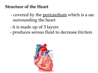
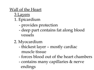
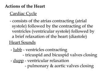
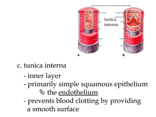
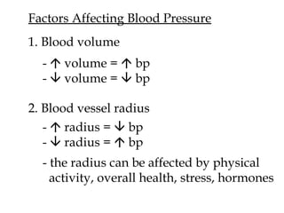
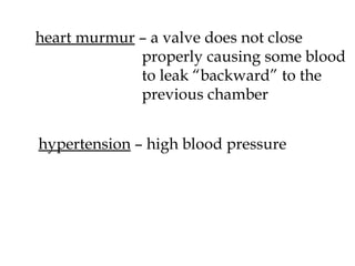
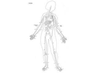
CHAPTER 13: CARDIOVASCULAR SYSTEM
Structure of the Heart - covered by the  pericardium  which is a sac surrounding the heart - it is made up of 3 layers - produces serous fluid to decrease friction
Wall of the Heart 3 Layers 1. Epicardium - provides protection - deep part contains fat along blood    vessels 2. Myocardium - thickest layer – mostly cardiac  muscle tissue - forces blood out of the heart chambers - contains many capillaries & nerve   endings
3. Endocardium - contains  Perkinje fibers  – specialized  nerve fibers used  during the heart beat
 
Flow of blood through the heart          
Blood supply to the heart a) coronary artery - supplies blood to heart muscles b) cardiac veins - takes blood away from heart   muscles – empties into the coronary   sinus The cardiac cycle movie
Actions of the Heart Cardiac Cycle - consists of the atrias contracting (atrial  systole) followed by the contracting of the ventricles (ventricular systole) followed by a brief relaxation of the heart (diastole) Heart Sounds -  lubb  - ventricles contracting   - tricuspid and bicuspid valves closing -  dupp  - ventricular relaxation   - pulmonary & aortic valves closing
Cardiac Conduction System - specialized cells initiate and conduct  impulses through the myocardium SA node AV node AV bundle Perkinje fibers
1. Sinoatrial node (SA node) - start impulses with no connection to    nervous system - stimulates atrias to contract - a.k.a. the pacemaker 2. Atrioventricular node (AV node) - causes a delay in signal - impulse passes into  AV bundle - causes impulse speed to increase - separates into perkinje fibers - ventricles contract - force blood out of ventricles
Electrocardiogram  (ECG or EKG)    recording of electrical changes during cardiac cycle Cardiac conduction movie
P wave  – impulse spreading   from SA node to AV   node - atria depolarize QRS Complex  – spreading of impulse from   AV node to ventricles - ventricles depolarize T wave  – ventricles repolarize
 
Regulation of the Cardiac Cycle - depends on: physical exercise body temp concentration of various ions - when heart rate needs to be changed, the cardiovascular center in the medulla  oblongata sends impulses to the SA node - secretes ACH (   heart rate) - secretes norepinephrine (   heart rate
Blood Vessels Structure a. tunica externa tunica externa - outer most layer - contains elastin and collagen fibers
b. tunica media tunica media - middle layer - contains the muscles to allow for  vasoconstriction  and  vasodilation
c. tunica interna tunica interna - inner layer - primarily simple squamous epithelium    the  endothelium - prevents blood clotting by providing a smooth surface
1. Arteries - have thick muscular walls - carry blood away from the heart 2. Arterioles - little arteries - help to regulate blood flow to capillaries
3. Capillaries - wall is made up of endothelium      1 cell thick - found near every living cell - high metabolic tissues have  a greater density of capillaries - functions in gas exchange and filtration
4. Venules - small veins - drain blood from capillaries 5. Veins - carry blood toward the heart - have thinner walls than arteries  (less muscle) - act as a blood reservoir - contains valves to prevent the  backflow of blood
Blood Pressure    the hydrostatic pressure exerted by blood on the walls of blood vessels - there is a high bp in the arteries and low bp in the veins - must have a pressure difference in order for blood to flow
Factors Affecting Blood Pressure 1. Blood volume -    volume =    bp -    volume =    bp 2. Blood vessel radius -    radius =    bp -    radius =    bp - the radius can be affected by physical activity, overall health, stress, hormones
3. Blood viscosity - the “thickness” of the blood - due to decrease water or anemia    viscosity =    bp  4. Total blood vessel length -    length =    bp 5. Heart action - heart forces blood into arteries -    force =    bp
-  stroke volume  – amt. of blood discharged   from ventricles   - ~ 70 mL -  cardiac output  – amt. of blood discharged   from ventricles per minute   - ~5,040 mL Measuring Blood Pressure - measured with a  sphygmomanometer -  systolic pressure  – maximum pressure -  diastolic pressure  – minimum pressure
Clinical Applications asystole  – failure of the myocardium to contract sinus rhythm  – the normal cardiac rhythm  regulated by the SA node myocardial infarction  – death of the heart muscle tissue (heart attack)
heart murmur  – a valve does not close  properly causing some blood to leak “backward” to the  previous chamber hypertension  – high blood pressure
spider veins  – a milder form  of varicose veins varicose veins  – enlarged, dilated veins that are seen through the skin
aneurysm  – a sac that forms in a blood    vessel due to a weakened artery atherosclerosis  – deposit of fatty materials (plaque) on the walls of blood vessels


Physiology ch 13 lecture notes

  • 1.
  • 2.
    Structure of theHeart - covered by the pericardium which is a sac surrounding the heart - it is made up of 3 layers - produces serous fluid to decrease friction
  • 3.
    Wall of theHeart 3 Layers 1. Epicardium - provides protection - deep part contains fat along blood vessels 2. Myocardium - thickest layer – mostly cardiac muscle tissue - forces blood out of the heart chambers - contains many capillaries & nerve endings
  • 4.
    3. Endocardium -contains Perkinje fibers – specialized nerve fibers used during the heart beat
  • 5.
  • 6.
    Flow of bloodthrough the heart          
  • 7.
    Blood supply tothe heart a) coronary artery - supplies blood to heart muscles b) cardiac veins - takes blood away from heart muscles – empties into the coronary sinus The cardiac cycle movie
  • 8.
    Actions of theHeart Cardiac Cycle - consists of the atrias contracting (atrial systole) followed by the contracting of the ventricles (ventricular systole) followed by a brief relaxation of the heart (diastole) Heart Sounds - lubb - ventricles contracting - tricuspid and bicuspid valves closing - dupp - ventricular relaxation - pulmonary & aortic valves closing
  • 9.
    Cardiac Conduction System- specialized cells initiate and conduct impulses through the myocardium SA node AV node AV bundle Perkinje fibers
  • 10.
    1. Sinoatrial node(SA node) - start impulses with no connection to nervous system - stimulates atrias to contract - a.k.a. the pacemaker 2. Atrioventricular node (AV node) - causes a delay in signal - impulse passes into AV bundle - causes impulse speed to increase - separates into perkinje fibers - ventricles contract - force blood out of ventricles
  • 11.
    Electrocardiogram (ECGor EKG)  recording of electrical changes during cardiac cycle Cardiac conduction movie
  • 12.
    P wave – impulse spreading from SA node to AV node - atria depolarize QRS Complex – spreading of impulse from AV node to ventricles - ventricles depolarize T wave – ventricles repolarize
  • 13.
  • 14.
    Regulation of theCardiac Cycle - depends on: physical exercise body temp concentration of various ions - when heart rate needs to be changed, the cardiovascular center in the medulla oblongata sends impulses to the SA node - secretes ACH (  heart rate) - secretes norepinephrine (  heart rate
  • 15.
    Blood Vessels Structurea. tunica externa tunica externa - outer most layer - contains elastin and collagen fibers
  • 16.
    b. tunica mediatunica media - middle layer - contains the muscles to allow for vasoconstriction and vasodilation
  • 17.
    c. tunica internatunica interna - inner layer - primarily simple squamous epithelium  the endothelium - prevents blood clotting by providing a smooth surface
  • 18.
    1. Arteries -have thick muscular walls - carry blood away from the heart 2. Arterioles - little arteries - help to regulate blood flow to capillaries
  • 19.
    3. Capillaries -wall is made up of endothelium  1 cell thick - found near every living cell - high metabolic tissues have a greater density of capillaries - functions in gas exchange and filtration
  • 20.
    4. Venules -small veins - drain blood from capillaries 5. Veins - carry blood toward the heart - have thinner walls than arteries (less muscle) - act as a blood reservoir - contains valves to prevent the backflow of blood
  • 21.
    Blood Pressure  the hydrostatic pressure exerted by blood on the walls of blood vessels - there is a high bp in the arteries and low bp in the veins - must have a pressure difference in order for blood to flow
  • 22.
    Factors Affecting BloodPressure 1. Blood volume -  volume =  bp -  volume =  bp 2. Blood vessel radius -  radius =  bp -  radius =  bp - the radius can be affected by physical activity, overall health, stress, hormones
  • 23.
    3. Blood viscosity- the “thickness” of the blood - due to decrease water or anemia  viscosity =  bp 4. Total blood vessel length -  length =  bp 5. Heart action - heart forces blood into arteries -  force =  bp
  • 24.
    - strokevolume – amt. of blood discharged from ventricles - ~ 70 mL - cardiac output – amt. of blood discharged from ventricles per minute - ~5,040 mL Measuring Blood Pressure - measured with a sphygmomanometer - systolic pressure – maximum pressure - diastolic pressure – minimum pressure
  • 25.
    Clinical Applications asystole – failure of the myocardium to contract sinus rhythm – the normal cardiac rhythm regulated by the SA node myocardial infarction – death of the heart muscle tissue (heart attack)
  • 26.
    heart murmur – a valve does not close properly causing some blood to leak “backward” to the previous chamber hypertension – high blood pressure
  • 27.
    spider veins – a milder form of varicose veins varicose veins – enlarged, dilated veins that are seen through the skin
  • 28.
    aneurysm –a sac that forms in a blood vessel due to a weakened artery atherosclerosis – deposit of fatty materials (plaque) on the walls of blood vessels
  • 29.
  • 30.