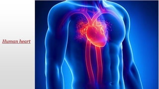
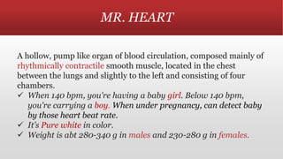
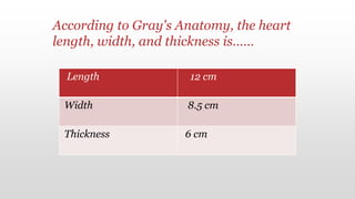
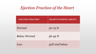
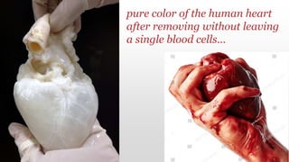
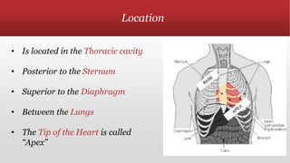
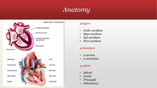
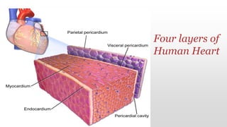
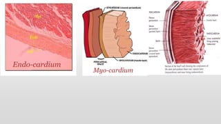
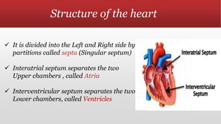
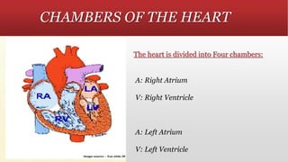
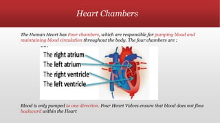
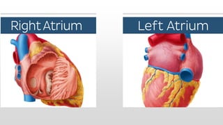
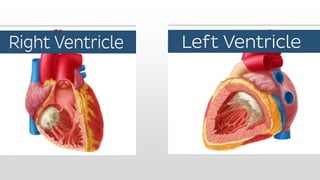
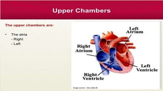
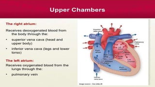
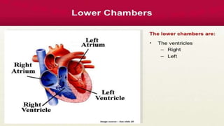
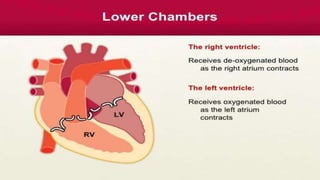
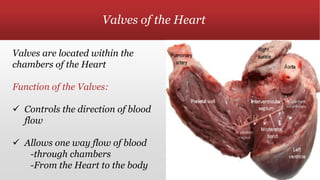
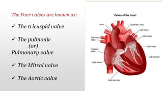
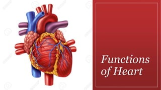
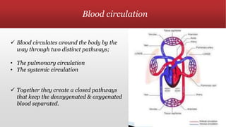
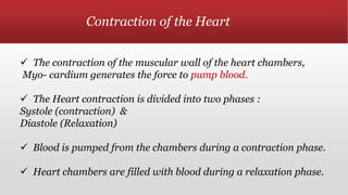
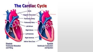
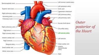
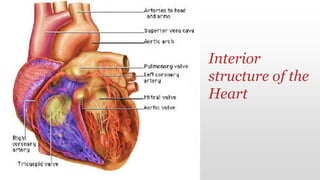
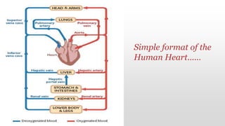
FUNCTIONS
OF HUMAN
HEART
A PRESENTATION BY
Ashritha s
Human heart
Table of contents
 Structure of the Heart
 Heart Chambers
 Valves
 Function of Heart
 Circulation of Blood
 Contraction of Heart
 Conduction system of the Heart
MR. HEART
A hollow, pump like organ of blood circulation, composed mainly of
rhythmically contractile smooth muscle, located in the chest
between the lungs and slightly to the left and consisting of four
chambers.
 When 140 bpm, you're having a baby girl. Below 140 bpm,
you're carrying a boy. When under pregnancy, can detect baby
by those heart beat rate.
 It’s Pure white in color.
 Weight is abt 280-340 g in males and 230-280 g in females.
Length 12 cm
Width 8.5 cm
Thickness 6 cm
According to Gray's Anatomy, the heart
length, width, and thickness is……
EJECTION FRACTION HEART'S PUMPING ABILITY
Normal 50-75 %
Below Normal 36-49 %
Low 35% and below
Ejection Fraction of the Heart
pure color of the human heart
after removing without leaving
a single blood cells…
Location
• Is located in the Thoracic cavity
• Posterior to the Sternum
• Superior to the Diaphragm
• Between the Lungs
• The Tip of the Heart is called
“Apex”
Anatomy
4 layers
• Endo-cardium
• Myo-cardium
• Epi-cardium
• Peri-cardium
4 chambers
• 2 atrium
• 2 ventricles
4 valves
• Mitral
• Aortic
• Tricuspid
• Pulmonary
The Heart consists of Four Cell Layers:
 The Endo Cardium (Inner layer of the Heart) is formed
by Endothelial cells, and it lines in the Interior of the
Heart chambers and Valves.
 The Myo Cardium (Heart Muscles) is the muscular
middle layer of the Heart that consists of Heart muscle
Cells
 The Epi- Cardium (Outer layer of the Heart )is formed by
Epithelial cells , and forms the outer cell layer of the Heart
 The Peri- Cardium (surrounding the Heart)is a membranous sac
that surrounds the Heart.
It consists of two layers:
• The Visceral pericardium(adheres to the epi- cardium)
• Parietal pericardium (outer coat)
Pericardial cavity(space between those two layers & it also
contains Peri- cardial fluid
Four layers of
Human Heart
Endo-cardium
Myo-cardium
Structure of the heart
 It is divided into the Left and Right side by
partitions called septa (Singular septum)
 Interatrial septum separates the two
Upper chambers , called Atria
 Interventricular septum separates the two
Lower chambers, called Ventricles
CHAMBERS OF THE HEART
The heart is divided into Four chambers:
A: Right Atrium
V: Right Ventricle
A: Left Atrium
V: Left Ventricle
Heart Chambers
The Human Heart has Four chambers, which are responsible for pumping blood and
maintaining blood circulation throughout the body. The four chambers are :
Blood is only pumped to one direction. Four Heart Valves ensure that blood does not flow
backward within the Heart
Valves of the Heart
Valves are located within the
chambers of the Heart
Function of the Valves:
 Controls the direction of blood
flow
 Allows one way flow of blood
-through chambers
-From the Heart to the body
The Four valves are known as:
 The tricuspid valve
 The pulmonic
(or)
Pulmonary valve
 The Mitral valve
 The Aortic valve
Functions
of Heart
 Heart functions to circulate blood around the body .The right and left side of the
Heart pump blood into two different circulations.
 Right side pumps deoxygenated (without oxygen)blood into pulmonary
circulation, while the left side pumps oxygenated blood into systematic
circulation.
 Blood enters the right atrium, which pumps the blood into the right ventricle.
The tricuspid valve prevents blood from flowing backward into right atrium.
The right ventricle pumps the blood into the pulmonary artery by the way
through pulmonary valve
 pulmonary artery will deliver the deoxygenated blood to the lungs, where gas
exchange occurs
 Oxygen is taken from the air into the blood (oxygenated blood) while carbon-
dioxide is expelled from the blood into the air. The oxygenated blood returns to
the left side of the Heart by the way through the pulmonary veins
 The oxygenated blood enters the left atrium.
 The left atrium pumps blood into the left ventricle. The mitral valve prevents
blood from flowing backward into the left atrium .
 The left ventricle pumps the blood into the aorta & systemic circulation. The
oxygenated blood is delivered everywhere in the body (besides the lungs )
Blood circulation
 Blood circulates around the body by the
way through two distinct pathways;
• The pulmonary circulation
• The systemic circulation
 Together they create a closed pathways
that keep the deoxygenated & oxygenated
blood separated.
Contraction of the Heart
 The contraction of the muscular wall of the heart chambers,
Myo- cardium generates the force to pump blood.
 The Heart contraction is divided into two phases :
Systole (contraction) &
Diastole (Relaxation)
 Blood is pumped from the chambers during a contraction phase.
 Heart chambers are filled with blood during a relaxation phase.
Outer
posterior of
the Heart
Interior
structure of the
Heart
Simple format of the
Human Heart……
Thank you…

Functions of human heart

  • 1.
  • 2.
  • 3.
    Table of contents Structure of the Heart  Heart Chambers  Valves  Function of Heart  Circulation of Blood  Contraction of Heart  Conduction system of the Heart
  • 4.
    MR. HEART A hollow,pump like organ of blood circulation, composed mainly of rhythmically contractile smooth muscle, located in the chest between the lungs and slightly to the left and consisting of four chambers.  When 140 bpm, you're having a baby girl. Below 140 bpm, you're carrying a boy. When under pregnancy, can detect baby by those heart beat rate.  It’s Pure white in color.  Weight is abt 280-340 g in males and 230-280 g in females.
  • 5.
    Length 12 cm Width8.5 cm Thickness 6 cm According to Gray's Anatomy, the heart length, width, and thickness is……
  • 6.
    EJECTION FRACTION HEART'SPUMPING ABILITY Normal 50-75 % Below Normal 36-49 % Low 35% and below Ejection Fraction of the Heart
  • 7.
    pure color ofthe human heart after removing without leaving a single blood cells…
  • 8.
    Location • Is locatedin the Thoracic cavity • Posterior to the Sternum • Superior to the Diaphragm • Between the Lungs • The Tip of the Heart is called “Apex”
  • 9.
    Anatomy 4 layers • Endo-cardium •Myo-cardium • Epi-cardium • Peri-cardium 4 chambers • 2 atrium • 2 ventricles 4 valves • Mitral • Aortic • Tricuspid • Pulmonary
  • 10.
    The Heart consistsof Four Cell Layers:  The Endo Cardium (Inner layer of the Heart) is formed by Endothelial cells, and it lines in the Interior of the Heart chambers and Valves.  The Myo Cardium (Heart Muscles) is the muscular middle layer of the Heart that consists of Heart muscle Cells
  • 11.
     The Epi-Cardium (Outer layer of the Heart )is formed by Epithelial cells , and forms the outer cell layer of the Heart  The Peri- Cardium (surrounding the Heart)is a membranous sac that surrounds the Heart. It consists of two layers: • The Visceral pericardium(adheres to the epi- cardium) • Parietal pericardium (outer coat) Pericardial cavity(space between those two layers & it also contains Peri- cardial fluid
  • 12.
  • 13.
  • 14.
    Structure of theheart  It is divided into the Left and Right side by partitions called septa (Singular septum)  Interatrial septum separates the two Upper chambers , called Atria  Interventricular septum separates the two Lower chambers, called Ventricles
  • 15.
    CHAMBERS OF THEHEART The heart is divided into Four chambers: A: Right Atrium V: Right Ventricle A: Left Atrium V: Left Ventricle
  • 16.
    Heart Chambers The HumanHeart has Four chambers, which are responsible for pumping blood and maintaining blood circulation throughout the body. The four chambers are : Blood is only pumped to one direction. Four Heart Valves ensure that blood does not flow backward within the Heart
  • 23.
    Valves of theHeart Valves are located within the chambers of the Heart Function of the Valves:  Controls the direction of blood flow  Allows one way flow of blood -through chambers -From the Heart to the body
  • 24.
    The Four valvesare known as:  The tricuspid valve  The pulmonic (or) Pulmonary valve  The Mitral valve  The Aortic valve
  • 25.
  • 26.
     Heart functionsto circulate blood around the body .The right and left side of the Heart pump blood into two different circulations.  Right side pumps deoxygenated (without oxygen)blood into pulmonary circulation, while the left side pumps oxygenated blood into systematic circulation.  Blood enters the right atrium, which pumps the blood into the right ventricle. The tricuspid valve prevents blood from flowing backward into right atrium. The right ventricle pumps the blood into the pulmonary artery by the way through pulmonary valve
  • 27.
     pulmonary arterywill deliver the deoxygenated blood to the lungs, where gas exchange occurs  Oxygen is taken from the air into the blood (oxygenated blood) while carbon- dioxide is expelled from the blood into the air. The oxygenated blood returns to the left side of the Heart by the way through the pulmonary veins  The oxygenated blood enters the left atrium.  The left atrium pumps blood into the left ventricle. The mitral valve prevents blood from flowing backward into the left atrium .  The left ventricle pumps the blood into the aorta & systemic circulation. The oxygenated blood is delivered everywhere in the body (besides the lungs )
  • 28.
    Blood circulation  Bloodcirculates around the body by the way through two distinct pathways; • The pulmonary circulation • The systemic circulation  Together they create a closed pathways that keep the deoxygenated & oxygenated blood separated.
  • 29.
    Contraction of theHeart  The contraction of the muscular wall of the heart chambers, Myo- cardium generates the force to pump blood.  The Heart contraction is divided into two phases : Systole (contraction) & Diastole (Relaxation)  Blood is pumped from the chambers during a contraction phase.  Heart chambers are filled with blood during a relaxation phase.
  • 31.
  • 32.
  • 33.
    Simple format ofthe Human Heart……
  • 34.