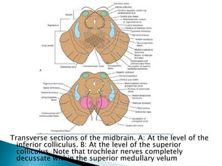
DR. JITHIN GEORGE
1. POSTERIOR CEREBRAL ARTERY
2. BASILAR ARTERY
3. VERTEBRAL ARTERY
Occlusion of PCA– P1 syndromes
P2 syndromes
POSTERIOR CEREBRAL ARTERY
P1 Syndromes : Central/perforating branch
occlusion
Dejerine-Roussy Synd : Thalamic Synd
Sensory loss
Spontaneous pain & dysesthesias
Choreoathetosis
Intention tremor
Spasm of hand
Mild hemiparesis
Claude’s Synd :
Ipsilateral lll CN palsy
Contralateral cerebellar ataxia
Weber’s Synd
Ipsilateral lll CN palsy
Contralateral hemiplegia
Contralateral Hemiballismus :
lesion in the Subthalamic nucleus
P2 Syndromes : Peripheral artery occlusion
 Homonymous hemianopia with macular
sparing
 Anomic aphasia
 Alexia without agraphia/Visual agnosia
 Cortical blindness
 Memory defect
 Prosapagnosia
 Anton’s Syndrome
 Balint’s Syndrome
Millard-Gubler Syndrome
•unilateral lesion of the ventrocaudal pons
•involve the basis pontis and the fascicles of cranial nerves VI
and VII.
•Contralateral hemiplegia (sparing the face) is due to
pyramidal tract involvement.
•Ipsilateral lateral rectus paresis (cranial nerve VI) with
diplopia that is accentuated when the patienT looks towards•
the lesion.
•Ipsilateral peripheral facial paresis (cranial nerve VII).
 A unilateral lesion of the ventral medial pons,
 affects the ipsilateral abducens nerve
fascicles and the corticospinal tract but
spares cranial nerve VII
 also called alternating abducens hemiplegia)
 Ipsilateral lateral rectus paresis (cranial nerve
VI)
 Contralateral hemiplegia, sparing the face,
due to pyramidal tract involvement
 Lesions (especially lacunar infarction)
involving the corticospinal tracts in the basis
pontis may produce a pure motor hemiplegia
with or without facial involvement
 Patients often have severe dysarthria and
dysphagia.
 Bouts of uncontrollable laughter may also
occur
 A combination of dysarthria and a history of
previous transient gait abnormality or vertigo
favor a pontine lesion as the cause of pure
motor hemiparesis rather than a more
common capsular lesion
 Vascular lesions in the basis pontis (especially
lacunar infarction) at the junction of the
upper one-third and lower two-thirds of the
pons may result in dysarthria clumsy hand
syndrome.
 In this syndrome facial weakness and severe
dysarthria and dysphagia occur along with
clumsiness, and paresis of the hand.
 Hyperreflexia and a Babinski's sign may occur
on the same side as the arm paresis, but
sensation is spared.
 A lesion (usually a lacunar infarction) the basis pontis
at the junction of the upper one-third and the lower
two-thirds of the pons may result in the ataxic
hemiparesis (homolateral ataxia and crural paresis)
syndrome.
 In this syndrome hemiparesis that is more severe in
the lower extremity, is associated with ipsilateral
hemiataxia and occasionally dysarthria, nystagmus,
and paresthesias.
 The lesion is located in the contralateral pons.
 The ataxia is unilateral, probably because transverse
fibers originating from the contralateral pontine
nuclei (and projecting to the contralateral
cerebellum) are spared
 Bilateral ventral pontine lesions
 This syndrome consists of the following signs:
◦ Quadriplegia due to bilateral corticospinal tract
involvement in the basis pontis
◦ Aphonia due to involvement of the corticobulbar
fibers innervating the lower cranial nerve nuclei
◦ Occasional impairment of horizontal eye movements
due to bilateral involvement of the fascicles of cranial
nerve VI
 Because the reticular formation is not injured,
the patient is fully awake.
 The supranuclear ocular motor pathways lie
dorsally and are therefore spared; therefore,
vertical eye movements and blinking are intact
Locked-in Syndrome
 Foville Syndrome
◦ lesions involving the dorsal pontine tegmentum in the
caudal third of the pons.
◦ Contralateral hemiplegia (with facial sparing) which is
due to interruption of the corticospinal tract.
◦ Ipsilateral peripheral-type facial palsy which is due to
involvement of the nucleus and fascicle (or both) of
cranial nerve VII.
◦ Inability to move the eyes conjugately to the
ipsilateral side due to involvement of the PPRF or
abducens nucleus, or both
 rostral lesions of the dorsal pons.
 Cerebellar signs (ataxia) with a coarse tremor
which is due to the involvement of the
cerebellum.
 Contralateral hypesthesia with reduction of all
sensory modalities (face and extremities) which
is due to the involvement of the medial
lemniscus and the spinothalamic tract.
 With ventral extension, there may be
contralateral hemiparesis (due to corticospinal
tract involvement) or paralysis of conjugate
gaze toward the side of the lesion (due to
involvement of the PPRF).
 Unilateral mediobasal infarcts.
 severe facio-brachio-crural hemiparesis,
dysarthria, and homolateral or bilateral ataxia.
 Presentations include dysarthria Clumsy hand
syndrome, ataxic hemiparesis with prominent
sensory or eye movement disorders, and
hemiparesis with contralateral facial or
abducens palsy.
 Unilateral mediobasal infarcts. These patients
have pseudobulbar palsy and bilateral
sensorimotor disturbances.
 The most common etiology for paramedian
pontine infarcts is small vessel disease;
 vertebrobasilar large vessel disease and
cardiac embolism are less common causes.
 Marie-Foix Syndrome
◦ lateral pontine lesions
◦ affecting the brachium pontis
 Ipsilateral cerebellar ataxia due to
involvement of cerebellar connections
 Contralateral hemiparesis due to involvement
of the corticospinal tract
 Variable contralateral hemihypesthesia for
pain and temperature due to involvement of
the spinothalamic tract
 combined right superior cerebellar artery
occlusion resulting in lateral superior pontine
infarction and
 left posterior inferior cerebellar artery occlusion,
resulting in a left Wallenberg lateral medullary
syndrome
 loss of pain and temperature sensation, whereas
light touch, vibration, position, and deep pain
sensation were preserved (dissociated sensory
loss).
 This interesting lesson in localization was due to
bilateral discrete interruption of spinothalamic
fibers and the spinal nucleus and tract of the
trigeminal nerve.
Medulla
oblongata
A- ant view
B- post view
Medulla Oblongata
Gross appearnse:
- Connect the pons sup to spinal cord inf
- About 2.5 cm in length
- The junction of the medulla and spinal cord is at the origin of
the anterior and posterior roots of the first cervical spinal
nerve at level of foramen magnum
- - It is conical in shape
- - central canal
- - cavity of fourth ventricle
- Anteriorly:
- - ant median fissure
- - pyramid
- - decussation of the pyramids
- - Posterolateral to the pyramids are the olives
- Posteriorly:
- - sup is the floor 4th ventricle
- - inf the median sulcus
- - gracile tubercle and lat to it the cuneate tubercle
The internal structure of the medulla oblongata is
considered at four levels:
1) level of decussation of pyramids
2) level of decussation of lemnisci
3) level of the olives
4) level just inferior to the pons.
Transverse section of the medulla oblongata
at the level of decussation of the pyramids
Transverse section of the medulla oblongata at
the level of decussation of the medial lemnisci
Transverse section of the medulla oblongata at
the level of the middle of the olivary nuclei
Blood supply of Medulla oblongata:
1) ventrally: branches from vertebral and basilar
arteries, Also branches from ant spinal artery
artery
2) dorsolaterally: by post inf cerebellar artery
- Venous drainage:
- 1)ventrally: basilar venous plexus
and inf petrosal sinus
2) Dorsally and dorsolaterally to occipital sinus
3) Medullary veins communicate with sinuses and
spinal veins
Lateral Medullary Synd:
Wallenberg’s synd
On the side of lesion
V, VII, VIII, IX, X CN &
desc. sympathetic tract
On the opp side
Impaired pain &thermal
sense over half of the
body
Medial Medullary Synd
On the side of lesion
Paralysis with atrophy of half of the
tongue
On the opp side
Paralysis of arm and leg sparing face
Impaired tactile & proprioceptive sense
over half of the body
Transverse section of the midbrain through the inferior
colliculi shows the division of the midbrain into the
tectum and the cerebral peduncles. Note that the
cerebral peduncles are subdivided by the substantia
nigra into the tegmentum and the crus cerebri
Transverse sections of the midbrain. A: At the level of the
inferior colliculus. B: At the level of the superior
colliculus. Note that trochlear nerves completely
decussate within the superior medullary velum
Brain stem syndromes

Brain stem syndromes

  • 1.
  • 3.
    1. POSTERIOR CEREBRALARTERY 2. BASILAR ARTERY 3. VERTEBRAL ARTERY Occlusion of PCA– P1 syndromes P2 syndromes
  • 4.
    POSTERIOR CEREBRAL ARTERY P1Syndromes : Central/perforating branch occlusion Dejerine-Roussy Synd : Thalamic Synd Sensory loss Spontaneous pain & dysesthesias Choreoathetosis Intention tremor Spasm of hand Mild hemiparesis
  • 5.
    Claude’s Synd : Ipsilaterallll CN palsy Contralateral cerebellar ataxia Weber’s Synd Ipsilateral lll CN palsy Contralateral hemiplegia Contralateral Hemiballismus : lesion in the Subthalamic nucleus
  • 6.
    P2 Syndromes :Peripheral artery occlusion  Homonymous hemianopia with macular sparing  Anomic aphasia  Alexia without agraphia/Visual agnosia  Cortical blindness  Memory defect  Prosapagnosia  Anton’s Syndrome  Balint’s Syndrome
  • 13.
    Millard-Gubler Syndrome •unilateral lesionof the ventrocaudal pons •involve the basis pontis and the fascicles of cranial nerves VI and VII. •Contralateral hemiplegia (sparing the face) is due to pyramidal tract involvement. •Ipsilateral lateral rectus paresis (cranial nerve VI) with diplopia that is accentuated when the patienT looks towards• the lesion. •Ipsilateral peripheral facial paresis (cranial nerve VII).
  • 14.
     A unilaterallesion of the ventral medial pons,  affects the ipsilateral abducens nerve fascicles and the corticospinal tract but spares cranial nerve VII  also called alternating abducens hemiplegia)  Ipsilateral lateral rectus paresis (cranial nerve VI)  Contralateral hemiplegia, sparing the face, due to pyramidal tract involvement
  • 15.
     Lesions (especiallylacunar infarction) involving the corticospinal tracts in the basis pontis may produce a pure motor hemiplegia with or without facial involvement  Patients often have severe dysarthria and dysphagia.  Bouts of uncontrollable laughter may also occur  A combination of dysarthria and a history of previous transient gait abnormality or vertigo favor a pontine lesion as the cause of pure motor hemiparesis rather than a more common capsular lesion
  • 16.
     Vascular lesionsin the basis pontis (especially lacunar infarction) at the junction of the upper one-third and lower two-thirds of the pons may result in dysarthria clumsy hand syndrome.  In this syndrome facial weakness and severe dysarthria and dysphagia occur along with clumsiness, and paresis of the hand.  Hyperreflexia and a Babinski's sign may occur on the same side as the arm paresis, but sensation is spared.
  • 17.
     A lesion(usually a lacunar infarction) the basis pontis at the junction of the upper one-third and the lower two-thirds of the pons may result in the ataxic hemiparesis (homolateral ataxia and crural paresis) syndrome.  In this syndrome hemiparesis that is more severe in the lower extremity, is associated with ipsilateral hemiataxia and occasionally dysarthria, nystagmus, and paresthesias.  The lesion is located in the contralateral pons.  The ataxia is unilateral, probably because transverse fibers originating from the contralateral pontine nuclei (and projecting to the contralateral cerebellum) are spared
  • 18.
     Bilateral ventralpontine lesions  This syndrome consists of the following signs: ◦ Quadriplegia due to bilateral corticospinal tract involvement in the basis pontis ◦ Aphonia due to involvement of the corticobulbar fibers innervating the lower cranial nerve nuclei ◦ Occasional impairment of horizontal eye movements due to bilateral involvement of the fascicles of cranial nerve VI  Because the reticular formation is not injured, the patient is fully awake.  The supranuclear ocular motor pathways lie dorsally and are therefore spared; therefore, vertical eye movements and blinking are intact Locked-in Syndrome
  • 19.
     Foville Syndrome ◦lesions involving the dorsal pontine tegmentum in the caudal third of the pons. ◦ Contralateral hemiplegia (with facial sparing) which is due to interruption of the corticospinal tract. ◦ Ipsilateral peripheral-type facial palsy which is due to involvement of the nucleus and fascicle (or both) of cranial nerve VII. ◦ Inability to move the eyes conjugately to the ipsilateral side due to involvement of the PPRF or abducens nucleus, or both
  • 20.
     rostral lesionsof the dorsal pons.  Cerebellar signs (ataxia) with a coarse tremor which is due to the involvement of the cerebellum.  Contralateral hypesthesia with reduction of all sensory modalities (face and extremities) which is due to the involvement of the medial lemniscus and the spinothalamic tract.  With ventral extension, there may be contralateral hemiparesis (due to corticospinal tract involvement) or paralysis of conjugate gaze toward the side of the lesion (due to involvement of the PPRF).
  • 21.
     Unilateral mediobasalinfarcts.  severe facio-brachio-crural hemiparesis, dysarthria, and homolateral or bilateral ataxia.  Presentations include dysarthria Clumsy hand syndrome, ataxic hemiparesis with prominent sensory or eye movement disorders, and hemiparesis with contralateral facial or abducens palsy.
  • 22.
     Unilateral mediobasalinfarcts. These patients have pseudobulbar palsy and bilateral sensorimotor disturbances.  The most common etiology for paramedian pontine infarcts is small vessel disease;  vertebrobasilar large vessel disease and cardiac embolism are less common causes.
  • 23.
     Marie-Foix Syndrome ◦lateral pontine lesions ◦ affecting the brachium pontis  Ipsilateral cerebellar ataxia due to involvement of cerebellar connections  Contralateral hemiparesis due to involvement of the corticospinal tract  Variable contralateral hemihypesthesia for pain and temperature due to involvement of the spinothalamic tract
  • 24.
     combined rightsuperior cerebellar artery occlusion resulting in lateral superior pontine infarction and  left posterior inferior cerebellar artery occlusion, resulting in a left Wallenberg lateral medullary syndrome  loss of pain and temperature sensation, whereas light touch, vibration, position, and deep pain sensation were preserved (dissociated sensory loss).  This interesting lesson in localization was due to bilateral discrete interruption of spinothalamic fibers and the spinal nucleus and tract of the trigeminal nerve.
  • 34.
  • 35.
    Medulla Oblongata Gross appearnse: -Connect the pons sup to spinal cord inf - About 2.5 cm in length - The junction of the medulla and spinal cord is at the origin of the anterior and posterior roots of the first cervical spinal nerve at level of foramen magnum - - It is conical in shape - - central canal - - cavity of fourth ventricle - Anteriorly: - - ant median fissure - - pyramid - - decussation of the pyramids - - Posterolateral to the pyramids are the olives - Posteriorly: - - sup is the floor 4th ventricle - - inf the median sulcus - - gracile tubercle and lat to it the cuneate tubercle
  • 36.
    The internal structureof the medulla oblongata is considered at four levels: 1) level of decussation of pyramids 2) level of decussation of lemnisci 3) level of the olives 4) level just inferior to the pons.
  • 37.
    Transverse section ofthe medulla oblongata at the level of decussation of the pyramids
  • 38.
    Transverse section ofthe medulla oblongata at the level of decussation of the medial lemnisci
  • 39.
    Transverse section ofthe medulla oblongata at the level of the middle of the olivary nuclei
  • 40.
    Blood supply ofMedulla oblongata: 1) ventrally: branches from vertebral and basilar arteries, Also branches from ant spinal artery artery 2) dorsolaterally: by post inf cerebellar artery - Venous drainage: - 1)ventrally: basilar venous plexus and inf petrosal sinus 2) Dorsally and dorsolaterally to occipital sinus 3) Medullary veins communicate with sinuses and spinal veins
  • 41.
    Lateral Medullary Synd: Wallenberg’ssynd On the side of lesion V, VII, VIII, IX, X CN & desc. sympathetic tract On the opp side Impaired pain &thermal sense over half of the body
  • 42.
    Medial Medullary Synd Onthe side of lesion Paralysis with atrophy of half of the tongue On the opp side Paralysis of arm and leg sparing face Impaired tactile & proprioceptive sense over half of the body
  • 44.
    Transverse section ofthe midbrain through the inferior colliculi shows the division of the midbrain into the tectum and the cerebral peduncles. Note that the cerebral peduncles are subdivided by the substantia nigra into the tegmentum and the crus cerebri
  • 45.
    Transverse sections ofthe midbrain. A: At the level of the inferior colliculus. B: At the level of the superior colliculus. Note that trochlear nerves completely decussate within the superior medullary velum