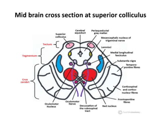
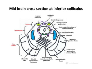
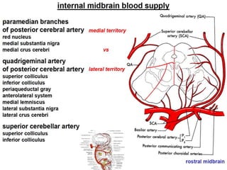
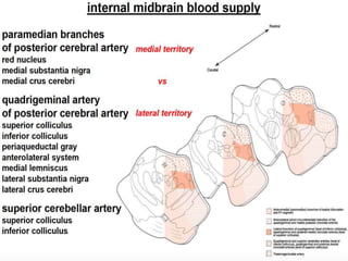
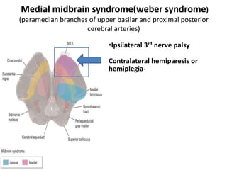
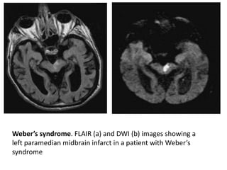
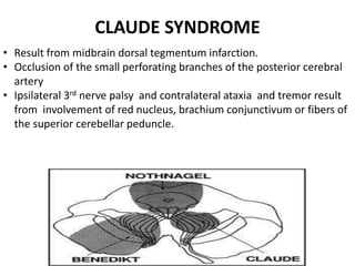
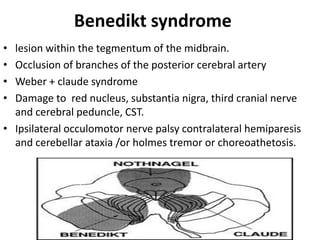
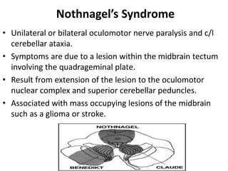
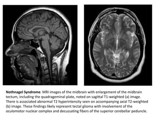
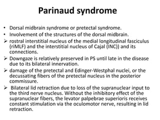
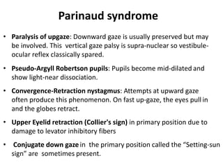
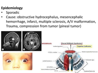
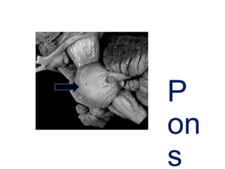
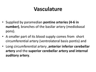
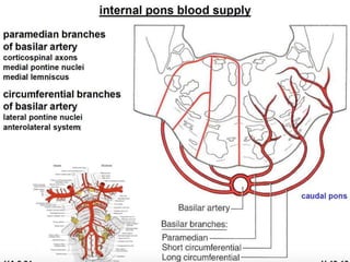
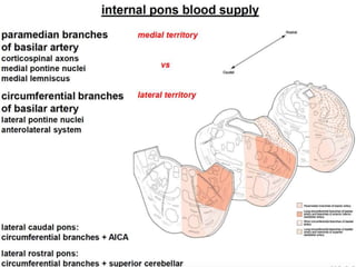
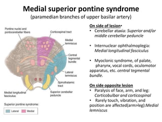
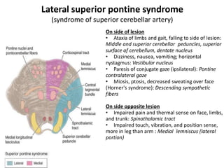
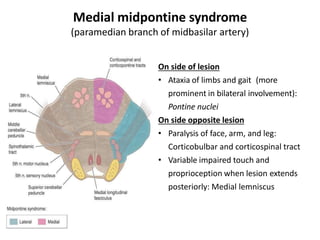
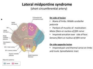
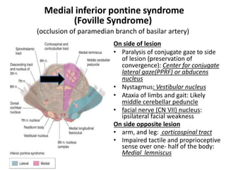
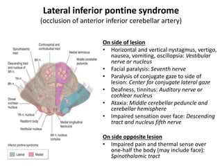
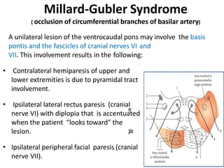
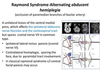
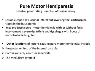
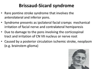
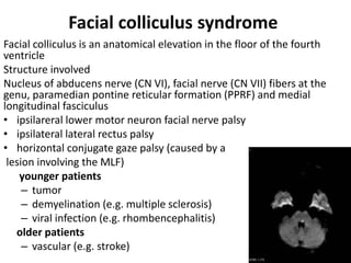
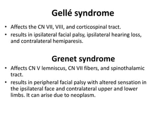
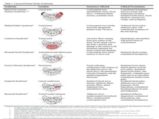
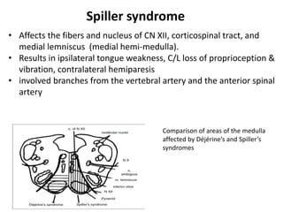
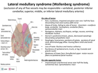
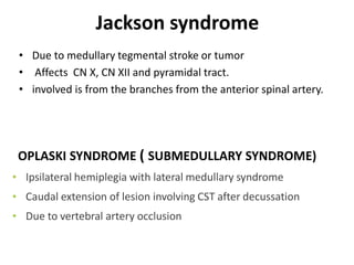
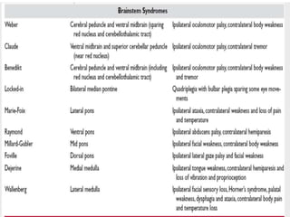
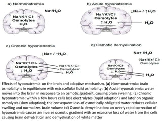
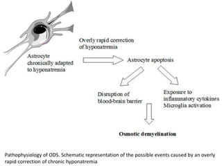
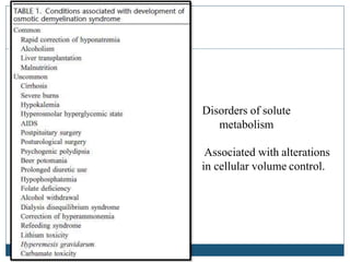
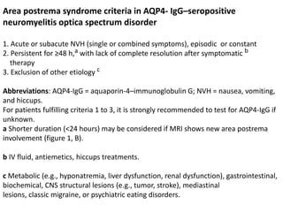
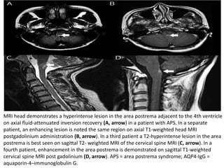
VASCULAR
AND
DEMYELINATING SYNDROME
OF BRAINSTEM
DR. VINOD SINGH JATAV
SR NEUROLOGY
GMC KOTA
Mid brain cross section at superior colliculus
Mid brain cross section at inferior colliculus
Medial midbrain syndrome(weber syndrome)
(paramedian branches of upper basilar and proximal posterior
cerebral arteries)
•Ipsilateral 3rd nerve palsy
Contralateral hemiparesis or
hemiplegia-
Weber’s syndrome. FLAIR (a) and DWI (b) images showing a
left paramedian midbrain infarct in a patient with Weber’s
syndrome
CLAUDE SYNDROME
• Result from midbrain dorsal tegmentum infarction.
• Occlusion of the small perforating branches of the posterior cerebral
artery
• Ipsilateral 3rd nerve palsy and contralateral ataxia and tremor result
from involvement of red nucleus, brachium conjunctivum or fibers of
the superior cerebellar peduncle.
Benedikt syndrome
• lesion within the tegmentum of the midbrain.
• Occlusion of branches of the posterior cerebral artery
• Weber + claude syndrome
• Damage to red nucleus, substantia nigra, third cranial nerve
and cerebral peduncle, CST.
• Ipsilateral occulomotor nerve palsy contralateral hemiparesis
and cerebellar ataxia /or holmes tremor or choreoathetosis.
Nothnagel’s Syndrome
• Unilateral or bilateral oculomotor nerve paralysis and c/l
cerebellar ataxia.
• Symptoms are due to a lesion within the midbrain tectum
involving the quadrageminal plate.
• Result from extension of the lesion to the oculomotor
nuclear complex and superior cerebellar peduncles.
• Associated with mass occupying lesions of the midbrain
such as a glioma or stroke.
Nothnagel Syndrome. MRI images of the midbrain with enlargement of the midbrain
tectum, including the quadrageminal plate, noted on sagittal T1-weighted (a) image.
There is associated abnormal T2 hyperintensity seen on accompanying axial T2-weighted
(b) image. These findings likely represent tectal glioma with involvement of the
oculomotor nuclear complex and decussating fibers of the superior cerebellar peduncle.
Parinaud syndrome
• Dorsal midbrain syndrome or pretectal syndrome.
• Involvement of the structures of the dorsal midbrain.
 rostral interstitial nucleus of the medial longitudinal fasciculus
(riMLF) and the interstitial nucleus of Cajal (INC)) and its
connections.
 Downgaze is relatively preserved in PS until late in the disease
due to its bilateral innervation.
 damage of the pretectal and Edinger-Westphal nuclei, or the
decussating fibers of the pretectal nucleus in the posterior
commissure.
 Bilateral lid retraction due to loss of the supranuclear input to
the third nerve nucleus. Without the inhibitory effect of the
supranuclear fibers, the levator palpebrae superioris receives
constant stimulation via the oculomotor nerve, resulting in lid
retraction.
Parinaud syndrome
• Paralysis of upgaze: Downward gaze is usually preserved but may
be involved. This vertical gaze palsy is supra-nuclear so vestibule-
ocular reflex classically spared.
• Pseudo-Argyll Robertson pupils: Pupils become mid-dilated and
show light-near dissociation.
• Convergence-Retraction nystagmus: Attempts at upward gaze
often produce this phenomenon. On fast up-gaze, the eyes pullin
and the globes retract.
• Upper Eyelid retraction (Collier's sign) in primary position due to
damage to levator inhibitory fibers
• Conjugate down gaze in the primary position called the “Setting-sun
sign” are sometimes present.
Epidemiology
• Sporadic
• Cause: obstructive hydrocephalus, mesencephalic
hemorrhage, infarct, multiple sclerosis, A/V malformation,
Trauma, compression from tumor (pineal tumor)
P
on
s
Vasculature
• Supplied by paramedian pontine arteries (4-6 in
number), branches of the basilar artery (mediobasal
pons).
• A smaller part of its blood supply comes from short
circumferential artery (ventrolateral basis pontis) and
• Long circumferential artery ,anterior inferior cerebellar
artery and the superior cerebellar artery and internal
auditory artery.
Medial superior pontine syndrome
(paramedian branches of upper basilar artery)
On side of lesion•
• Cerebellar ataxia: Superior and/or
middle cerebellar peduncle
• Internuclear ophthalmoplegia:
Medial longitudinal fasciculus
• Myoclonic syndrome, of palate,
pharynx, vocal cords, oculomotor
apparatus, etc. central tegmental
bundle.
On side opposite lesion
• Paralysis of face, arm, and leg:
Corticobulbar and corticospinal
• Rarely touch, vibration, and
position are affected(arm>leg):Medial
lemniscus
Lateral superior pontine syndrome
(syndrome of superior cerebellar artery)
On side of lesion
• Ataxia of limbs and gait, falling to side of lesion:
Middle and superior cerebellar peduncles, superior
surface of cerebellum, dentate nucleus
• Dizziness, nausea, vomiting; horizontal
nystagmus: Vestibular nucleus
• Paresis of conjugate gaze (ipsilateral): Pontine
contralateral gaze
• Miosis, ptosis, decreased sweating over face
(Horner's syndrome): Descending sympathetic
fibers
On side opposite lesion
• Impaired pain and thermal sense on face, limbs,
and trunk: Spinothalamic tract
• Impaired touch, vibration, and position sense,
more in leg than arm : Medial lemniscus (lateral
portion)
Medial midpontine syndrome
(paramedian branch of midbasilar artery)
On side of lesion
• Ataxia of limbs and gait (more
prominent in bilateral involvement):
Pontine nuclei
On side opposite lesion
• Paralysis of face, arm, and leg:
Corticobulbar and corticospinal tract
• Variable impaired touch and
proprioception when lesion extends
posteriorly: Medial lemniscus
Lateral midpontine syndrome
(short circumferential artery)
• On side of lesion
On side of lesion
• Ataxia of limbs: Middle cerebellar
peduncle
• Paralysis of muscles of mastication:
Motor fibers or nucleus of fifth nerve
• Impaired sensation over side of face:
Sensory fibers or nucleus of fifth nerve
On side opposite lesion
• Impaired pain and thermal sense on limbs
and trunk: Spinothalamic tract
Medial inferior pontine syndrome
(Foville Syndrome)
(occlusion of paramedian branch of basilar artery)
On side of lesion
• Paralysis of conjugate gaze to side
of lesion (preservation of
convergence): Center for conjugate
lateral gaze(PPRF) or abducens
nucleus
• Nystagmus: Vestibular nucleus
• Ataxia of limbs and gait: Likely
middle cerebellar peduncle
• facial nerve (CN VII) nucleus:
ipsilateral facial weakness
On side opposite lesion
• arm, and leg: corticospinal tract
• Impaired tactile and proprioceptive
sense over one- half of the body:
Medial lemniscus
Lateral inferior pontine syndrome
(occlusion of anterior inferior cerebellar artery)
On side of lesion
• Horizontal and vertical nystagmus, vertigo,
nausea, vomiting, oscillopsia: Vestibular
nerve or nucleus
• Facial paralysis: Seventh nerve
• Paralysis of conjugate gaze to side of
lesion: Center for conjugate lateral gaze
• Deafness, tinnitus: Auditory nerve or
cochlear nucleus
• Ataxia: Middle cerebellar peduncle and
cerebellar hemisphere
• Impaired sensation over face: Descending
tract and nucleus fifth nerve
On side opposite lesion
• Impaired pain and thermal sense over
one-half the body (may include face):
Spinothalamic tract
1. Ventral pontine syndrome
• Millard-gubler syndrome
• Raymond syndrome (alternating abducence hemiplegia)
• Pure motor hemiparesis
• Dysarthria clumsy hand syndrome
• Ataxic hemiparesis
• Locked-in syndrome
2. Dorsal pontine syndrome
• Foville syndrome
• Raymond cestan syndrome
3. Lateral pontine syndrome
• Marie-foix syndrome
• Gasperini syndrome
4. Paramedian pontine syndrome
• Unilateral mediobasal infarct
• Unilateral mediolateral basal infarct
• Unilateral mediocentral or mediotegmental infarct
• Bilateral centrobasal infarct
Millard-Gubler Syndrome
( occlusion of circumferential branches of basilar artery)
A unilateral lesion of the ventrocaudal pons may involve the basis
pontis and the fascicles of cranial nerves VI and
VII. This involvement results in the following:
• Contralateral hemiparesis of upper and
lower extremities is due to pyramidal tract
involvement.
• Ipsilateral lateral rectus paresis (cranial
nerve VI) with diplopia that is accentuated
when the patient “looks toward” the
lesion.
• Ipsilateral peripheral facial paresis (cranial
nerve VII).
Raymond Syndrome-Alternating abducent
hemiplegia
(occlusion of paramedian branches of basilar artery)
A unilateral lesion of the ventral medial
pons, which affects the ipsilateral abducens
nerve fascicles and the corticospinal tract
but spares cranial nerve VII in common
type.
• Ipsilateral lateral rectus paresis (cranial
nerve VI).
• Contralateral hemiplegia, sparing the
face, due to pyramidal tract involvement.
• In classical raymond syndrome c/l central
facial paresis may occur.
Pure Motor Hemiparesis
(ventral penetrating branches of basilar artery)
• Lesions (especially lacunar infarction) involving the corticospinal
tracts in the basis pontis
• may produce a pure motor hemiplegia with or without facial
involvement severe dysarthria and dysphagia with Bouts of
uncontrollable laughter.
• Other locations of lesions causing pure motor hemiplegia include
 the posterior limb of the internal capsule,
 Corona radiate/ centrum semiovale
 The medullary pyramid
Dysarthria Clumsy Hand Syndrome
 Vascular lesions in the basis pontis (especially lacunar
infarction) at the junction of the upper one-third and lower
two-thirds of the pons
 Occlusion of paramedian pontine branches of basilar artery
 In this syndrome facial weakness and severe dysarthria and
dysphagia occur along with clumsiness, and paresis of the
hand.
 Hyperreflexia and a Babinski's sign may occur on the same
side as the arm paresis, but sensation is spared.
 A lesion (usually a lacunar infarction) the basis pontis at the junction
of the upper one-third and the lower two-thirds of the pons may
result in the ataxic hemiparesis (homolateral ataxia and crural
paresis) syndrome.
 In this syndrome hemiparesis that is more severe in the lower
extremity, is associated with ipsilateral hemiataxia and occasionally
dysarthria, nystagmus, and paresthesias.
 The lesion is located in the contralateral pons or posterior limb of
internal capsule or c/l red nucleus or c/l thalamocapsular lesion .
 The ataxia is unilateral, probably because transverse fibers
originating from the contralateral pontine nuclei (and projecting to
the contralateral cerebellum) are spared.
Ataxic hemiparesis
(perforating branch of basilar artery)
Locked-in Syndrome
 Synonymous – “de-efferented state” or “pseudocoma” or “coma
vigilante.”
 Bilateral ventral pontine lesions
 This syndrome consists of the following signs:
◦ Quadriplegia due to bilateral corticospinal tract involvement in the
basis pontis.
◦ Aphonia and dysphagia due to involvement of the
corticobulbar fibers innervating the lower cranial nerve nuclei.
◦ Occasional impairment of horizontal eye movements due to bilateral
involvement of the fascicles of cranial nerve VI or PPRF.
 Because the reticular formation is not injured, the patient is fully
awake.
 The supranuclear ocular motor pathways lie dorsally and are
therefore spared; therefore, vertical eye movements and blinking are
intact
cause-infarction, tumor, hemorrhage, trauma, cervical manipulation,
pontine abscess, encephalitis, arteritis, neuro-Behcet’s, multiple
sclerosis, air embolism, diazepam toxicity, or central pontine
myelinolysis
Raymond-Céstan syndrome
(upper dorsal pontine syndrome)
• Caused by blockage of the long circumferential branches of
the basilar artery
• Ipsilateral internuclear opthalmoplegia
• c/l cerebellar sign (ataxia) and coarse rubral tremor (damage to
superior and middle cerebellar peduncle)
• Contralateral loss of all sensory modalities in the body (damage
to spinothalamic tract and medial lemniscus)
• Contralateral hemiparesis of face and body (damage
to corticospinal tract) may occur with ventral extension of lesion
• paralysis of conjugate gaze toward the side of the lesion (due to
involvement of the PPRF)
Marie-Foix Syndrome
 lateral pontine lesions affecting the brachium pontis.
 Due to involvement of long circumferential branch of
basilar artery or AICA
 Ipsilateral cerebellar ataxia due to involvement of cerebellar
tracts.
 Contralateral hemiparesis due to involvement of the
corticospinal tract
 Variable contralateral hemihypesthesia for pain and
temperature due to involvement of the spinothalamic tract
Gasperini syndrome
• Rare lateral pontine stroke syndrome that involves the caudal pontine
tegmentum.
Classically, the syndrome presents with
• Involvement of the CN V nucleus: ipsilateral facial sensory loss
• Involvement of the CN VI nucleus: ipsilateral impaired eye abduction
• Involvement of the CN VII nucleus: ipsilateral facial palsy
• contralateral or ipsilateral hypacusis : lateral leminiscus
• Occasionally Involvement of the CN VIII nucleus: vertigo and
ipsilateral or c/l nystagmus
• Involvement of the spinothalamic tract: contralateral hemi-sensory
impairment
• Usually due to occlusion of either pontine branches of the basilar
artery or the long circumferential branch of the anterior inferior
cerebellar artery, but can also be seen due to demyelination or
hemorrhage
Transverse section through the caudal pons showing: 1 - Facial nerve and nucleus;
2 - Abducent nerve and nucleus; 3 - Lateral lemniscus; 4 - Spinal trigeminal tract;
5 - Spinothalamic tract; 6 - Fourth ventricle
Brissaud-Sicard syndrome
• Rare pontine stroke syndrome that involves the
anterolateral and inferior pons.
• Syndrome presents as ipsilateral facial cramps mechanical
irritation of facial nerve and contralateral hemiparesis
• Due to damage to the pons involving the corticospinal
tract and irritation of CN VII nucleus or nerve root
• Caused by a posterior circulation ischemic stroke, neoplasm
(e.g. brainstem glioma)
Facial colliculus syndrome
Facial colliculus is an anatomical elevation in the floor of the fourth
ventricle
Structure involved
Nucleus of abducens nerve (CN VI), facial nerve (CN VII) fibers at the
genu, paramedian pontine reticular formation (PPRF) and medial
longitudinal fasciculus
• ipsilareral lower motor neuron facial nerve palsy
• ipsilateral lateral rectus palsy
• horizontal conjugate gaze palsy (caused by a
lesion involving the MLF)
younger patients
– tumor
– demyelination (e.g. multiple sclerosis)
– viral infection (e.g. rhombencephalitis)
older patients
– vascular (e.g. stroke)
Gellé syndrome
• Affects the CN VII, VIII, and corticospinal tract.
• results in ipsilateral facial palsy, ipsilateral hearing loss,
and contralateral hemiparesis.
Grenet syndrome
• Affects CN V lemniscus, CN VII fibers, and spinothalamic
tract.
• results in peripheral facial palsy with altered sensation in
the ipsilateral face and contralateral upper and lower
limbs. It can arise due to neoplasm.
ME
DUL
LA
OBL
ONG
ATA
Vasculature
Medial medullary syndrome
(DEJERINE SYNDROME)
(occlusion of vertebral artery or paramedian branches of anterior spinal artery or
basilar artery, vertebral artery)
On side of lesion
•Paralysis with atrophy of one-half
the tongue: Ipsilateral twelfth nerve
On side opposite lesion
• Paralysis of arm and leg, sparing
face; pyramidal tract
Comparison of areas of the medulla
affected by Déjérine’s and Spiller’s
syndromes
• Affects the fibers and nucleus of CN XII, corticospinal tract, and
medial lemniscus (medial hemi-medulla).
• Results in ipsilateral tongue weakness, C/L loss of proprioception &
vibration, contralateral hemiparesis
• involved branches from the vertebral artery and the anterior spinal
artery
Spiller syndrome
Lateral medullary syndrome (Wallenberg syndrome)
(occlusion of any of five vessels may be responsible—vertebral, posterior inferior
cerebellar, superior, middle, or inferior lateral medullary arteries)
On side of lesion
• Pain, numbness, impaired sensation over one- half the face:
Descending tract and nucleus fifth nerve
• Ataxia of limbs, falling to side of lesion: Uncertain—restiform
body, cerebellar hemisphere, cerebellar fibers,
spinocerebellar tract
• Nystagmus, diplopia, oscillopsia, vertigo, nausea, vomiting:
Vestibular nucleus
• Horner's syndrome (miosis, ptosis, decreased sweating):
Descending sympathetic tract
• Dysphagia, hoarseness, paralysis of palate, paralysis of vocal
cord, diminished gag reflex: Issuing fibers ninth and tenth
nerves
• Loss of taste: Nucleus and tractus solitarius
• Numbness of ipsilateral arm, trunk, or leg: Cuneate and
gracile nuclei
• Weakness of lower face: Genuflected upper motor neuron
fibers to ipsilateral facial nucleus
On side opposite lesion
• Impaired pain and thermal sense over half the body,
sometimes face: Spinothalamic tract
Avellis syndrome
• Ipsilateral palatopharyngeal palsy and c/l hemiparesis
and/or hypesthesia.
• Horner syndrome may be associated.
• Involved lesions are nucleus ambiguous, lateral
spinothalamic tract and descending sympathetic fibre of
medulla oblongata
• occlusion of vertebral artery or tumor
The picture shows a T2 weighted MRI in a patient with Avellis' syndrome due to infarction. A 47
year old woman developed sudden onset of hoarseness and dysphagia. Neurological
examination disclosed palsies of the left vocal cord and left soft palate, hypalgesia, and
thermohypaesthesia on the right side of the trunk and right arm, and bilateral horizontal
transient gaze dependent nystagmus, which improved within one month.
Jackson syndrome
• Due to medullary tegmental stroke or tumor
• Affects CN X, CN XII and pyramidal tract.
• involved is from the branches from the anterior spinal artery.
OPLASKI SYNDROME ( SUBMEDULLARY SYNDROME)
• Ipsilateral hemiplegia with lateral medullary syndrome
• Caudal extension of lesion involving CST after decussation
• Due to vertebral artery occlusion
Fig. 1 T2-weighted MRI of Babinski-
Nageotte syndrome shows an area
of increased signal intensity in the
lateral portion of the left medulla
oblongata with spreading of the
lesion to the pyramidal tract (a);
Schematic drawing of the MRI
findings (b)
Fig. 2 T2-weighted MRI of Cestan-
Chenais syndrome shows an area of
increased signal intensity in the
lateral portion of the left medulla
oblongata with spreading of the
lesion to the pyramidal tract and
sparing of the posterior
spinocerebellar tract (a); Schematic
drawing of the MRI findings (b)
Fig. 5 T2-weighted MRI of the
hemimedullary (Reinhold)
syndrome shows an area of
increased signal intensity in the
left hemimedulla (a);Schematic
drawing of the MRI findings(b)
Fig. 4 T2-weighted MRI of the
medial (Dejerine) syndrome
shows an area of increased
signal intensity in the medial
portion of the right medulla
oblongata (a); Schematic
drawing of the MRI findings (b)
Fig. 3 T2-weighted MRI of the
lateral (Wallenberg) syndrome
shows an area of increased signal
intensity in the lateral portion of
the right medulla oblongata (a);
Schematic drawing of the MRI
findings (b)
Schmidt syndrome
• Affects the fibers and nuclei of CN IX, X, XI, and pyramidal
system.
• results in ipsilateral palsy of the vocal cords, soft palate,
trapezius, & sternocleidomastoid muscle, and contralateral
spastic hemiparesis.
• involved branches from the vertebral artery, the posterior
inferior cerebellar artery the anterior spinal artery.
Vernet syndrome
• Affects the CN IX, X, and XI. It occurs due to compression
in the jugular foramen
Internuclear ophthalmoplegia
• Disorder of conjugate lateral gaze in which the affected
eye shows impairment of adduction.
• The contralateral eye abducts, however with
nystagmus.
• Divergence of the eyes leads to horizontal diplopia.
• Caused by injury or dysfunction in the medial
longitudinal fasciculus(MLF).
• Cause in Young patients- multiple sclerosis, pontine
glioma.
old age- stroke
One and a half syndrome
• Results from single unilateral lesion of the paramedian pontine
reticular formation and the ipsilateral medial longitudinal
fasciculus
• Characterized by "a conjugate horizontal gaze palsy in one
direction and an internuclear ophthalmoplegia in the other.”
• Nystagmus is also present when the eye on the opposite side of
the lesion is abducted
• Convergence is classically spared as cranial nerve III (oculomotor
nerve) and its nucleus is spared bilaterally.
• Cause include pontine hemorrhage, ischemia, tumors, infective
mass lesions such as tuberculomas, and demyelinating conditions
like multiple sclerosis
Wall-Eyed bilateral internuclear
ophthalmoplaegia (WEBINO) syndrome
• A rare syndrome characterized by bilateral exotropia on primary
gaze, bilateral inter nuclear ophthalmoplegia (INO) leads to
bilateral adduction deficits, bilateral abducting nystagmus and
impaired convergence due to bilateral medial longitudinal
fasciculus (MLF) damage with abnormalities of the medial rectus
sub nuclei (MRSN) of the ventral oculomotor nuclear complex.
• It may also be associated with vertical gaze palsy, up-beat
nystagmus and skew deviation involvement of rostral interstitial
nucleus of MLF or the interstitial nucleus of Cajal in the midbrain-
thalamic region.
• Common etiology includes inflammatory, toxic, infectious,
degenerative, traumatic, postsurgical, demyelinating and
neoplastic conditions, but midbrain infarction is the most
common reported etiology
(a) Clinical photograph showing (in a clockwise pattern) impaired adduction of left eye with
restricted abduction of right eye on attempted right horizontal gaze, impaired adduction of right
eye with restricted abduction of left eye on attempted left horizontal gaze. Both upward and
downward gaze paresis is evident on attempted vertical gaze as is bilateral exotropia. (b) Cranial
MRI showing acute infarct in midline mesencephalic tegmentum with hyperintensity on
diffusion weighted sequence and hypointensity on apparent diffusion coefficient sequence
Eight-and-a-Half syndrome
• One-and-a-half syndrome with ipsilateral peripheral facial paralysis
• lesion is easily located in the ipsilateral pontine tegmentum.
• common cause of eight-and-a-half syndrome is pontine infarction
caused by the perforating artery occlusion
• The rare causes include infection, tumor, demyelination, or brain
stem vasculitis.
Nine syndrome
• Due to the expansion of the pontine tegmentum lesion to the
surrounding tissue
• Eight-and-a-half syndrome + hemiparesis and/or
hemihypesthesia/or ipsilateral or contralateral ataxia (ipsilateral
tegmental and ventromedial pons)
• The etiological factors cerebrovascular disease and demyelinating
disease.
Thirteen-and-a-Half syndrome
• Eight-and-a-half syndrome plus the ipsilateral fifth cranial
nerve (trigeminal nerve) damaged.
• localizes the disease process to the pons
• Commonly as a result of stroke but also from tumor,
demyelination, and vasculitis
Fifteen-and-a-half syndrome
• One-and-a-half syndrome + damaged bilateral seventh
cranial nerve (7+7+1½=15½)
• Bilateral pontine tegmentum lesion
• Most frequent causes include multiple sclerosis and
brainstem infarction
Sixteen-and-a-half syndrome
• One-and-a-half syndrome + 7th cranial nerve + 8th
cranial nerve
• Cause- demyelination, tumor, stroke
Sixteen syndrome
• Bilateral 7th + one and half syndrome + hemiparesis
• Cause - demyelination, stroke
Twenty-and-a-half syndrome
• Characterized by one-and-a-half syndrome with
bilateral seventh and right fifth nerve palsy
(1½ + 7 + 7 + 5 = 20½)
Twenty-four-and-a-half’ syndrome
• Characterised by ‘one-and-a-half’ syndrome ipsilateral
seventh and bilateral eighth cranial nerve palsies
(1½+7+8+8=24½)
• Caused by a pontine carvernoma
Schematic diagram of the lesion of one-and-a-half syndrome spectrum. (A) Eight-and-a-half
syndrome; (B-F) nine syndrome; (G) thirteen-and-a-half syndrome; (H) fifteen-and-a-half
syndrome; (I) sixteen-and-a-half syndrome. Abd. nucl., abducens nucleus; IV, IV ventricle; ML,
medial lemniscus; MLF, medial longitudinal fascicle; 7th nucl., facial nerve nucleus; 7th n., facial
nerve; CST, corticospinal tract; PPRF, parapontine reticular formation; ICP, inferior cerebellar
peduncle; spinal trigeminal nucl., spinal trigeminal nucleus; cochlear nucl., cochlear nucleus
Osmotic demyelination syndrome
• CPM is defined as concentrated, frequently symmetrical,
non-inflammatory demyelination within the central pons
• isolated pons in about 50% of cases,
• 30% both pontine and extrapontine areas are involved
• in 20% of cases demyelination also occurs in extrapontine
regions including the midbrain, thalamus, basal nuclei and
cerebellum
Pathology
 Predominantly - basis pontis, sparing the tegmentum
 may extend up to midbrain, very rarely down to medulla
 Pathologically, loss of myelin sheath with relative sparing
of axons and neurons in sharply demarcated lesion
 Absence of an inflammatory infiltrate in these lesions
Role of organic osmolytes
• Hyponatremia – causes loss of osmotically active, protective,
organic osmolytes within few hours to days of hyponatremia i.e.
Glycine, taurine, myoinositol, glutamate, glutamine from
astrocytes
• However, not as quickly replaced when brain volume begins to
shrink due to correction of hyponatremia
• As a result, brain volume falls below normal with rapid correction
of hyponatremia.
• The cells most involved in swelling are the astocytes with sparing
of neurons suggesting the presence of specific water channels
localised in astrocytes AQP1 and AQP4 subtypes
Effects of hyponatremia on the brain and adaptive mechanism. (a) Normonatremia: brain
osmolality is in equilibrium with extracellular fluid osmolality; (b) Acute hyponatremia: water
moves into the brain in response to an osmotic gradient, causing brain swelling; (c) Chronic
hyponatremia: within a few hours cells loss electrolytes (rapid adaption) and later on organic
osmolytes (slow adaption); the consequent loss of osmotically obligated water reduces cellular
swelling and normalizes brain volume (d) Osmotic demyelination: an overly rapid correction of
hyponatremia causes an inverse osmotic gradient with an excessive loss of water from the cells
causing brain dehydration and demyelination of white matter
Pathophysiology of ODS. Schematic representation of the possible events caused by an overly
rapid correction of chronic hyponatremia
Disorders of solute
metabolism
Associated with alterations
in cellular volume control.
Duration of Hyponatremia to cause
ODS
 Brain damage does not occur when hyponatremia < 1
day duration is rapidly corrected
 If persists for > 2 to 3 days same treatment results in
ODS
 The clinical manifestations of ODS are typically delayed
for 2-6 days after rapid overcorrection of severe
hyponatremia
Clinical features
 Appear 2 to 6 days after rapid correction of sodium
 Include:
 Central pontine myelinolysis (CPM)
 Extra pontine myelinolysis (EPM)
 Movement disorders in EPM
Central pontine myelinolysis (CPM)
 Biphasic clinical course,
 initially encephalopathic or seizures from hyponatraemia.
 then recovering rapidly as normonatraemia is restored.
 deteriorate several days later- s/o CPM.
 dysarthria and dysphagia (secondary to corticobulbar fibre
involvement).
 a flaccid quadriparesis (corticospinal tract) later becomes spastic.
 hyperreflexia and bilateral Babinski signs.
 if tegmentum of pons is involved pupillary, oculomotor abnormalities
and apparent change in conscious level -‘‘locked-in syndrome’.’
the piglet sign is evident at the level of the middle cerebellar peduncles (A). Increased T2-signal
in the central pons, sparing the corticospinal and corticobulbar tracts, constitute the “snout”
while the internal carotid arteries (the “eyes”), the fourth ventricle (the “mouth”), and temporal
lobes (the “ears”) make up the rest of this conspicuous sign (B).
Central pontine T2 hyperintensity and diffusion restriction with "trident" configuration, with
peripheral sparing, consistent with osmotic demyelination
Area Postrema syndrome
• NMOSD typical brain MRI lesion occur in AQP4- enriched
area, including area postrema (AP)
• Emetic reflex centre at floor of fourth ventricle in dorsal
medulla
• AP regulate fluid balance, osmoregulation, immuno-
modulation and have chemosensitive neurons mediating
hiccups
• Commonly encountered in AQP4 positive NMOSD
• Core clinical criteria in NMOSD
• Symptoms- intractable nausea, vomitting, hiccuogh
Area postrema syndrome criteria in AQP4- IgG–seropositive
neuromyelitis optica spectrum disorder
1. Acute or subacute NVH (single or combined symptoms), episodic or constant
2. Persistent for ≥48 h,a with lack of complete resolution after symptomatic b
therapy
3. Exclusion of other etiology c
Abbreviations: AQP4-IgG = aquaporin-4–immunoglobulin G; NVH = nausea, vomiting,
and hiccups.
For patients fulfilling criteria 1 to 3, it is strongly recommended to test for AQP4-IgG if
unknown.
a Shorter duration (<24 hours) may be considered if MRI shows new area postrema
involvement (figure 1, B).
b IV fluid, antiemetics, hiccups treatments.
c Metabolic (e.g., hyponatremia, liver dysfunction, renal dysfunction), gastrointestinal,
biochemical, CNS structural lesions (e.g., tumor, stroke), mediastinal
lesions, classic migraine, or psychiatric eating disorders.
MRI head demonstrates a hyperintense lesion in the area postrema adjacent to the 4th ventricle
on axial fluid-attenuated inversion recovery (A, arrow) in a patient with APS. In a separate
patient, an enhancing lesion is noted the same region on axial T1-weighted head MRI
postgadolinium administration (B, arrow). In a third patient a T2-hyperintense lesion in the area
postrema is best seen on sagittal T2- weighted MRI of the cervical spine MRI (C, arrow). In a
fourth patient, enhancement in the area postrema is demonstrated on sagittal T1-weighted
cervical spine MRI post gadolinium (D, arrow). APS = area postrema syndrome; AQP4-IgG =
aquaporin-4–immunoglobulin G.
References
• Cuoco JA, Hitscherich K, Hoehmann CL. Brainstem vascular
syndromes: A practical guide for medical students. Edorium J
Neurol 2016;3:4–16.
• Xue F, Zhang L, Zhang L, Ying Z, Sha O, Ding Y. One-and-a-half
syndrome with its spectrum disorders. Quant Imaging Med Surg.
2017;7(6):691‐697. doi:10.21037/qims.2017.12.04
• Giuliani C, Peri A. Effects of Hyponatremia on the Brain. J Clin Med.
2014;3(4):1163‐1177. Published 2014 Oct 28.
• 71.Atchaneeyasakul K, Tipirneni A, Gloria S, Berry AC, Shah K,
Yavagal DR. Osmotic demyelination syndrome: plasmapheresis
versus intravenous immunoglobulin. Intern Emerg Med. 2016.
• Shosha E, Dubey D, Palace J, et al. Area postrema syndrome:
Frequency, criteria, and severity in AQP4-IgG-positive
NMOSD. Neurology. 2018;91(17):e1642‐e1651.
Thank you

Brainstem syndrome vinod (1)

  • 1.
    VASCULAR AND DEMYELINATING SYNDROME OF BRAINSTEM DR.VINOD SINGH JATAV SR NEUROLOGY GMC KOTA
  • 2.
    Mid brain crosssection at superior colliculus
  • 3.
    Mid brain crosssection at inferior colliculus
  • 6.
    Medial midbrain syndrome(webersyndrome) (paramedian branches of upper basilar and proximal posterior cerebral arteries) •Ipsilateral 3rd nerve palsy Contralateral hemiparesis or hemiplegia-
  • 7.
    Weber’s syndrome. FLAIR(a) and DWI (b) images showing a left paramedian midbrain infarct in a patient with Weber’s syndrome
  • 8.
    CLAUDE SYNDROME • Resultfrom midbrain dorsal tegmentum infarction. • Occlusion of the small perforating branches of the posterior cerebral artery • Ipsilateral 3rd nerve palsy and contralateral ataxia and tremor result from involvement of red nucleus, brachium conjunctivum or fibers of the superior cerebellar peduncle.
  • 9.
    Benedikt syndrome • lesionwithin the tegmentum of the midbrain. • Occlusion of branches of the posterior cerebral artery • Weber + claude syndrome • Damage to red nucleus, substantia nigra, third cranial nerve and cerebral peduncle, CST. • Ipsilateral occulomotor nerve palsy contralateral hemiparesis and cerebellar ataxia /or holmes tremor or choreoathetosis.
  • 10.
    Nothnagel’s Syndrome • Unilateralor bilateral oculomotor nerve paralysis and c/l cerebellar ataxia. • Symptoms are due to a lesion within the midbrain tectum involving the quadrageminal plate. • Result from extension of the lesion to the oculomotor nuclear complex and superior cerebellar peduncles. • Associated with mass occupying lesions of the midbrain such as a glioma or stroke.
  • 11.
    Nothnagel Syndrome. MRIimages of the midbrain with enlargement of the midbrain tectum, including the quadrageminal plate, noted on sagittal T1-weighted (a) image. There is associated abnormal T2 hyperintensity seen on accompanying axial T2-weighted (b) image. These findings likely represent tectal glioma with involvement of the oculomotor nuclear complex and decussating fibers of the superior cerebellar peduncle.
  • 12.
    Parinaud syndrome • Dorsalmidbrain syndrome or pretectal syndrome. • Involvement of the structures of the dorsal midbrain.  rostral interstitial nucleus of the medial longitudinal fasciculus (riMLF) and the interstitial nucleus of Cajal (INC)) and its connections.  Downgaze is relatively preserved in PS until late in the disease due to its bilateral innervation.  damage of the pretectal and Edinger-Westphal nuclei, or the decussating fibers of the pretectal nucleus in the posterior commissure.  Bilateral lid retraction due to loss of the supranuclear input to the third nerve nucleus. Without the inhibitory effect of the supranuclear fibers, the levator palpebrae superioris receives constant stimulation via the oculomotor nerve, resulting in lid retraction.
  • 13.
    Parinaud syndrome • Paralysisof upgaze: Downward gaze is usually preserved but may be involved. This vertical gaze palsy is supra-nuclear so vestibule- ocular reflex classically spared. • Pseudo-Argyll Robertson pupils: Pupils become mid-dilated and show light-near dissociation. • Convergence-Retraction nystagmus: Attempts at upward gaze often produce this phenomenon. On fast up-gaze, the eyes pullin and the globes retract. • Upper Eyelid retraction (Collier's sign) in primary position due to damage to levator inhibitory fibers • Conjugate down gaze in the primary position called the “Setting-sun sign” are sometimes present.
  • 14.
    Epidemiology • Sporadic • Cause:obstructive hydrocephalus, mesencephalic hemorrhage, infarct, multiple sclerosis, A/V malformation, Trauma, compression from tumor (pineal tumor)
  • 16.
  • 17.
    Vasculature • Supplied byparamedian pontine arteries (4-6 in number), branches of the basilar artery (mediobasal pons). • A smaller part of its blood supply comes from short circumferential artery (ventrolateral basis pontis) and • Long circumferential artery ,anterior inferior cerebellar artery and the superior cerebellar artery and internal auditory artery.
  • 20.
    Medial superior pontinesyndrome (paramedian branches of upper basilar artery) On side of lesion• • Cerebellar ataxia: Superior and/or middle cerebellar peduncle • Internuclear ophthalmoplegia: Medial longitudinal fasciculus • Myoclonic syndrome, of palate, pharynx, vocal cords, oculomotor apparatus, etc. central tegmental bundle. On side opposite lesion • Paralysis of face, arm, and leg: Corticobulbar and corticospinal • Rarely touch, vibration, and position are affected(arm>leg):Medial lemniscus
  • 21.
    Lateral superior pontinesyndrome (syndrome of superior cerebellar artery) On side of lesion • Ataxia of limbs and gait, falling to side of lesion: Middle and superior cerebellar peduncles, superior surface of cerebellum, dentate nucleus • Dizziness, nausea, vomiting; horizontal nystagmus: Vestibular nucleus • Paresis of conjugate gaze (ipsilateral): Pontine contralateral gaze • Miosis, ptosis, decreased sweating over face (Horner's syndrome): Descending sympathetic fibers On side opposite lesion • Impaired pain and thermal sense on face, limbs, and trunk: Spinothalamic tract • Impaired touch, vibration, and position sense, more in leg than arm : Medial lemniscus (lateral portion)
  • 22.
    Medial midpontine syndrome (paramedianbranch of midbasilar artery) On side of lesion • Ataxia of limbs and gait (more prominent in bilateral involvement): Pontine nuclei On side opposite lesion • Paralysis of face, arm, and leg: Corticobulbar and corticospinal tract • Variable impaired touch and proprioception when lesion extends posteriorly: Medial lemniscus
  • 23.
    Lateral midpontine syndrome (shortcircumferential artery) • On side of lesion On side of lesion • Ataxia of limbs: Middle cerebellar peduncle • Paralysis of muscles of mastication: Motor fibers or nucleus of fifth nerve • Impaired sensation over side of face: Sensory fibers or nucleus of fifth nerve On side opposite lesion • Impaired pain and thermal sense on limbs and trunk: Spinothalamic tract
  • 24.
    Medial inferior pontinesyndrome (Foville Syndrome) (occlusion of paramedian branch of basilar artery) On side of lesion • Paralysis of conjugate gaze to side of lesion (preservation of convergence): Center for conjugate lateral gaze(PPRF) or abducens nucleus • Nystagmus: Vestibular nucleus • Ataxia of limbs and gait: Likely middle cerebellar peduncle • facial nerve (CN VII) nucleus: ipsilateral facial weakness On side opposite lesion • arm, and leg: corticospinal tract • Impaired tactile and proprioceptive sense over one- half of the body: Medial lemniscus
  • 25.
    Lateral inferior pontinesyndrome (occlusion of anterior inferior cerebellar artery) On side of lesion • Horizontal and vertical nystagmus, vertigo, nausea, vomiting, oscillopsia: Vestibular nerve or nucleus • Facial paralysis: Seventh nerve • Paralysis of conjugate gaze to side of lesion: Center for conjugate lateral gaze • Deafness, tinnitus: Auditory nerve or cochlear nucleus • Ataxia: Middle cerebellar peduncle and cerebellar hemisphere • Impaired sensation over face: Descending tract and nucleus fifth nerve On side opposite lesion • Impaired pain and thermal sense over one-half the body (may include face): Spinothalamic tract
  • 26.
    1. Ventral pontinesyndrome • Millard-gubler syndrome • Raymond syndrome (alternating abducence hemiplegia) • Pure motor hemiparesis • Dysarthria clumsy hand syndrome • Ataxic hemiparesis • Locked-in syndrome 2. Dorsal pontine syndrome • Foville syndrome • Raymond cestan syndrome 3. Lateral pontine syndrome • Marie-foix syndrome • Gasperini syndrome 4. Paramedian pontine syndrome • Unilateral mediobasal infarct • Unilateral mediolateral basal infarct • Unilateral mediocentral or mediotegmental infarct • Bilateral centrobasal infarct
  • 27.
    Millard-Gubler Syndrome ( occlusionof circumferential branches of basilar artery) A unilateral lesion of the ventrocaudal pons may involve the basis pontis and the fascicles of cranial nerves VI and VII. This involvement results in the following: • Contralateral hemiparesis of upper and lower extremities is due to pyramidal tract involvement. • Ipsilateral lateral rectus paresis (cranial nerve VI) with diplopia that is accentuated when the patient “looks toward” the lesion. • Ipsilateral peripheral facial paresis (cranial nerve VII).
  • 28.
    Raymond Syndrome-Alternating abducent hemiplegia (occlusionof paramedian branches of basilar artery) A unilateral lesion of the ventral medial pons, which affects the ipsilateral abducens nerve fascicles and the corticospinal tract but spares cranial nerve VII in common type. • Ipsilateral lateral rectus paresis (cranial nerve VI). • Contralateral hemiplegia, sparing the face, due to pyramidal tract involvement. • In classical raymond syndrome c/l central facial paresis may occur.
  • 29.
    Pure Motor Hemiparesis (ventralpenetrating branches of basilar artery) • Lesions (especially lacunar infarction) involving the corticospinal tracts in the basis pontis • may produce a pure motor hemiplegia with or without facial involvement severe dysarthria and dysphagia with Bouts of uncontrollable laughter. • Other locations of lesions causing pure motor hemiplegia include  the posterior limb of the internal capsule,  Corona radiate/ centrum semiovale  The medullary pyramid
  • 30.
    Dysarthria Clumsy HandSyndrome  Vascular lesions in the basis pontis (especially lacunar infarction) at the junction of the upper one-third and lower two-thirds of the pons  Occlusion of paramedian pontine branches of basilar artery  In this syndrome facial weakness and severe dysarthria and dysphagia occur along with clumsiness, and paresis of the hand.  Hyperreflexia and a Babinski's sign may occur on the same side as the arm paresis, but sensation is spared.
  • 31.
     A lesion(usually a lacunar infarction) the basis pontis at the junction of the upper one-third and the lower two-thirds of the pons may result in the ataxic hemiparesis (homolateral ataxia and crural paresis) syndrome.  In this syndrome hemiparesis that is more severe in the lower extremity, is associated with ipsilateral hemiataxia and occasionally dysarthria, nystagmus, and paresthesias.  The lesion is located in the contralateral pons or posterior limb of internal capsule or c/l red nucleus or c/l thalamocapsular lesion .  The ataxia is unilateral, probably because transverse fibers originating from the contralateral pontine nuclei (and projecting to the contralateral cerebellum) are spared. Ataxic hemiparesis (perforating branch of basilar artery)
  • 32.
    Locked-in Syndrome  Synonymous– “de-efferented state” or “pseudocoma” or “coma vigilante.”  Bilateral ventral pontine lesions  This syndrome consists of the following signs: ◦ Quadriplegia due to bilateral corticospinal tract involvement in the basis pontis. ◦ Aphonia and dysphagia due to involvement of the corticobulbar fibers innervating the lower cranial nerve nuclei. ◦ Occasional impairment of horizontal eye movements due to bilateral involvement of the fascicles of cranial nerve VI or PPRF.  Because the reticular formation is not injured, the patient is fully awake.  The supranuclear ocular motor pathways lie dorsally and are therefore spared; therefore, vertical eye movements and blinking are intact
  • 33.
    cause-infarction, tumor, hemorrhage,trauma, cervical manipulation, pontine abscess, encephalitis, arteritis, neuro-Behcet’s, multiple sclerosis, air embolism, diazepam toxicity, or central pontine myelinolysis
  • 34.
    Raymond-Céstan syndrome (upper dorsalpontine syndrome) • Caused by blockage of the long circumferential branches of the basilar artery • Ipsilateral internuclear opthalmoplegia • c/l cerebellar sign (ataxia) and coarse rubral tremor (damage to superior and middle cerebellar peduncle) • Contralateral loss of all sensory modalities in the body (damage to spinothalamic tract and medial lemniscus) • Contralateral hemiparesis of face and body (damage to corticospinal tract) may occur with ventral extension of lesion • paralysis of conjugate gaze toward the side of the lesion (due to involvement of the PPRF)
  • 35.
    Marie-Foix Syndrome  lateralpontine lesions affecting the brachium pontis.  Due to involvement of long circumferential branch of basilar artery or AICA  Ipsilateral cerebellar ataxia due to involvement of cerebellar tracts.  Contralateral hemiparesis due to involvement of the corticospinal tract  Variable contralateral hemihypesthesia for pain and temperature due to involvement of the spinothalamic tract
  • 36.
    Gasperini syndrome • Rarelateral pontine stroke syndrome that involves the caudal pontine tegmentum. Classically, the syndrome presents with • Involvement of the CN V nucleus: ipsilateral facial sensory loss • Involvement of the CN VI nucleus: ipsilateral impaired eye abduction • Involvement of the CN VII nucleus: ipsilateral facial palsy • contralateral or ipsilateral hypacusis : lateral leminiscus • Occasionally Involvement of the CN VIII nucleus: vertigo and ipsilateral or c/l nystagmus • Involvement of the spinothalamic tract: contralateral hemi-sensory impairment • Usually due to occlusion of either pontine branches of the basilar artery or the long circumferential branch of the anterior inferior cerebellar artery, but can also be seen due to demyelination or hemorrhage
  • 37.
    Transverse section throughthe caudal pons showing: 1 - Facial nerve and nucleus; 2 - Abducent nerve and nucleus; 3 - Lateral lemniscus; 4 - Spinal trigeminal tract; 5 - Spinothalamic tract; 6 - Fourth ventricle
  • 38.
    Brissaud-Sicard syndrome • Rarepontine stroke syndrome that involves the anterolateral and inferior pons. • Syndrome presents as ipsilateral facial cramps mechanical irritation of facial nerve and contralateral hemiparesis • Due to damage to the pons involving the corticospinal tract and irritation of CN VII nucleus or nerve root • Caused by a posterior circulation ischemic stroke, neoplasm (e.g. brainstem glioma)
  • 39.
    Facial colliculus syndrome Facialcolliculus is an anatomical elevation in the floor of the fourth ventricle Structure involved Nucleus of abducens nerve (CN VI), facial nerve (CN VII) fibers at the genu, paramedian pontine reticular formation (PPRF) and medial longitudinal fasciculus • ipsilareral lower motor neuron facial nerve palsy • ipsilateral lateral rectus palsy • horizontal conjugate gaze palsy (caused by a lesion involving the MLF) younger patients – tumor – demyelination (e.g. multiple sclerosis) – viral infection (e.g. rhombencephalitis) older patients – vascular (e.g. stroke)
  • 40.
    Gellé syndrome • Affectsthe CN VII, VIII, and corticospinal tract. • results in ipsilateral facial palsy, ipsilateral hearing loss, and contralateral hemiparesis. Grenet syndrome • Affects CN V lemniscus, CN VII fibers, and spinothalamic tract. • results in peripheral facial palsy with altered sensation in the ipsilateral face and contralateral upper and lower limbs. It can arise due to neoplasm.
  • 42.
  • 43.
  • 45.
    Medial medullary syndrome (DEJERINESYNDROME) (occlusion of vertebral artery or paramedian branches of anterior spinal artery or basilar artery, vertebral artery) On side of lesion •Paralysis with atrophy of one-half the tongue: Ipsilateral twelfth nerve On side opposite lesion • Paralysis of arm and leg, sparing face; pyramidal tract
  • 46.
    Comparison of areasof the medulla affected by Déjérine’s and Spiller’s syndromes • Affects the fibers and nucleus of CN XII, corticospinal tract, and medial lemniscus (medial hemi-medulla). • Results in ipsilateral tongue weakness, C/L loss of proprioception & vibration, contralateral hemiparesis • involved branches from the vertebral artery and the anterior spinal artery Spiller syndrome
  • 47.
    Lateral medullary syndrome(Wallenberg syndrome) (occlusion of any of five vessels may be responsible—vertebral, posterior inferior cerebellar, superior, middle, or inferior lateral medullary arteries) On side of lesion • Pain, numbness, impaired sensation over one- half the face: Descending tract and nucleus fifth nerve • Ataxia of limbs, falling to side of lesion: Uncertain—restiform body, cerebellar hemisphere, cerebellar fibers, spinocerebellar tract • Nystagmus, diplopia, oscillopsia, vertigo, nausea, vomiting: Vestibular nucleus • Horner's syndrome (miosis, ptosis, decreased sweating): Descending sympathetic tract • Dysphagia, hoarseness, paralysis of palate, paralysis of vocal cord, diminished gag reflex: Issuing fibers ninth and tenth nerves • Loss of taste: Nucleus and tractus solitarius • Numbness of ipsilateral arm, trunk, or leg: Cuneate and gracile nuclei • Weakness of lower face: Genuflected upper motor neuron fibers to ipsilateral facial nucleus On side opposite lesion • Impaired pain and thermal sense over half the body, sometimes face: Spinothalamic tract
  • 48.
    Avellis syndrome • Ipsilateralpalatopharyngeal palsy and c/l hemiparesis and/or hypesthesia. • Horner syndrome may be associated. • Involved lesions are nucleus ambiguous, lateral spinothalamic tract and descending sympathetic fibre of medulla oblongata • occlusion of vertebral artery or tumor
  • 49.
    The picture showsa T2 weighted MRI in a patient with Avellis' syndrome due to infarction. A 47 year old woman developed sudden onset of hoarseness and dysphagia. Neurological examination disclosed palsies of the left vocal cord and left soft palate, hypalgesia, and thermohypaesthesia on the right side of the trunk and right arm, and bilateral horizontal transient gaze dependent nystagmus, which improved within one month.
  • 50.
    Jackson syndrome • Dueto medullary tegmental stroke or tumor • Affects CN X, CN XII and pyramidal tract. • involved is from the branches from the anterior spinal artery. OPLASKI SYNDROME ( SUBMEDULLARY SYNDROME) • Ipsilateral hemiplegia with lateral medullary syndrome • Caudal extension of lesion involving CST after decussation • Due to vertebral artery occlusion
  • 52.
    Fig. 1 T2-weightedMRI of Babinski- Nageotte syndrome shows an area of increased signal intensity in the lateral portion of the left medulla oblongata with spreading of the lesion to the pyramidal tract (a); Schematic drawing of the MRI findings (b) Fig. 2 T2-weighted MRI of Cestan- Chenais syndrome shows an area of increased signal intensity in the lateral portion of the left medulla oblongata with spreading of the lesion to the pyramidal tract and sparing of the posterior spinocerebellar tract (a); Schematic drawing of the MRI findings (b)
  • 53.
    Fig. 5 T2-weightedMRI of the hemimedullary (Reinhold) syndrome shows an area of increased signal intensity in the left hemimedulla (a);Schematic drawing of the MRI findings(b) Fig. 4 T2-weighted MRI of the medial (Dejerine) syndrome shows an area of increased signal intensity in the medial portion of the right medulla oblongata (a); Schematic drawing of the MRI findings (b) Fig. 3 T2-weighted MRI of the lateral (Wallenberg) syndrome shows an area of increased signal intensity in the lateral portion of the right medulla oblongata (a); Schematic drawing of the MRI findings (b)
  • 54.
    Schmidt syndrome • Affectsthe fibers and nuclei of CN IX, X, XI, and pyramidal system. • results in ipsilateral palsy of the vocal cords, soft palate, trapezius, & sternocleidomastoid muscle, and contralateral spastic hemiparesis. • involved branches from the vertebral artery, the posterior inferior cerebellar artery the anterior spinal artery. Vernet syndrome • Affects the CN IX, X, and XI. It occurs due to compression in the jugular foramen
  • 56.
    Internuclear ophthalmoplegia • Disorderof conjugate lateral gaze in which the affected eye shows impairment of adduction. • The contralateral eye abducts, however with nystagmus. • Divergence of the eyes leads to horizontal diplopia. • Caused by injury or dysfunction in the medial longitudinal fasciculus(MLF). • Cause in Young patients- multiple sclerosis, pontine glioma. old age- stroke
  • 58.
    One and ahalf syndrome • Results from single unilateral lesion of the paramedian pontine reticular formation and the ipsilateral medial longitudinal fasciculus • Characterized by "a conjugate horizontal gaze palsy in one direction and an internuclear ophthalmoplegia in the other.” • Nystagmus is also present when the eye on the opposite side of the lesion is abducted • Convergence is classically spared as cranial nerve III (oculomotor nerve) and its nucleus is spared bilaterally. • Cause include pontine hemorrhage, ischemia, tumors, infective mass lesions such as tuberculomas, and demyelinating conditions like multiple sclerosis
  • 59.
    Wall-Eyed bilateral internuclear ophthalmoplaegia(WEBINO) syndrome • A rare syndrome characterized by bilateral exotropia on primary gaze, bilateral inter nuclear ophthalmoplegia (INO) leads to bilateral adduction deficits, bilateral abducting nystagmus and impaired convergence due to bilateral medial longitudinal fasciculus (MLF) damage with abnormalities of the medial rectus sub nuclei (MRSN) of the ventral oculomotor nuclear complex. • It may also be associated with vertical gaze palsy, up-beat nystagmus and skew deviation involvement of rostral interstitial nucleus of MLF or the interstitial nucleus of Cajal in the midbrain- thalamic region. • Common etiology includes inflammatory, toxic, infectious, degenerative, traumatic, postsurgical, demyelinating and neoplastic conditions, but midbrain infarction is the most common reported etiology
  • 60.
    (a) Clinical photographshowing (in a clockwise pattern) impaired adduction of left eye with restricted abduction of right eye on attempted right horizontal gaze, impaired adduction of right eye with restricted abduction of left eye on attempted left horizontal gaze. Both upward and downward gaze paresis is evident on attempted vertical gaze as is bilateral exotropia. (b) Cranial MRI showing acute infarct in midline mesencephalic tegmentum with hyperintensity on diffusion weighted sequence and hypointensity on apparent diffusion coefficient sequence
  • 61.
    Eight-and-a-Half syndrome • One-and-a-halfsyndrome with ipsilateral peripheral facial paralysis • lesion is easily located in the ipsilateral pontine tegmentum. • common cause of eight-and-a-half syndrome is pontine infarction caused by the perforating artery occlusion • The rare causes include infection, tumor, demyelination, or brain stem vasculitis. Nine syndrome • Due to the expansion of the pontine tegmentum lesion to the surrounding tissue • Eight-and-a-half syndrome + hemiparesis and/or hemihypesthesia/or ipsilateral or contralateral ataxia (ipsilateral tegmental and ventromedial pons) • The etiological factors cerebrovascular disease and demyelinating disease.
  • 62.
    Thirteen-and-a-Half syndrome • Eight-and-a-halfsyndrome plus the ipsilateral fifth cranial nerve (trigeminal nerve) damaged. • localizes the disease process to the pons • Commonly as a result of stroke but also from tumor, demyelination, and vasculitis Fifteen-and-a-half syndrome • One-and-a-half syndrome + damaged bilateral seventh cranial nerve (7+7+1½=15½) • Bilateral pontine tegmentum lesion • Most frequent causes include multiple sclerosis and brainstem infarction
  • 63.
    Sixteen-and-a-half syndrome • One-and-a-halfsyndrome + 7th cranial nerve + 8th cranial nerve • Cause- demyelination, tumor, stroke Sixteen syndrome • Bilateral 7th + one and half syndrome + hemiparesis • Cause - demyelination, stroke
  • 64.
    Twenty-and-a-half syndrome • Characterizedby one-and-a-half syndrome with bilateral seventh and right fifth nerve palsy (1½ + 7 + 7 + 5 = 20½) Twenty-four-and-a-half’ syndrome • Characterised by ‘one-and-a-half’ syndrome ipsilateral seventh and bilateral eighth cranial nerve palsies (1½+7+8+8=24½) • Caused by a pontine carvernoma
  • 65.
    Schematic diagram ofthe lesion of one-and-a-half syndrome spectrum. (A) Eight-and-a-half syndrome; (B-F) nine syndrome; (G) thirteen-and-a-half syndrome; (H) fifteen-and-a-half syndrome; (I) sixteen-and-a-half syndrome. Abd. nucl., abducens nucleus; IV, IV ventricle; ML, medial lemniscus; MLF, medial longitudinal fascicle; 7th nucl., facial nerve nucleus; 7th n., facial nerve; CST, corticospinal tract; PPRF, parapontine reticular formation; ICP, inferior cerebellar peduncle; spinal trigeminal nucl., spinal trigeminal nucleus; cochlear nucl., cochlear nucleus
  • 66.
    Osmotic demyelination syndrome •CPM is defined as concentrated, frequently symmetrical, non-inflammatory demyelination within the central pons • isolated pons in about 50% of cases, • 30% both pontine and extrapontine areas are involved • in 20% of cases demyelination also occurs in extrapontine regions including the midbrain, thalamus, basal nuclei and cerebellum
  • 68.
    Pathology  Predominantly -basis pontis, sparing the tegmentum  may extend up to midbrain, very rarely down to medulla  Pathologically, loss of myelin sheath with relative sparing of axons and neurons in sharply demarcated lesion  Absence of an inflammatory infiltrate in these lesions
  • 69.
    Role of organicosmolytes • Hyponatremia – causes loss of osmotically active, protective, organic osmolytes within few hours to days of hyponatremia i.e. Glycine, taurine, myoinositol, glutamate, glutamine from astrocytes • However, not as quickly replaced when brain volume begins to shrink due to correction of hyponatremia • As a result, brain volume falls below normal with rapid correction of hyponatremia. • The cells most involved in swelling are the astocytes with sparing of neurons suggesting the presence of specific water channels localised in astrocytes AQP1 and AQP4 subtypes
  • 70.
    Effects of hyponatremiaon the brain and adaptive mechanism. (a) Normonatremia: brain osmolality is in equilibrium with extracellular fluid osmolality; (b) Acute hyponatremia: water moves into the brain in response to an osmotic gradient, causing brain swelling; (c) Chronic hyponatremia: within a few hours cells loss electrolytes (rapid adaption) and later on organic osmolytes (slow adaption); the consequent loss of osmotically obligated water reduces cellular swelling and normalizes brain volume (d) Osmotic demyelination: an overly rapid correction of hyponatremia causes an inverse osmotic gradient with an excessive loss of water from the cells causing brain dehydration and demyelination of white matter
  • 71.
    Pathophysiology of ODS.Schematic representation of the possible events caused by an overly rapid correction of chronic hyponatremia
  • 72.
    Disorders of solute metabolism Associatedwith alterations in cellular volume control.
  • 73.
    Duration of Hyponatremiato cause ODS  Brain damage does not occur when hyponatremia < 1 day duration is rapidly corrected  If persists for > 2 to 3 days same treatment results in ODS  The clinical manifestations of ODS are typically delayed for 2-6 days after rapid overcorrection of severe hyponatremia
  • 74.
    Clinical features  Appear2 to 6 days after rapid correction of sodium  Include:  Central pontine myelinolysis (CPM)  Extra pontine myelinolysis (EPM)  Movement disorders in EPM
  • 75.
    Central pontine myelinolysis(CPM)  Biphasic clinical course,  initially encephalopathic or seizures from hyponatraemia.  then recovering rapidly as normonatraemia is restored.  deteriorate several days later- s/o CPM.  dysarthria and dysphagia (secondary to corticobulbar fibre involvement).  a flaccid quadriparesis (corticospinal tract) later becomes spastic.  hyperreflexia and bilateral Babinski signs.  if tegmentum of pons is involved pupillary, oculomotor abnormalities and apparent change in conscious level -‘‘locked-in syndrome’.’
  • 77.
    the piglet signis evident at the level of the middle cerebellar peduncles (A). Increased T2-signal in the central pons, sparing the corticospinal and corticobulbar tracts, constitute the “snout” while the internal carotid arteries (the “eyes”), the fourth ventricle (the “mouth”), and temporal lobes (the “ears”) make up the rest of this conspicuous sign (B).
  • 78.
    Central pontine T2hyperintensity and diffusion restriction with "trident" configuration, with peripheral sparing, consistent with osmotic demyelination
  • 79.
    Area Postrema syndrome •NMOSD typical brain MRI lesion occur in AQP4- enriched area, including area postrema (AP) • Emetic reflex centre at floor of fourth ventricle in dorsal medulla • AP regulate fluid balance, osmoregulation, immuno- modulation and have chemosensitive neurons mediating hiccups • Commonly encountered in AQP4 positive NMOSD • Core clinical criteria in NMOSD • Symptoms- intractable nausea, vomitting, hiccuogh
  • 80.
    Area postrema syndromecriteria in AQP4- IgG–seropositive neuromyelitis optica spectrum disorder 1. Acute or subacute NVH (single or combined symptoms), episodic or constant 2. Persistent for ≥48 h,a with lack of complete resolution after symptomatic b therapy 3. Exclusion of other etiology c Abbreviations: AQP4-IgG = aquaporin-4–immunoglobulin G; NVH = nausea, vomiting, and hiccups. For patients fulfilling criteria 1 to 3, it is strongly recommended to test for AQP4-IgG if unknown. a Shorter duration (<24 hours) may be considered if MRI shows new area postrema involvement (figure 1, B). b IV fluid, antiemetics, hiccups treatments. c Metabolic (e.g., hyponatremia, liver dysfunction, renal dysfunction), gastrointestinal, biochemical, CNS structural lesions (e.g., tumor, stroke), mediastinal lesions, classic migraine, or psychiatric eating disorders.
  • 81.
    MRI head demonstratesa hyperintense lesion in the area postrema adjacent to the 4th ventricle on axial fluid-attenuated inversion recovery (A, arrow) in a patient with APS. In a separate patient, an enhancing lesion is noted the same region on axial T1-weighted head MRI postgadolinium administration (B, arrow). In a third patient a T2-hyperintense lesion in the area postrema is best seen on sagittal T2- weighted MRI of the cervical spine MRI (C, arrow). In a fourth patient, enhancement in the area postrema is demonstrated on sagittal T1-weighted cervical spine MRI post gadolinium (D, arrow). APS = area postrema syndrome; AQP4-IgG = aquaporin-4–immunoglobulin G.
  • 82.
    References • Cuoco JA,Hitscherich K, Hoehmann CL. Brainstem vascular syndromes: A practical guide for medical students. Edorium J Neurol 2016;3:4–16. • Xue F, Zhang L, Zhang L, Ying Z, Sha O, Ding Y. One-and-a-half syndrome with its spectrum disorders. Quant Imaging Med Surg. 2017;7(6):691‐697. doi:10.21037/qims.2017.12.04 • Giuliani C, Peri A. Effects of Hyponatremia on the Brain. J Clin Med. 2014;3(4):1163‐1177. Published 2014 Oct 28. • 71.Atchaneeyasakul K, Tipirneni A, Gloria S, Berry AC, Shah K, Yavagal DR. Osmotic demyelination syndrome: plasmapheresis versus intravenous immunoglobulin. Intern Emerg Med. 2016. • Shosha E, Dubey D, Palace J, et al. Area postrema syndrome: Frequency, criteria, and severity in AQP4-IgG-positive NMOSD. Neurology. 2018;91(17):e1642‐e1651.
  • 83.