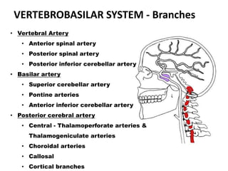
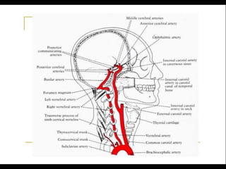
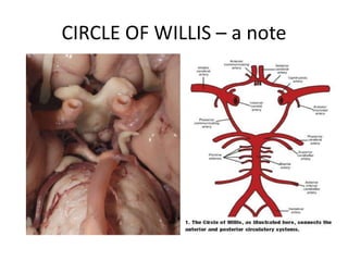
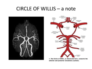
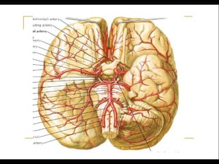
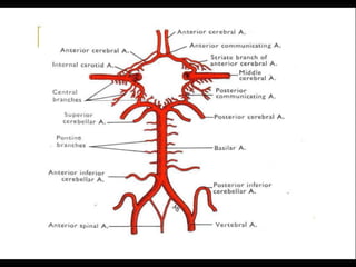
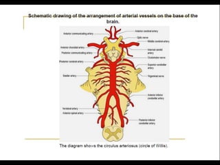
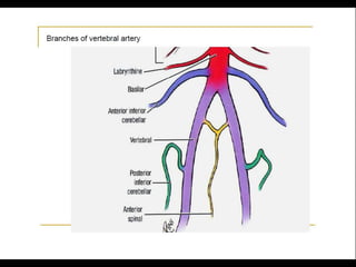
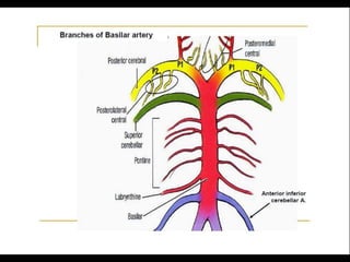
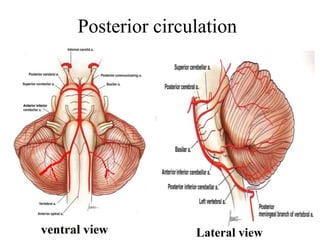
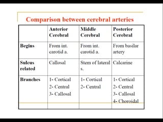
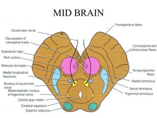
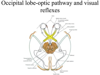
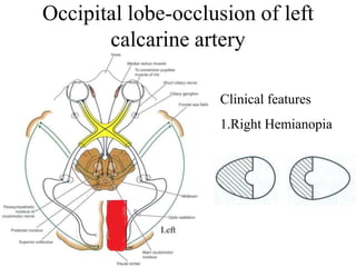
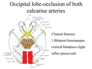
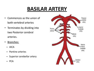
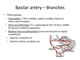
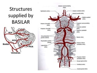
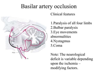
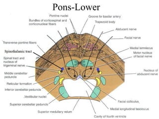
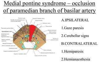
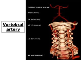
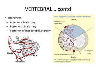
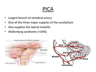
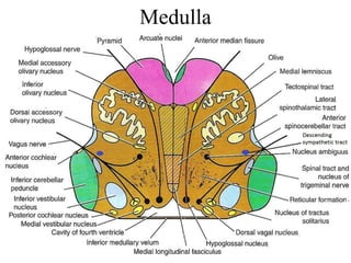
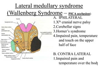
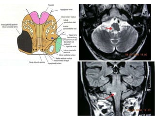
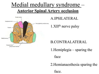
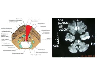
Posterior circulation strokes can be differentiated from anterior circulation strokes based on clinical features. Posterior circulation strokes often present with vertigo, unsteadiness, crossed hemiplegia, bilateral deficits, cerebellar signs, ocular findings, dissociated sensory loss, and Horner's syndrome. The vertebrobasilar system supplies structures such as the cerebellum, medulla, pons, midbrain, thalamus, and occipital and temporal lobes. Common syndromes include lateral medullary syndrome and superior cerebellar artery occlusion. Infarctions in different vascular territories can produce characteristic clinical deficits.
localization of stroke, CVS, stroke, for post graduates Kurian Joseph
New localization of stroke syndromes
1.Clinical localization of the site of the lesion.
2.Identifying the vascular territory and the vessel involved.
3.Correlating with the imaging findings.
Lecture by Prof. Osama Shukir Muhammed Amin FRCP(Edin), FRCP(Glasg), FRCP(Ire), FRCP(Lond), FACP, FAHA, to consolidate information pre-Task Based Learning about Limb Weakness. This lecture addresses upper motor neuron signs, their localization, and rationale for choosing diagnostic investigations. The next lecture will be about lower motor neuron lesions.
Please find the power point on Brainsteam stroke. I tried to present it on understandable way and all the contents are reviewed by experts and from very reliable references. Thank you
Cerebrovascular Diseases - Strokes and Their Etiology with Cerebral Blood SupplySudheera Semasinghe
Cerebrovascular accidents/strokes and their etiology (cerebral blood supply) by O. D. Roshan Indika. REFERENCES :
HARRISON’S Principles of Internal Medicine
20th edition
STROKE is cerebrovascular event with rapidly developing clinical signs of focal or global disturbances of cerebral functions with signs lasting 24 hours or longer or leading to death ,with no apparent cause other than of vascular origin (by WHO)
These simplified slides by Dr. Sidra Arshad present an overview of the non-respiratory functions of the respiratory tract.
Learning objectives:
1. Enlist the non-respiratory functions of the respiratory tract
2. Briefly explain how these functions are carried out
3. Discuss the significance of dead space
4. Differentiate between minute ventilation and alveolar ventilation
5. Describe the cough and sneeze reflexes
Study Resources:
1. Chapter 39, Guyton and Hall Textbook of Medical Physiology, 14th edition
2. Chapter 34, Ganong’s Review of Medical Physiology, 26th edition
3. Chapter 17, Human Physiology by Lauralee Sherwood, 9th edition
4. Non-respiratory functions of the lungs https://academic.oup.com/bjaed/article/13/3/98/278874
The prostate is an exocrine gland of the male mammalian reproductive system
It is a walnut-sized gland that forms part of the male reproductive system and is located in front of the rectum and just below the urinary bladder
Function is to store and secrete a clear, slightly alkaline fluid that constitutes 10-30% of the volume of the seminal fluid that along with the spermatozoa, constitutes semen
A healthy human prostate measures (4cm-vertical, by 3cm-horizontal, 2cm ant-post ).
It surrounds the urethra just below the urinary bladder. It has anterior, median, posterior and two lateral lobes
It’s work is regulated by androgens which are responsible for male sex characteristics
Generalised disease of the prostate due to hormonal derangement which leads to non malignant enlargement of the gland (increase in the number of epithelial cells and stromal tissue)to cause compression of the urethra leading to symptoms (LUTS
HOT NEW PRODUCT! BIG SALES FAST SHIPPING NOW FROM CHINA!! EU KU DB BK substit...GL Anaacs
Contact us if you are interested:
Email / Skype : kefaya1771@gmail.com
Threema: PXHY5PDH
New BATCH Ku !!! MUCH IN DEMAND FAST SALE EVERY BATCH HAPPY GOOD EFFECT BIG BATCH !
Contact me on Threema or skype to start big business!!
Hot-sale products:
NEW HOT EUTYLONE WHITE CRYSTAL!!
5cl-adba precursor (semi finished )
5cl-adba raw materials
ADBB precursor (semi finished )
ADBB raw materials
APVP powder
5fadb/4f-adb
Jwh018 / Jwh210
Eutylone crystal
Protonitazene (hydrochloride) CAS: 119276-01-6
Flubrotizolam CAS: 57801-95-3
Metonitazene CAS: 14680-51-4
Payment terms: Western Union,MoneyGram,Bitcoin or USDT.
Deliver Time: Usually 7-15days
Shipping method: FedEx, TNT, DHL,UPS etc.Our deliveries are 100% safe, fast, reliable and discreet.
Samples will be sent for your evaluation!If you are interested in, please contact me, let's talk details.
We specializes in exporting high quality Research chemical, medical intermediate, Pharmaceutical chemicals and so on. Products are exported to USA, Canada, France, Korea, Japan,Russia, Southeast Asia and other countries.
These lecture slides, by Dr Sidra Arshad, offer a quick overview of physiological basis of a normal electrocardiogram.
Learning objectives:
1. Define an electrocardiogram (ECG) and electrocardiography
2. Describe how dipoles generated by the heart produce the waveforms of the ECG
3. Describe the components of a normal electrocardiogram of a typical bipolar leads (limb II)
4. Differentiate between intervals and segments
5. Enlist some common indications for obtaining an ECG
Study Resources:
1. Chapter 11, Guyton and Hall Textbook of Medical Physiology, 14th edition
2. Chapter 9, Human Physiology - From Cells to Systems, Lauralee Sherwood, 9th edition
3. Chapter 29, Ganong’s Review of Medical Physiology, 26th edition
4. Electrocardiogram, StatPearls - https://www.ncbi.nlm.nih.gov/books/NBK549803/
5. ECG in Medical Practice by ABM Abdullah, 4th edition
6. ECG Basics, http://www.nataliescasebook.com/tag/e-c-g-basics
Tom Selleck Health: A Comprehensive Look at the Iconic Actor’s Wellness Journeygreendigital
Tom Selleck, an enduring figure in Hollywood. has captivated audiences for decades with his rugged charm, iconic moustache. and memorable roles in television and film. From his breakout role as Thomas Magnum in Magnum P.I. to his current portrayal of Frank Reagan in Blue Bloods. Selleck's career has spanned over 50 years. But beyond his professional achievements. fans have often been curious about Tom Selleck Health. especially as he has aged in the public eye.
Follow us on: Pinterest
Introduction
Many have been interested in Tom Selleck health. not only because of his enduring presence on screen but also because of the challenges. and lifestyle choices he has faced and made over the years. This article delves into the various aspects of Tom Selleck health. exploring his fitness regimen, diet, mental health. and the challenges he has encountered as he ages. We'll look at how he maintains his well-being. the health issues he has faced, and his approach to ageing .
Early Life and Career
Childhood and Athletic Beginnings
Tom Selleck was born on January 29, 1945, in Detroit, Michigan, and grew up in Sherman Oaks, California. From an early age, he was involved in sports, particularly basketball. which played a significant role in his physical development. His athletic pursuits continued into college. where he attended the University of Southern California (USC) on a basketball scholarship. This early involvement in sports laid a strong foundation for his physical health and disciplined lifestyle.
Transition to Acting
Selleck's transition from an athlete to an actor came with its physical demands. His first significant role in "Magnum P.I." required him to perform various stunts and maintain a fit appearance. This role, which he played from 1980 to 1988. necessitated a rigorous fitness routine to meet the show's demands. setting the stage for his long-term commitment to health and wellness.
Fitness Regimen
Workout Routine
Tom Selleck health and fitness regimen has evolved. adapting to his changing roles and age. During his "Magnum, P.I." days. Selleck's workouts were intense and focused on building and maintaining muscle mass. His routine included weightlifting, cardiovascular exercises. and specific training for the stunts he performed on the show.
Selleck adjusted his fitness routine as he aged to suit his body's needs. Today, his workouts focus on maintaining flexibility, strength, and cardiovascular health. He incorporates low-impact exercises such as swimming, walking, and light weightlifting. This balanced approach helps him stay fit without putting undue strain on his joints and muscles.
Importance of Flexibility and Mobility
In recent years, Selleck has emphasized the importance of flexibility and mobility in his fitness regimen. Understanding the natural decline in muscle mass and joint flexibility with age. he includes stretching and yoga in his routine. These practices help prevent injuries, improve posture, and maintain mobilit
New Directions in Targeted Therapeutic Approaches for Older Adults With Mantl...i3 Health
i3 Health is pleased to make the speaker slides from this activity available for use as a non-accredited self-study or teaching resource.
This slide deck presented by Dr. Kami Maddocks, Professor-Clinical in the Division of Hematology and
Associate Division Director for Ambulatory Operations
The Ohio State University Comprehensive Cancer Center, will provide insight into new directions in targeted therapeutic approaches for older adults with mantle cell lymphoma.
STATEMENT OF NEED
Mantle cell lymphoma (MCL) is a rare, aggressive B-cell non-Hodgkin lymphoma (NHL) accounting for 5% to 7% of all lymphomas. Its prognosis ranges from indolent disease that does not require treatment for years to very aggressive disease, which is associated with poor survival (Silkenstedt et al, 2021). Typically, MCL is diagnosed at advanced stage and in older patients who cannot tolerate intensive therapy (NCCN, 2022). Although recent advances have slightly increased remission rates, recurrence and relapse remain very common, leading to a median overall survival between 3 and 6 years (LLS, 2021). Though there are several effective options, progress is still needed towards establishing an accepted frontline approach for MCL (Castellino et al, 2022). Treatment selection and management of MCL are complicated by the heterogeneity of prognosis, advanced age and comorbidities of patients, and lack of an established standard approach for treatment, making it vital that clinicians be familiar with the latest research and advances in this area. In this activity chaired by Michael Wang, MD, Professor in the Department of Lymphoma & Myeloma at MD Anderson Cancer Center, expert faculty will discuss prognostic factors informing treatment, the promising results of recent trials in new therapeutic approaches, and the implications of treatment resistance in therapeutic selection for MCL.
Target Audience
Hematology/oncology fellows, attending faculty, and other health care professionals involved in the treatment of patients with mantle cell lymphoma (MCL).
Learning Objectives
1.) Identify clinical and biological prognostic factors that can guide treatment decision making for older adults with MCL
2.) Evaluate emerging data on targeted therapeutic approaches for treatment-naive and relapsed/refractory MCL and their applicability to older adults
3.) Assess mechanisms of resistance to targeted therapies for MCL and their implications for treatment selection
NVBDCP.pptx Nation vector borne disease control programSapna Thakur
NVBDCP was launched in 2003-2004 . Vector-Borne Disease: Disease that results from an infection transmitted to humans and other animals by blood-feeding arthropods, such as mosquitoes, ticks, and fleas. Examples of vector-borne diseases include Dengue fever, West Nile Virus, Lyme disease, and malaria.
- Video recording of this lecture in English language: https://youtu.be/lK81BzxMqdo
- Video recording of this lecture in Arabic language: https://youtu.be/Ve4P0COk9OI
- Link to download the book free: https://nephrotube.blogspot.com/p/nephrotube-nephrology-books.html
- Link to NephroTube website: www.NephroTube.com
- Link to NephroTube social media accounts: https://nephrotube.blogspot.com/p/join-nephrotube-on-social-media.html
Report Back from SGO 2024: What’s the Latest in Cervical Cancer?bkling
Are you curious about what’s new in cervical cancer research or unsure what the findings mean? Join Dr. Emily Ko, a gynecologic oncologist at Penn Medicine, to learn about the latest updates from the Society of Gynecologic Oncology (SGO) 2024 Annual Meeting on Women’s Cancer. Dr. Ko will discuss what the research presented at the conference means for you and answer your questions about the new developments.
Knee anatomy and clinical tests 2024.pdfvimalpl1234
This includes all relevant anatomy and clinical tests compiled from standard textbooks, Campbell,netter etc..It is comprehensive and best suited for orthopaedicians and orthopaedic residents.
Title: Sense of Smell
Presenter: Dr. Faiza, Assistant Professor of Physiology
Qualifications:
MBBS (Best Graduate, AIMC Lahore)
FCPS Physiology
ICMT, CHPE, DHPE (STMU)
MPH (GC University, Faisalabad)
MBA (Virtual University of Pakistan)
Learning Objectives:
Describe the primary categories of smells and the concept of odor blindness.
Explain the structure and location of the olfactory membrane and mucosa, including the types and roles of cells involved in olfaction.
Describe the pathway and mechanisms of olfactory signal transmission from the olfactory receptors to the brain.
Illustrate the biochemical cascade triggered by odorant binding to olfactory receptors, including the role of G-proteins and second messengers in generating an action potential.
Identify different types of olfactory disorders such as anosmia, hyposmia, hyperosmia, and dysosmia, including their potential causes.
Key Topics:
Olfactory Genes:
3% of the human genome accounts for olfactory genes.
400 genes for odorant receptors.
Olfactory Membrane:
Located in the superior part of the nasal cavity.
Medially: Folds downward along the superior septum.
Laterally: Folds over the superior turbinate and upper surface of the middle turbinate.
Total surface area: 5-10 square centimeters.
Olfactory Mucosa:
Olfactory Cells: Bipolar nerve cells derived from the CNS (100 million), with 4-25 olfactory cilia per cell.
Sustentacular Cells: Produce mucus and maintain ionic and molecular environment.
Basal Cells: Replace worn-out olfactory cells with an average lifespan of 1-2 months.
Bowman’s Gland: Secretes mucus.
Stimulation of Olfactory Cells:
Odorant dissolves in mucus and attaches to receptors on olfactory cilia.
Involves a cascade effect through G-proteins and second messengers, leading to depolarization and action potential generation in the olfactory nerve.
Quality of a Good Odorant:
Small (3-20 Carbon atoms), volatile, water-soluble, and lipid-soluble.
Facilitated by odorant-binding proteins in mucus.
Membrane Potential and Action Potential:
Resting membrane potential: -55mV.
Action potential frequency in the olfactory nerve increases with odorant strength.
Adaptation Towards the Sense of Smell:
Rapid adaptation within the first second, with further slow adaptation.
Psychological adaptation greater than receptor adaptation, involving feedback inhibition from the central nervous system.
Primary Sensations of Smell:
Camphoraceous, Musky, Floral, Pepperminty, Ethereal, Pungent, Putrid.
Odor Detection Threshold:
Examples: Hydrogen sulfide (0.0005 ppm), Methyl-mercaptan (0.002 ppm).
Some toxic substances are odorless at lethal concentrations.
Characteristics of Smell:
Odor blindness for single substances due to lack of appropriate receptor protein.
Behavioral and emotional influences of smell.
Transmission of Olfactory Signals:
From olfactory cells to glomeruli in the olfactory bulb, involving lateral inhibition.
Primitive, less old, and new olfactory systems with different path
4. • Comprises of:
(Tracing from Below Upwards of VertebroBasilar System)
1) Paired vertebral arteries
2) Basilar artery
3) Paired posterior cerebral arteries
• Vertebrals join to form basilar at the pontomedullary junction
• Basilar divides into two posterior cerebrals in the interpeduncular
fossa.
• These 3 give rise to long & short circumferential branches and to
smaller deep penetrating branches.
• Supply: Cerebellum, Medulla, Pons, midbrain, subthalamus,
thalamus, hippocampus and medial temporal & occipital lobes
POSTERIOR CIRCULATION
25. Posterior Cerebral Artery:- (PCA)
• Terminal branch of the basilar artery
• Paired
• At the interpeduncular fossa
• Branches:
• P 1 segment: Proximal PCA prior to junction of PCA with posterior
communicating (=Precommunal segment)
• Penetrating branches of P1:Thalamogeneculate, Percheron, posterior
choroidal)
• P 2 segment: Distal PCA (distal to junction of PCA and posterior
communicating)
PCA
26. • 75% cases: from bifurcation of basilar artery
• 20% cases: One PCA arises from ipsilateral ICA via
posterior communicating artery
• 5% cases: BOTH PCAs originate from respective ipsilateral
ICAs. The P1 segment (precommunal) of the true PCA is
atretic in such cases.
PCA - ORIGINS
27. • The artery of Percheron is a rare variant of the posterior
cerebral circulation.
• The term is used to refer to a solitary arterial trunk that
branches from one of the proximal segments of either
posterior cerebral artery.
• It supplies blood to the paramedian thalami and the
rostral midbrain bilaterally.
• Percheron infarct: bilateral thalamic and mesencephalic
infarctions ; clinically, often obtunded, comatose, or
agitated, with associated hemiplegia or hemisensory loss
PERCHERON???
28. • Supplies posterior cranial fossa structures:
– Medial area of occipital lobe
– Inferior temporal lobe
– Midbrain
– Thalamus
• Lesion causes:
– Visual agnosia
– Hemianopsia
– Alexia
– Loss of smell
POSTERIOR CEREBRAL ARTERY (PCA)
29. • Causes:
– Atheroma/Emboli @ Basilar
– Dissection @ Vertebral
– Fibromuscular dysplasia
• Two syndromes
– P 1 Syndrome
– P 2 Syndrome
PCA Syndromes:
30. • Area infarcted:
– Ipsilateral subthalamus
– Medial thalamus
– Ipsilateral cerebral peduncle
– Midbrain
• Weber’s/Claude’s syndrome can occur
• Contralateral hemiballismus +/-
• Art. of Percheron occlusion: Upward gaze paresis,
drowsiness, abulia
P 1 syndrome:
31. • B/L Prox PCA occlusion: Extensive infarction:
– Coma, Unreactive pupils, b/l pyramidal signs, decerebrate rigidity
• Penetrating branches of thalamic and thalamogeniculate
arteries if occluded:
– Less extensive syndromes
• Thalamic Dejerine-Roussy syndrome:
– Contralateral hemisensory loss
– Followed by agonising, searing, burning pain
– Persistent, poor response to analgesics
– Anticonvulsants (Carbamazepine, gabapentin) & TCAs used.
P 1 syndrome… contd:
32. • Infarction of:
– Medial temporal and occipital lobes
• Contralateral homonymous hemianopia with macular
sparing
• Occasional only the upper quadrant is involved.
• Dominant medial temporal lobe and hippocampal lesions:
Acute disturbances in memory – usually recovers
• Alexia without Agraphia
• Visual agnosia
• Amnestic aphasia
• Peduncular hallucinosis
P 2 syndrome
33. • Anton’s blindness
• Gun barrel vision
• Balint’s syndrome
• Palinopsia
• Asimultanagnosia
• Embolic occulsion of top of basilar:
– HALLMARK is sudden onset of bilateral signs, including ptosis,
pupillary asymmetry or lack of reaction to light, somnolence.
P 2 syndrome… contd:
41. Occipital lobe-occlusion of both
calcarine arteries
Clinical features
1.Bilateral hemianopia-
cortical blindness (light
reflex preserved)
42.
43. Left occipital lobe with corpus
callosum infarction
Left
Clinical features
1.Right hemianopia
2.Alexia without
agraphia
44.
45. • Commences as the union of
both vertebral arteries
• Terminates by dividing into
two Posterior cerebral
arteries.
• Branches:
– AICA
– Pontine arteries
– Superior cerebellar artery
– PCA
BASILAR ARTERY
46. • Three groups:
– Paramedian, 7-10 in number, supply a wedge of pons on
either side of midline
– Short circumferential, 5-7, supply lateral 2/3rd of Pons, middle
& superior cerebellar peduncles.
– Bilateral long circumferentials (curve around pons to supply
cerebellum):
• Superior cerebellar art
• Anterior inferior cerebellar art
Basilar artery – Branches
48. • Complete basilar occlusion
– Constellation of bilateral long tract signs (sensory & motor)
with signs of cranial nerve & cerebellar dysfunction.
• “Locked-in” state:
– Preserved consciousness with quadriplegia & cranial nerve
signs.
Basilar syndromes
49. Basilar artery occlusion
Clinical features
1.Paralysis of all four limbs
2.Bulbar paralysis
3.Eye movements
abnormalities
4.Nystagmus
5.Coma
Note: The neurological
deficit is variable depending
upon the ischemia –
modifying factors.
50. • Severe ipsilateral cerebellar ataxia
• Nausea & vomitings
• Dysarthria
• Contralateral loss of pain & temperature over extremities,
body & face.
• Partial deafness, ataxic tremor of ipsilateral UL, Horner’s
syndrome & Palatal myoclonus rare
Superior cerebellar artery
occlusion
52. Medial pontine syndrome – occlusion
of paramedian branch of basilar artery
A.IPSILATERAL
1.Gaze paresis
2.Cerebellar signs
B.CONTRALATERAL
1.Hemiparesis
2.Hemianaesthesia
53. Lateral pontine syndrome-occlusion of
anterior inferior cerebellar artery
A.IPSILATERAL
1.LMN VIIth nerve palsy
2.Gaze palsy
3.Deafness,tinnitus
4.Cerebellar signs
B.CONTRALATERAL
1.Impairment of pain and
temperature on the
body
55. • Commences as a branch of the subclavian on left and
brachiocephalic on right and terminates by joining to form
the basilar artery
• Four parts:
– V-1: Preforaminal- origin to entrance into C5 or C6 foramen
– V-2: Foraminal- vertebral foramina C6 to C2
– V-3: C2 to dura- passes through transverse foramen and
circles around the arch of the atlas to pierce the atlas at the
formen magnum
– V-4: Intradural-courses upwards and joins other to form
basilar. Gives branches that supply BS & cerebellum.
VERTEBRAL ARTERY
57. • Largest branch of vertebral artery
• One of the three major supplies of the cerebellum
• Also supplies the lateral medulla
• Wallenberg syndrome (=LMS)
PICA
58. • Posterior meningeal branch
• Arises from opposite the formen magnum
• Supplies Falx cerebri
MENINGEAL BRANCHES OF VERTEBRAL a.
59. • Predilection for V1 and V4
• Usually lesion of one vertebral does not cause TIAs.
• TIAs occur if one is atretic and other is developing
occlusion.
• Symptoms:
– Syncope
– Vertigo
– Alternating hemiplegia
– ‘Sets the stage for thrombosis’
• Stenosis proximal to origin of PICA can threaten lateral
medulla & posterior inferior surface of cerebellum.
ATHEROTHROMBOTIC LESIONS – V1 & V4
60. • Atheromatous disease is rare.
• Fibromuscular dysplasia, dissection common here
• Rarely due to encroachment from osteophytic spurs
within vertebral foramina
LESIONS OF V2 & V3
61. • Subclavian occluded proximal to
origin of vertebral.
• Leads of reversal in the direction of
blood flow in the ipsilateral vertebral
artery.
• Exercise of ipsilateral arm may
increase demand on vertebral flow,
leading to posterior circulation TIAs.
“SUBCLAVIAN STEAL”
63. Lateral medullary syndrome
(Wallenberg Syndrome – PICA occlusion)
A. IPSILATERAL
1.Xth cranial nerve palsy
2.Cerebellar signs
3.Horner’s syndrome
4.Impaired pain, temperature
and touch on the upper
half of face
B. CONTRA LATERAL
1.Impaired pain and
temperature over the body
64.
65. Medial medullary syndrome –
Anterior Spinal Artery occlusion
A.IPSILATERAL
1.XIIth nerve palsy
B.CONTRALATERAL
1.Hemiplegia – sparing the
face
2.Hemianaesthesia sparing the
face.
66.
67. • Can lead to sudden respiratory arrest
• Due to raised ICP in the posterior fossa
• Symptoms:
– Drowsiness
– Babinski signs
– Dysarthria
– Bifacial weakness maybe absent, or present only briefly,
before respiratory arrest ensues.
– Gait unsteadiness, headache, dizziness, nausea and vomiting
maybe the only early symptoms and signs and should arouse
suspicion.
• D/D: Viral labrynthitis (Headache, neck stiffness &
unilateral dysmetria favor stroke)
CEREBELLAR INFARCTION