SlideShare a Scribd company logo
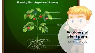
Anatomy of
plant parts
By Mr Biswanath prusty
ANATOMY OF
ROOT
Distinct Anatomical Characteristics of the
Root:
The outermost layer is termed as epiblema.
Cuticle and stomata are absent.
Cortex is formed of parenchymatous cells.
Endodermis is well developed.
Pericycle is distinct.
Vascular bundles are radial.
Xylem is exarch.
Phloem consists of sieve tubes, companion
cells and phloem parenchyma. (In
monocots however, the phloem parenchyma
is absent).
Anatomy of Dicot Root
The anatomy of a dicot root exhibits following structures (Fig. 2.52).
a. Epiblema
• It is the outermost one cell thick layer in which cells are compactly arranged.
• The cells usually have fine tubular elongations, called root hairs.
• These help in the absorption of water.
b. Cortex:
• It lies between epiblema and endodermis.
• It consists of circular or polygonal; parenchymatous cells with intercellular spaces.
c. Endodermis:
It is innermost layer of cortex and consists of a layer of compactly arranged barrel shaped cells.
The cells have casparian bands (special thickenings) on radial walls. The cells lying opposite to xylem, do not have
casparian strips and are called passage cells.
d. Pericycle:
It is one or two layered, thin walled parenchymatous structure that is present below the endodermis.
It forms the outermost layer of stele.
It takes part in the formation of secondary roots (branches) and cambium for secondary growth.
e. Vascular Bundles:
• These are arranged in a ring i.e., radial.
• Xylem and phloem are situated at separate radii. The
number of xylem and phloem groups vary from 2 to 6.
• Xylem consists of tracheids, vessels and xylem
parenchyma. Phloem consists of sieve tubes, companion
cells and phloem parenchyma.
• Xylem is exarch i.e., the protoxylem towards outside and
metaxylem towards centre.
• The parenchymatous cells lying between them form
conjuctive tissue.
f. Pith:
• It is parenchymatous with large intercellular spaces and
lies in the centre but is poorly developed or absent due to
more development of metaxylem towards the centre.
Anatomy of Monocot Root
The anatomy of a monocot root exhibits following structures (Fig. 2.53).
a. Epiblema:
• It is the outermost one cell thick layer in which cells are compactly arranged
• The cells usually have fine tubular elongations, called root hairs & These help
in the absorption of water.
b. Cortex:
• It lies between epiblema and endodermis.
• It consists of circular or polygonal; parenchymatous cells with intercellular
spaces.
c. Endodermis:
• It is innermost layer of cortex and consists of a layer of compactly arranged
barrel shaped cells.
• The cells have Casparian bands (special thickenings) on radial walls. The cells
lying opposite to xylem, do not have casparian strips and are called passage
cells.
d. Pericycle:
• It is one layered, thin walled parenchymatous structure that is present below the endodermis.
• It forms the outermost layer of stele.
• It takes part in the formation of secondary roots (branches).
• In old roots the cells become lignified and thick walled.
e. Vascular bundles:
• Vascular bundles are polyarch, radial and exarch.
• Xylem and phloem are situated at separate radii. The number of xylem and phloem groups is more
than 6.
• Xylem consists of tracheids and xylem parenchyma. Phloem consists of sieve tubes and companion
cells. Phloem parenchyma is absent.
• Xylem is exarch i.e., the protoxylem towards outside and metaxylem towards centre.
• The parenchymatous cells lying between them form conjunctive tissue.
f. Pith:
• It is well defined and lies in the center.
• It consists of loosely arranged parenchymatous cells.
Table 2.1 summarizes major differences between anatomy of dicot and
monocot roots.
Sr. No Characters Dicotyledon root Monocotyledon root
1 Vascular bundle • The number of
vascular bundle vary
from 2 to 6.
• Phloem parenchyma is
present.
• The number of
vascular bundle is
more than 6.
• Phloem parenchyma is
absent.
2 Pericycle • Give out lateral roots,
vascular cambium and
cork cambium.
• Give out only lateral
roots.
3 Cambium • Develops later on
during secondary
growth.
• Does not develop at
any stage.
4 Pith • Absent or poorly
developed.
• Large and well
developed.
ANATOMY OF STEM
Anomy is the general term used for the
study of internal structure.
Distinct Anatomical Characters of the
Stem:
• Epidermis is covered by cuticle.
• Hairs or trichomes are present on
epidermis that are unicellular or
multicellular.
• The vascular bundles are mainly
conjoint, collateral or bicollateral and in
some cases may be concentric.
Anatomy of Dicot Stem
The anatomy of a dicot stem exhibits following structures (Fig. 2.54).
a. Epidermis:
• It is the outermost one cell thick layer in which cells are compactly arranged.
• Chloroplast, multicellular hairs and stomata are present.
• The outer wall of the cells is cuticularised.
b. Cortex:
• It is present below the epidermis.
• It is differentiated into three regions hypodermis, middle cortex and endodermis.
1. i. Hypodermis:
• It is made up of collenchymatous cells and is 4-6 layered thick.
• These cells contain chloroplast.
• The intercellular spaces are absent.
• It provides mechanical support.
2. ii. Middle cortex:
• It is present between hypodermis and endodermis.
• It is made up of parenchymatous cells and is several layers thick.
• The cells are oval or spherical in shape with intercellular spaces.
• It mainly serves for food storage, besides providing mechanical strength.
1. iii. Endodermis:
• It is the innermost layer and present upto the stele.
• The cells are barrel-shaped and due to the starch accumulation of starch it is also called sheath (in young dicot
stem).
• Casparian strips are present in these cells.
C. Pericycle:
• It is formed of alternate bands of parenchymatous and sclerenchymatous cells.
• The sclerenchymatous cells are present between endodermis and phloem cells. The patches of
sclerenchymatous cells is also known as bast.
• The parenchymatous cells are present above the medullary rays.
d. Vascular bundles:
• The vascular bundles are conjoint, collateral and open.
• These are arranged in a ring.
• Each vascular bundle contains xylem, phloem and cambium.
• Phloem is present in the outer region, and it consists of sieve tubes, companion cells and phloem
parenchyma.
• Xylem forms the inner part of the vascular bundle. It consists of vessels, tracheids. fibres and xylem
parenchyma. Protoxylem consists of narrow elements where as metaxylem consists of relatively
wide elements.
e. Cambium:
• It is present between xylem and phloem.
• It is made up of 2 to 3 layers of thin-walled
meristematic cell, which appear rectangular in
transverse section.
f. Medulla or Pith:
• Large and well-developed pith is present in the center.
• It is made of parenchymatous cells with large
intercellular spaces.
• The pith is connected with the cortex through radiating
regions of parenchyma between vascular bundles.
These are called medullary rays.
Anatomy of Monocot Stem
The anatomy of a monocot stem exhibits following structures (Fig. 2.55).
a. Epidermis:
• It is the outermost one cell thick layer in which cells are compactly arranged.
• Chloroplast and stomata are present but multicellular hairs are absent.
• The outer wall of the cells is cuticularized.
b. Hypodermis:
• It is made up of sclerenchymatous cells and is 2-4 layers thick.
• The intercellular spaces are absent.
C. Ground tissues:
• All the parenchymatous tissues from below the hypodermis to the center are called ground tissues.
• There is no differentiation between cortex, endodermis, pericycle and pith.
• The cells contain reserve food material.
• Vascular bundles are embedded in the ground tissues.
d. Vascular bundles:
• These are oval and scattered throughout the ground tissue. Large and loosely arranged vascular bundles are present
towards center while numerous, small and densely arranged vascular bundles are present towards periphery.
• Each bundle is surrounded by a sclerenchymatous sheath.
• The vascular bundles are conjoint, collateral, endarch and closed.
• In each vascular bundle, xylem and phloem is present but cambium is absent.
i. Xylem:
• It is Y shaped and consists of four distinct
vessels surrounded by many tracheids.
• Mataxylem consists of two wide and pitted
vessels which form the two arms or Y.
• Protoxylem consists of one or two vessels
with spiral or annular thickening which form
the base of 'Y'.
ii. Phloem:
• Meta phloem is present between the forked
arms of Y shaped xylem.
• Protophloem is present near the periphery of
vascular bundle.
• Phloem consists of sieve tubes and
companion cells, but phloem parenchyma is
absent.
• The outer layer of vascular bundle and
sclerenchymatous cells of hypodermis often
coalesces, thus vascular bundle appear
embedded in the sheath.
Anatomy of Dicot Leaf (Dorsiventral Leaf)
The anatomy of a dicot leaf exhibits following
structures (Fig. 2.57).
a. Upper Epidermis:
• It is the upper outermost, single layer thick
and made up of parenchymatous cells.
• Its outer walls are cuticularised. Stomata and
chloroplasts are absent.
b. Lower Epidermis:
• It is like the upper epidermis, but stomata are
present on this surface. Chloroplasts are
absent in the cells, but the guard cells of the
stomata contain chloroplast.
c. Mesophyll:
It is present between the upper and lower epidermis, and it is divided
into two regions; Palisade parenchyma and spongy parenchyma.
1. i. Palisade parenchyma:
• The cells are elongated, mostly two layered and perpendicular
to the upper epidermis and have no intercellular spaces.
• These cells contain chloroplasts and actively participate in
photosynthesis.
2. Spongy parenchyma:
• It is present below the palisade parenchyma.
• The cells are spherical or oval and are irregularly arranged.
They possess chloroplasts.
• Intercellular spaces are interconnected and open to outside
through stomata that help In diffusion of gases.
Vascular bundles:
• These are scattered in spongy parenchyma.
• The vascular bundle of midrib is the largest.
• The vascular bundles are collateral and closed.
• Around each bundle is present a parenchymatous sheath, called
bundle sheath.
• Xylem consists of tracheae, wood fibers and wood parenchyma.
Anatomy of Monocot Leaf (lso-bilateral Leaf)
The anatomy of a Monocot leaf exhibits following structures
(Fig. 2.58).
a. Epidermis:
• The upper and lower epidermises of leaf are similar, and
they consist of a single layer of cells.
• The leaf is bounded by thick cuticularized epidermis on
both sides.
• Stomata are present on both upper and lower epidermis.
• Some cells in the upper epidermis get enlarged in sized and
are called bulliform cells or motor cells. It helps in leaf
curling in grasses to check transpiration.
b. Mesophyll:
• It is present between the upper and lower epidermis and is
not differentiated into palisade and spongy tissues.
• The cells are spherical, irregularly arranged and enclose
intercellular spaces .
• The cells contain chloroplasts.
Vascular bundles:
• Many large and small vascular bundles
are present in the leaf.
• Each bundle is surrounded by a layer of
thin-walled cells, called bundle sheath.
• The vascular bundles are conjoint,
collateral and closed. Vascular bundle is
surrounded by a parenchymatous bundle
sheath.
• A patch of sclerenchyma occurs on both
the ends of each vascular bundle, which
extends upto the epidermis on their
respective sides.
• Xylem is located towards the upper side
while the phloem is present on lower
side.
Anatomy of plant parts by BNP.pdf

More Related Content

What's hot (20)

Complex permanent tissue
Complex permanent tissueComplex permanent tissue
Complex permanent tissue
 
periderm, new 2023.pptx
periderm, new 2023.pptxperiderm, new 2023.pptx
periderm, new 2023.pptx
 
Plant tissue systems
Plant tissue systemsPlant tissue systems
Plant tissue systems
 
Plant tissues
Plant  tissuesPlant  tissues
Plant tissues
 
Inflorescence
InflorescenceInflorescence
Inflorescence
 
Anatomy of dicot and monocot stem
Anatomy of dicot and monocot stemAnatomy of dicot and monocot stem
Anatomy of dicot and monocot stem
 
Sexual Reproduction in Flowering Plants (Part I)
Sexual Reproduction in Flowering Plants (Part I)Sexual Reproduction in Flowering Plants (Part I)
Sexual Reproduction in Flowering Plants (Part I)
 
Alternation of generation in archegoniates
Alternation of generation in archegoniatesAlternation of generation in archegoniates
Alternation of generation in archegoniates
 
Lesson 7 bio101 (c)Dr. Evangelista
Lesson 7 bio101 (c)Dr.  EvangelistaLesson 7 bio101 (c)Dr.  Evangelista
Lesson 7 bio101 (c)Dr. Evangelista
 
Structural variations in reproductive structures of division Bryophyta
Structural variations in reproductive structures of division BryophytaStructural variations in reproductive structures of division Bryophyta
Structural variations in reproductive structures of division Bryophyta
 
Funaria
FunariaFunaria
Funaria
 
Microsporangium
MicrosporangiumMicrosporangium
Microsporangium
 
3-APICAL MERISTEM.pptx
3-APICAL MERISTEM.pptx3-APICAL MERISTEM.pptx
3-APICAL MERISTEM.pptx
 
Selaginella zk
Selaginella  zkSelaginella  zk
Selaginella zk
 
Anatomy of Dicot and monocot Root.pptx
Anatomy of Dicot and monocot Root.pptxAnatomy of Dicot and monocot Root.pptx
Anatomy of Dicot and monocot Root.pptx
 
Morphology of flowering plants
Morphology of flowering plantsMorphology of flowering plants
Morphology of flowering plants
 
Phloem
PhloemPhloem
Phloem
 
Marchantia
MarchantiaMarchantia
Marchantia
 
Riccia
RicciaRiccia
Riccia
 
Plant tissue 1
Plant tissue 1Plant tissue 1
Plant tissue 1
 

Similar to Anatomy of plant parts by BNP.pdf

Internal structure of stem
Internal structure of stemInternal structure of stem
Internal structure of stemManojDas87
 
Unit1 part 1 (1).pptx dicot anatomy in which it will show the anatomical stru...
Unit1 part 1 (1).pptx dicot anatomy in which it will show the anatomical stru...Unit1 part 1 (1).pptx dicot anatomy in which it will show the anatomical stru...
Unit1 part 1 (1).pptx dicot anatomy in which it will show the anatomical stru...prekshanalwaya
 
Tissue System in plant
Tissue System in plantTissue System in plant
Tissue System in plantvidan biology
 
1a- Primary Plant Body.pptx
1a- Primary Plant Body.pptx1a- Primary Plant Body.pptx
1a- Primary Plant Body.pptxMUNAWARSHAH26
 
Anatomy of flowering plants
Anatomy of flowering plantsAnatomy of flowering plants
Anatomy of flowering plantsShahina Akhter
 
Anatomy Of Coniferales.pptx
Anatomy Of Coniferales.pptxAnatomy Of Coniferales.pptx
Anatomy Of Coniferales.pptxKavan Chaudhari
 
Primary structure Monocot Root
Primary structure Monocot RootPrimary structure Monocot Root
Primary structure Monocot RootDr. T. A. Gitte
 
meristmatic tissue.pptx
meristmatic tissue.pptxmeristmatic tissue.pptx
meristmatic tissue.pptxmanoj Joshi
 
EPITHELIA TISSUES.ppt
EPITHELIA TISSUES.pptEPITHELIA TISSUES.ppt
EPITHELIA TISSUES.pptPharmTecM
 
PlantTissueTissueSystempptx__2021_02_11_16_10_26.pptx
PlantTissueTissueSystempptx__2021_02_11_16_10_26.pptxPlantTissueTissueSystempptx__2021_02_11_16_10_26.pptx
PlantTissueTissueSystempptx__2021_02_11_16_10_26.pptxSelestineSalema
 
Anatomy of dicot and monocot stem
Anatomy of dicot and monocot stemAnatomy of dicot and monocot stem
Anatomy of dicot and monocot stemanuragpandey304
 

Similar to Anatomy of plant parts by BNP.pdf (20)

Dicot stem
Dicot  stemDicot  stem
Dicot stem
 
Plant anatomy
Plant anatomyPlant anatomy
Plant anatomy
 
Internal structure of stem
Internal structure of stemInternal structure of stem
Internal structure of stem
 
Anatomy of leaves
Anatomy of leavesAnatomy of leaves
Anatomy of leaves
 
Tissue system
Tissue systemTissue system
Tissue system
 
Unit1 part 1 (1).pptx dicot anatomy in which it will show the anatomical stru...
Unit1 part 1 (1).pptx dicot anatomy in which it will show the anatomical stru...Unit1 part 1 (1).pptx dicot anatomy in which it will show the anatomical stru...
Unit1 part 1 (1).pptx dicot anatomy in which it will show the anatomical stru...
 
Tissue System in plant
Tissue System in plantTissue System in plant
Tissue System in plant
 
PLANT TISSUES.pptx
PLANT TISSUES.pptxPLANT TISSUES.pptx
PLANT TISSUES.pptx
 
1a- Primary Plant Body.pptx
1a- Primary Plant Body.pptx1a- Primary Plant Body.pptx
1a- Primary Plant Body.pptx
 
Std 11 biology chapter 06
Std 11 biology chapter 06Std 11 biology chapter 06
Std 11 biology chapter 06
 
Anatomy of flowering plants
Anatomy of flowering plantsAnatomy of flowering plants
Anatomy of flowering plants
 
6. anatomy of flowering plants
6. anatomy of flowering plants6. anatomy of flowering plants
6. anatomy of flowering plants
 
Tissues
TissuesTissues
Tissues
 
Anatomy Of Coniferales.pptx
Anatomy Of Coniferales.pptxAnatomy Of Coniferales.pptx
Anatomy Of Coniferales.pptx
 
Primary structure Monocot Root
Primary structure Monocot RootPrimary structure Monocot Root
Primary structure Monocot Root
 
meristmatic tissue.pptx
meristmatic tissue.pptxmeristmatic tissue.pptx
meristmatic tissue.pptx
 
EPITHELIA TISSUES.ppt
EPITHELIA TISSUES.pptEPITHELIA TISSUES.ppt
EPITHELIA TISSUES.ppt
 
PlantTissueTissueSystempptx__2021_02_11_16_10_26.pptx
PlantTissueTissueSystempptx__2021_02_11_16_10_26.pptxPlantTissueTissueSystempptx__2021_02_11_16_10_26.pptx
PlantTissueTissueSystempptx__2021_02_11_16_10_26.pptx
 
Anatomy of dicot and monocot stem
Anatomy of dicot and monocot stemAnatomy of dicot and monocot stem
Anatomy of dicot and monocot stem
 
Plant cells and tissues
Plant cells and tissuesPlant cells and tissues
Plant cells and tissues
 

More from College of pharmaceutical sciences

Synthesis and significance of biological substances by BNP.pdf
Synthesis and significance of biological substances by BNP.pdfSynthesis and significance of biological substances by BNP.pdf
Synthesis and significance of biological substances by BNP.pdfCollege of pharmaceutical sciences
 
Catabolism of phenylalanine and tyrosine and their metabolic disorders by BNP...
Catabolism of phenylalanine and tyrosine and their metabolic disorders by BNP...Catabolism of phenylalanine and tyrosine and their metabolic disorders by BNP...
Catabolism of phenylalanine and tyrosine and their metabolic disorders by BNP...College of pharmaceutical sciences
 

More from College of pharmaceutical sciences (20)

Prescribed medication order and communication skills.pdf
Prescribed medication order and communication skills.pdfPrescribed medication order and communication skills.pdf
Prescribed medication order and communication skills.pdf
 
Education & training program in the hospital by BNP.pdf
Education & training program in the hospital by BNP.pdfEducation & training program in the hospital by BNP.pdf
Education & training program in the hospital by BNP.pdf
 
Drug information services by BNP.pdf
Drug information services by BNP.pdfDrug information services by BNP.pdf
Drug information services by BNP.pdf
 
Pateient counseling by BNP.pdf
Pateient counseling by BNP.pdfPateient counseling by BNP.pdf
Pateient counseling by BNP.pdf
 
Pharmacy and therapeutic committee by BNP.pdf
Pharmacy and therapeutic committee by BNP.pdfPharmacy and therapeutic committee by BNP.pdf
Pharmacy and therapeutic committee by BNP.pdf
 
Plant growth n development by BNP.pdf
Plant growth n development by BNP.pdfPlant growth n development by BNP.pdf
Plant growth n development by BNP.pdf
 
Virus, Viroid, Bacteriophage by BNP.pdf
Virus, Viroid, Bacteriophage by BNP.pdfVirus, Viroid, Bacteriophage by BNP.pdf
Virus, Viroid, Bacteriophage by BNP.pdf
 
6.Plant kingdom by BNP.pdf
6.Plant kingdom by BNP.pdf6.Plant kingdom by BNP.pdf
6.Plant kingdom by BNP.pdf
 
Human reproduction by BNP.pdf
Human reproduction by BNP.pdfHuman reproduction by BNP.pdf
Human reproduction by BNP.pdf
 
Nucleic acid and Nucleotide By BNP.pptx
Nucleic acid and Nucleotide By BNP.pptxNucleic acid and Nucleotide By BNP.pptx
Nucleic acid and Nucleotide By BNP.pptx
 
Synthesis and significance of biological substances by BNP.pdf
Synthesis and significance of biological substances by BNP.pdfSynthesis and significance of biological substances by BNP.pdf
Synthesis and significance of biological substances by BNP.pdf
 
Catabolism of Heme and jaundice by BNP.pdf
Catabolism of Heme and jaundice by BNP.pdfCatabolism of Heme and jaundice by BNP.pdf
Catabolism of Heme and jaundice by BNP.pdf
 
Catabolism of phenylalanine and tyrosine and their metabolic disorders by BNP...
Catabolism of phenylalanine and tyrosine and their metabolic disorders by BNP...Catabolism of phenylalanine and tyrosine and their metabolic disorders by BNP...
Catabolism of phenylalanine and tyrosine and their metabolic disorders by BNP...
 
Amino acid metabolism BY BISWANATH PRUSTY
Amino acid metabolism BY BISWANATH PRUSTYAmino acid metabolism BY BISWANATH PRUSTY
Amino acid metabolism BY BISWANATH PRUSTY
 
Diabetes notes by BISWANATH PRUSTY
Diabetes notes by BISWANATH PRUSTYDiabetes notes by BISWANATH PRUSTY
Diabetes notes by BISWANATH PRUSTY
 
CARBOHYDRATE METABOLISM BY BISWANATH PRUSTY
CARBOHYDRATE METABOLISM BY BISWANATH PRUSTYCARBOHYDRATE METABOLISM BY BISWANATH PRUSTY
CARBOHYDRATE METABOLISM BY BISWANATH PRUSTY
 
Bioenergetics by lecture Biswanath.pdf
Bioenergetics by lecture Biswanath.pdfBioenergetics by lecture Biswanath.pdf
Bioenergetics by lecture Biswanath.pdf
 
Biochemistry by Lecture Biswanath.pdf
Biochemistry by Lecture Biswanath.pdfBiochemistry by Lecture Biswanath.pdf
Biochemistry by Lecture Biswanath.pdf
 
Animal kingdom by BNP
Animal kingdom by BNPAnimal kingdom by BNP
Animal kingdom by BNP
 
Kingdom of fungi by Bnp
Kingdom of fungi by BnpKingdom of fungi by Bnp
Kingdom of fungi by Bnp
 

Recently uploaded

Gliese 12 b: A Temperate Earth-sized Planet at 12 pc Ideal for Atmospheric Tr...
Gliese 12 b: A Temperate Earth-sized Planet at 12 pc Ideal for Atmospheric Tr...Gliese 12 b: A Temperate Earth-sized Planet at 12 pc Ideal for Atmospheric Tr...
Gliese 12 b: A Temperate Earth-sized Planet at 12 pc Ideal for Atmospheric Tr...Sérgio Sacani
 
Mammalian Pineal Body Structure and Also Functions
Mammalian Pineal Body Structure and Also FunctionsMammalian Pineal Body Structure and Also Functions
Mammalian Pineal Body Structure and Also FunctionsYOGESH DOGRA
 
Aerodynamics. flippatterncn5tm5ttnj6nmnynyppt
Aerodynamics. flippatterncn5tm5ttnj6nmnynypptAerodynamics. flippatterncn5tm5ttnj6nmnynyppt
Aerodynamics. flippatterncn5tm5ttnj6nmnynypptsreddyrahul
 
GBSN - Microbiology (Lab 2) Compound Microscope
GBSN - Microbiology (Lab 2) Compound MicroscopeGBSN - Microbiology (Lab 2) Compound Microscope
GBSN - Microbiology (Lab 2) Compound MicroscopeAreesha Ahmad
 
biotech-regenration of plants, pharmaceutical applications.pptx
biotech-regenration of plants, pharmaceutical applications.pptxbiotech-regenration of plants, pharmaceutical applications.pptx
biotech-regenration of plants, pharmaceutical applications.pptxANONYMOUS
 
THYROID-PARATHYROID medical surgical nursing
THYROID-PARATHYROID medical surgical nursingTHYROID-PARATHYROID medical surgical nursing
THYROID-PARATHYROID medical surgical nursingJocelyn Atis
 
Constraints on Neutrino Natal Kicks from Black-Hole Binary VFTS 243
Constraints on Neutrino Natal Kicks from Black-Hole Binary VFTS 243Constraints on Neutrino Natal Kicks from Black-Hole Binary VFTS 243
Constraints on Neutrino Natal Kicks from Black-Hole Binary VFTS 243Sérgio Sacani
 
The importance of continents, oceans and plate tectonics for the evolution of...
The importance of continents, oceans and plate tectonics for the evolution of...The importance of continents, oceans and plate tectonics for the evolution of...
The importance of continents, oceans and plate tectonics for the evolution of...Sérgio Sacani
 
RNA INTERFERENCE: UNRAVELING GENETIC SILENCING
RNA INTERFERENCE: UNRAVELING GENETIC SILENCINGRNA INTERFERENCE: UNRAVELING GENETIC SILENCING
RNA INTERFERENCE: UNRAVELING GENETIC SILENCINGAADYARAJPANDEY1
 
word2vec, node2vec, graph2vec, X2vec: Towards a Theory of Vector Embeddings o...
word2vec, node2vec, graph2vec, X2vec: Towards a Theory of Vector Embeddings o...word2vec, node2vec, graph2vec, X2vec: Towards a Theory of Vector Embeddings o...
word2vec, node2vec, graph2vec, X2vec: Towards a Theory of Vector Embeddings o...Subhajit Sahu
 
Detectability of Solar Panels as a Technosignature
Detectability of Solar Panels as a TechnosignatureDetectability of Solar Panels as a Technosignature
Detectability of Solar Panels as a TechnosignatureSérgio Sacani
 
GBSN - Biochemistry (Unit 5) Chemistry of Lipids
GBSN - Biochemistry (Unit 5) Chemistry of LipidsGBSN - Biochemistry (Unit 5) Chemistry of Lipids
GBSN - Biochemistry (Unit 5) Chemistry of LipidsAreesha Ahmad
 
The ASGCT Annual Meeting was packed with exciting progress in the field advan...
The ASGCT Annual Meeting was packed with exciting progress in the field advan...The ASGCT Annual Meeting was packed with exciting progress in the field advan...
The ASGCT Annual Meeting was packed with exciting progress in the field advan...Health Advances
 
GBSN - Microbiology (Lab 1) Microbiology Lab Safety Procedures
GBSN -  Microbiology (Lab  1) Microbiology Lab Safety ProceduresGBSN -  Microbiology (Lab  1) Microbiology Lab Safety Procedures
GBSN - Microbiology (Lab 1) Microbiology Lab Safety ProceduresAreesha Ahmad
 
Astronomy Update- Curiosity’s exploration of Mars _ Local Briefs _ leadertele...
Astronomy Update- Curiosity’s exploration of Mars _ Local Briefs _ leadertele...Astronomy Update- Curiosity’s exploration of Mars _ Local Briefs _ leadertele...
Astronomy Update- Curiosity’s exploration of Mars _ Local Briefs _ leadertele...NathanBaughman3
 
Jet reorientation in central galaxies of clusters and groups: insights from V...
Jet reorientation in central galaxies of clusters and groups: insights from V...Jet reorientation in central galaxies of clusters and groups: insights from V...
Jet reorientation in central galaxies of clusters and groups: insights from V...Sérgio Sacani
 
Microbial Type Culture Collection (MTCC)
Microbial Type Culture Collection (MTCC)Microbial Type Culture Collection (MTCC)
Microbial Type Culture Collection (MTCC)abhishekdhamu51
 
Gliese 12 b, a temperate Earth-sized planet at 12 parsecs discovered with TES...
Gliese 12 b, a temperate Earth-sized planet at 12 parsecs discovered with TES...Gliese 12 b, a temperate Earth-sized planet at 12 parsecs discovered with TES...
Gliese 12 b, a temperate Earth-sized planet at 12 parsecs discovered with TES...Sérgio Sacani
 
Multi-source connectivity as the driver of solar wind variability in the heli...
Multi-source connectivity as the driver of solar wind variability in the heli...Multi-source connectivity as the driver of solar wind variability in the heli...
Multi-source connectivity as the driver of solar wind variability in the heli...Sérgio Sacani
 
Richard's entangled aventures in wonderland
Richard's entangled aventures in wonderlandRichard's entangled aventures in wonderland
Richard's entangled aventures in wonderlandRichard Gill
 

Recently uploaded (20)

Gliese 12 b: A Temperate Earth-sized Planet at 12 pc Ideal for Atmospheric Tr...
Gliese 12 b: A Temperate Earth-sized Planet at 12 pc Ideal for Atmospheric Tr...Gliese 12 b: A Temperate Earth-sized Planet at 12 pc Ideal for Atmospheric Tr...
Gliese 12 b: A Temperate Earth-sized Planet at 12 pc Ideal for Atmospheric Tr...
 
Mammalian Pineal Body Structure and Also Functions
Mammalian Pineal Body Structure and Also FunctionsMammalian Pineal Body Structure and Also Functions
Mammalian Pineal Body Structure and Also Functions
 
Aerodynamics. flippatterncn5tm5ttnj6nmnynyppt
Aerodynamics. flippatterncn5tm5ttnj6nmnynypptAerodynamics. flippatterncn5tm5ttnj6nmnynyppt
Aerodynamics. flippatterncn5tm5ttnj6nmnynyppt
 
GBSN - Microbiology (Lab 2) Compound Microscope
GBSN - Microbiology (Lab 2) Compound MicroscopeGBSN - Microbiology (Lab 2) Compound Microscope
GBSN - Microbiology (Lab 2) Compound Microscope
 
biotech-regenration of plants, pharmaceutical applications.pptx
biotech-regenration of plants, pharmaceutical applications.pptxbiotech-regenration of plants, pharmaceutical applications.pptx
biotech-regenration of plants, pharmaceutical applications.pptx
 
THYROID-PARATHYROID medical surgical nursing
THYROID-PARATHYROID medical surgical nursingTHYROID-PARATHYROID medical surgical nursing
THYROID-PARATHYROID medical surgical nursing
 
Constraints on Neutrino Natal Kicks from Black-Hole Binary VFTS 243
Constraints on Neutrino Natal Kicks from Black-Hole Binary VFTS 243Constraints on Neutrino Natal Kicks from Black-Hole Binary VFTS 243
Constraints on Neutrino Natal Kicks from Black-Hole Binary VFTS 243
 
The importance of continents, oceans and plate tectonics for the evolution of...
The importance of continents, oceans and plate tectonics for the evolution of...The importance of continents, oceans and plate tectonics for the evolution of...
The importance of continents, oceans and plate tectonics for the evolution of...
 
RNA INTERFERENCE: UNRAVELING GENETIC SILENCING
RNA INTERFERENCE: UNRAVELING GENETIC SILENCINGRNA INTERFERENCE: UNRAVELING GENETIC SILENCING
RNA INTERFERENCE: UNRAVELING GENETIC SILENCING
 
word2vec, node2vec, graph2vec, X2vec: Towards a Theory of Vector Embeddings o...
word2vec, node2vec, graph2vec, X2vec: Towards a Theory of Vector Embeddings o...word2vec, node2vec, graph2vec, X2vec: Towards a Theory of Vector Embeddings o...
word2vec, node2vec, graph2vec, X2vec: Towards a Theory of Vector Embeddings o...
 
Detectability of Solar Panels as a Technosignature
Detectability of Solar Panels as a TechnosignatureDetectability of Solar Panels as a Technosignature
Detectability of Solar Panels as a Technosignature
 
GBSN - Biochemistry (Unit 5) Chemistry of Lipids
GBSN - Biochemistry (Unit 5) Chemistry of LipidsGBSN - Biochemistry (Unit 5) Chemistry of Lipids
GBSN - Biochemistry (Unit 5) Chemistry of Lipids
 
The ASGCT Annual Meeting was packed with exciting progress in the field advan...
The ASGCT Annual Meeting was packed with exciting progress in the field advan...The ASGCT Annual Meeting was packed with exciting progress in the field advan...
The ASGCT Annual Meeting was packed with exciting progress in the field advan...
 
GBSN - Microbiology (Lab 1) Microbiology Lab Safety Procedures
GBSN -  Microbiology (Lab  1) Microbiology Lab Safety ProceduresGBSN -  Microbiology (Lab  1) Microbiology Lab Safety Procedures
GBSN - Microbiology (Lab 1) Microbiology Lab Safety Procedures
 
Astronomy Update- Curiosity’s exploration of Mars _ Local Briefs _ leadertele...
Astronomy Update- Curiosity’s exploration of Mars _ Local Briefs _ leadertele...Astronomy Update- Curiosity’s exploration of Mars _ Local Briefs _ leadertele...
Astronomy Update- Curiosity’s exploration of Mars _ Local Briefs _ leadertele...
 
Jet reorientation in central galaxies of clusters and groups: insights from V...
Jet reorientation in central galaxies of clusters and groups: insights from V...Jet reorientation in central galaxies of clusters and groups: insights from V...
Jet reorientation in central galaxies of clusters and groups: insights from V...
 
Microbial Type Culture Collection (MTCC)
Microbial Type Culture Collection (MTCC)Microbial Type Culture Collection (MTCC)
Microbial Type Culture Collection (MTCC)
 
Gliese 12 b, a temperate Earth-sized planet at 12 parsecs discovered with TES...
Gliese 12 b, a temperate Earth-sized planet at 12 parsecs discovered with TES...Gliese 12 b, a temperate Earth-sized planet at 12 parsecs discovered with TES...
Gliese 12 b, a temperate Earth-sized planet at 12 parsecs discovered with TES...
 
Multi-source connectivity as the driver of solar wind variability in the heli...
Multi-source connectivity as the driver of solar wind variability in the heli...Multi-source connectivity as the driver of solar wind variability in the heli...
Multi-source connectivity as the driver of solar wind variability in the heli...
 
Richard's entangled aventures in wonderland
Richard's entangled aventures in wonderlandRichard's entangled aventures in wonderland
Richard's entangled aventures in wonderland
 

Anatomy of plant parts by BNP.pdf

  • 1. Anatomy of plant parts By Mr Biswanath prusty
  • 2. ANATOMY OF ROOT Distinct Anatomical Characteristics of the Root: The outermost layer is termed as epiblema. Cuticle and stomata are absent. Cortex is formed of parenchymatous cells. Endodermis is well developed. Pericycle is distinct. Vascular bundles are radial. Xylem is exarch. Phloem consists of sieve tubes, companion cells and phloem parenchyma. (In monocots however, the phloem parenchyma is absent).
  • 3.
  • 4. Anatomy of Dicot Root The anatomy of a dicot root exhibits following structures (Fig. 2.52). a. Epiblema • It is the outermost one cell thick layer in which cells are compactly arranged. • The cells usually have fine tubular elongations, called root hairs. • These help in the absorption of water. b. Cortex: • It lies between epiblema and endodermis. • It consists of circular or polygonal; parenchymatous cells with intercellular spaces. c. Endodermis: It is innermost layer of cortex and consists of a layer of compactly arranged barrel shaped cells. The cells have casparian bands (special thickenings) on radial walls. The cells lying opposite to xylem, do not have casparian strips and are called passage cells. d. Pericycle: It is one or two layered, thin walled parenchymatous structure that is present below the endodermis. It forms the outermost layer of stele. It takes part in the formation of secondary roots (branches) and cambium for secondary growth.
  • 5. e. Vascular Bundles: • These are arranged in a ring i.e., radial. • Xylem and phloem are situated at separate radii. The number of xylem and phloem groups vary from 2 to 6. • Xylem consists of tracheids, vessels and xylem parenchyma. Phloem consists of sieve tubes, companion cells and phloem parenchyma. • Xylem is exarch i.e., the protoxylem towards outside and metaxylem towards centre. • The parenchymatous cells lying between them form conjuctive tissue. f. Pith: • It is parenchymatous with large intercellular spaces and lies in the centre but is poorly developed or absent due to more development of metaxylem towards the centre.
  • 6.
  • 7. Anatomy of Monocot Root The anatomy of a monocot root exhibits following structures (Fig. 2.53). a. Epiblema: • It is the outermost one cell thick layer in which cells are compactly arranged • The cells usually have fine tubular elongations, called root hairs & These help in the absorption of water. b. Cortex: • It lies between epiblema and endodermis. • It consists of circular or polygonal; parenchymatous cells with intercellular spaces. c. Endodermis: • It is innermost layer of cortex and consists of a layer of compactly arranged barrel shaped cells. • The cells have Casparian bands (special thickenings) on radial walls. The cells lying opposite to xylem, do not have casparian strips and are called passage cells.
  • 8. d. Pericycle: • It is one layered, thin walled parenchymatous structure that is present below the endodermis. • It forms the outermost layer of stele. • It takes part in the formation of secondary roots (branches). • In old roots the cells become lignified and thick walled. e. Vascular bundles: • Vascular bundles are polyarch, radial and exarch. • Xylem and phloem are situated at separate radii. The number of xylem and phloem groups is more than 6. • Xylem consists of tracheids and xylem parenchyma. Phloem consists of sieve tubes and companion cells. Phloem parenchyma is absent. • Xylem is exarch i.e., the protoxylem towards outside and metaxylem towards centre. • The parenchymatous cells lying between them form conjunctive tissue. f. Pith: • It is well defined and lies in the center. • It consists of loosely arranged parenchymatous cells.
  • 9. Table 2.1 summarizes major differences between anatomy of dicot and monocot roots. Sr. No Characters Dicotyledon root Monocotyledon root 1 Vascular bundle • The number of vascular bundle vary from 2 to 6. • Phloem parenchyma is present. • The number of vascular bundle is more than 6. • Phloem parenchyma is absent. 2 Pericycle • Give out lateral roots, vascular cambium and cork cambium. • Give out only lateral roots. 3 Cambium • Develops later on during secondary growth. • Does not develop at any stage. 4 Pith • Absent or poorly developed. • Large and well developed.
  • 10. ANATOMY OF STEM Anomy is the general term used for the study of internal structure. Distinct Anatomical Characters of the Stem: • Epidermis is covered by cuticle. • Hairs or trichomes are present on epidermis that are unicellular or multicellular. • The vascular bundles are mainly conjoint, collateral or bicollateral and in some cases may be concentric.
  • 11. Anatomy of Dicot Stem The anatomy of a dicot stem exhibits following structures (Fig. 2.54). a. Epidermis: • It is the outermost one cell thick layer in which cells are compactly arranged. • Chloroplast, multicellular hairs and stomata are present. • The outer wall of the cells is cuticularised. b. Cortex: • It is present below the epidermis. • It is differentiated into three regions hypodermis, middle cortex and endodermis. 1. i. Hypodermis: • It is made up of collenchymatous cells and is 4-6 layered thick. • These cells contain chloroplast. • The intercellular spaces are absent. • It provides mechanical support. 2. ii. Middle cortex: • It is present between hypodermis and endodermis. • It is made up of parenchymatous cells and is several layers thick. • The cells are oval or spherical in shape with intercellular spaces. • It mainly serves for food storage, besides providing mechanical strength.
  • 12. 1. iii. Endodermis: • It is the innermost layer and present upto the stele. • The cells are barrel-shaped and due to the starch accumulation of starch it is also called sheath (in young dicot stem). • Casparian strips are present in these cells. C. Pericycle: • It is formed of alternate bands of parenchymatous and sclerenchymatous cells. • The sclerenchymatous cells are present between endodermis and phloem cells. The patches of sclerenchymatous cells is also known as bast. • The parenchymatous cells are present above the medullary rays. d. Vascular bundles: • The vascular bundles are conjoint, collateral and open. • These are arranged in a ring. • Each vascular bundle contains xylem, phloem and cambium. • Phloem is present in the outer region, and it consists of sieve tubes, companion cells and phloem parenchyma. • Xylem forms the inner part of the vascular bundle. It consists of vessels, tracheids. fibres and xylem parenchyma. Protoxylem consists of narrow elements where as metaxylem consists of relatively wide elements.
  • 13. e. Cambium: • It is present between xylem and phloem. • It is made up of 2 to 3 layers of thin-walled meristematic cell, which appear rectangular in transverse section. f. Medulla or Pith: • Large and well-developed pith is present in the center. • It is made of parenchymatous cells with large intercellular spaces. • The pith is connected with the cortex through radiating regions of parenchyma between vascular bundles. These are called medullary rays.
  • 14. Anatomy of Monocot Stem The anatomy of a monocot stem exhibits following structures (Fig. 2.55). a. Epidermis: • It is the outermost one cell thick layer in which cells are compactly arranged. • Chloroplast and stomata are present but multicellular hairs are absent. • The outer wall of the cells is cuticularized. b. Hypodermis: • It is made up of sclerenchymatous cells and is 2-4 layers thick. • The intercellular spaces are absent. C. Ground tissues: • All the parenchymatous tissues from below the hypodermis to the center are called ground tissues. • There is no differentiation between cortex, endodermis, pericycle and pith. • The cells contain reserve food material. • Vascular bundles are embedded in the ground tissues. d. Vascular bundles: • These are oval and scattered throughout the ground tissue. Large and loosely arranged vascular bundles are present towards center while numerous, small and densely arranged vascular bundles are present towards periphery. • Each bundle is surrounded by a sclerenchymatous sheath. • The vascular bundles are conjoint, collateral, endarch and closed. • In each vascular bundle, xylem and phloem is present but cambium is absent.
  • 15. i. Xylem: • It is Y shaped and consists of four distinct vessels surrounded by many tracheids. • Mataxylem consists of two wide and pitted vessels which form the two arms or Y. • Protoxylem consists of one or two vessels with spiral or annular thickening which form the base of 'Y'. ii. Phloem: • Meta phloem is present between the forked arms of Y shaped xylem. • Protophloem is present near the periphery of vascular bundle. • Phloem consists of sieve tubes and companion cells, but phloem parenchyma is absent. • The outer layer of vascular bundle and sclerenchymatous cells of hypodermis often coalesces, thus vascular bundle appear embedded in the sheath.
  • 16.
  • 17.
  • 18. Anatomy of Dicot Leaf (Dorsiventral Leaf) The anatomy of a dicot leaf exhibits following structures (Fig. 2.57). a. Upper Epidermis: • It is the upper outermost, single layer thick and made up of parenchymatous cells. • Its outer walls are cuticularised. Stomata and chloroplasts are absent. b. Lower Epidermis: • It is like the upper epidermis, but stomata are present on this surface. Chloroplasts are absent in the cells, but the guard cells of the stomata contain chloroplast.
  • 19. c. Mesophyll: It is present between the upper and lower epidermis, and it is divided into two regions; Palisade parenchyma and spongy parenchyma. 1. i. Palisade parenchyma: • The cells are elongated, mostly two layered and perpendicular to the upper epidermis and have no intercellular spaces. • These cells contain chloroplasts and actively participate in photosynthesis. 2. Spongy parenchyma: • It is present below the palisade parenchyma. • The cells are spherical or oval and are irregularly arranged. They possess chloroplasts. • Intercellular spaces are interconnected and open to outside through stomata that help In diffusion of gases. Vascular bundles: • These are scattered in spongy parenchyma. • The vascular bundle of midrib is the largest. • The vascular bundles are collateral and closed. • Around each bundle is present a parenchymatous sheath, called bundle sheath. • Xylem consists of tracheae, wood fibers and wood parenchyma.
  • 20. Anatomy of Monocot Leaf (lso-bilateral Leaf) The anatomy of a Monocot leaf exhibits following structures (Fig. 2.58). a. Epidermis: • The upper and lower epidermises of leaf are similar, and they consist of a single layer of cells. • The leaf is bounded by thick cuticularized epidermis on both sides. • Stomata are present on both upper and lower epidermis. • Some cells in the upper epidermis get enlarged in sized and are called bulliform cells or motor cells. It helps in leaf curling in grasses to check transpiration. b. Mesophyll: • It is present between the upper and lower epidermis and is not differentiated into palisade and spongy tissues. • The cells are spherical, irregularly arranged and enclose intercellular spaces . • The cells contain chloroplasts.
  • 21. Vascular bundles: • Many large and small vascular bundles are present in the leaf. • Each bundle is surrounded by a layer of thin-walled cells, called bundle sheath. • The vascular bundles are conjoint, collateral and closed. Vascular bundle is surrounded by a parenchymatous bundle sheath. • A patch of sclerenchyma occurs on both the ends of each vascular bundle, which extends upto the epidermis on their respective sides. • Xylem is located towards the upper side while the phloem is present on lower side.