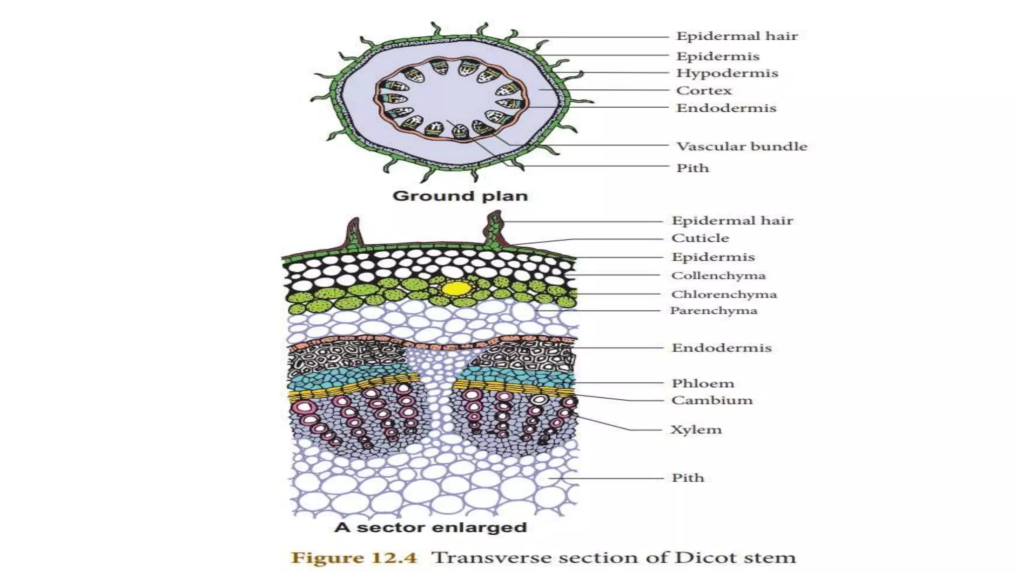
1. The internal structure of a dicot stem features an epidermis, hypodermis, cortex, endodermis, pericycle, vascular bundles arranged in rings, pith, and medullary rays.
2. Vascular bundles are conjoint, collateral or bicollateral, and open type, containing xylem, phloem, and cambium.
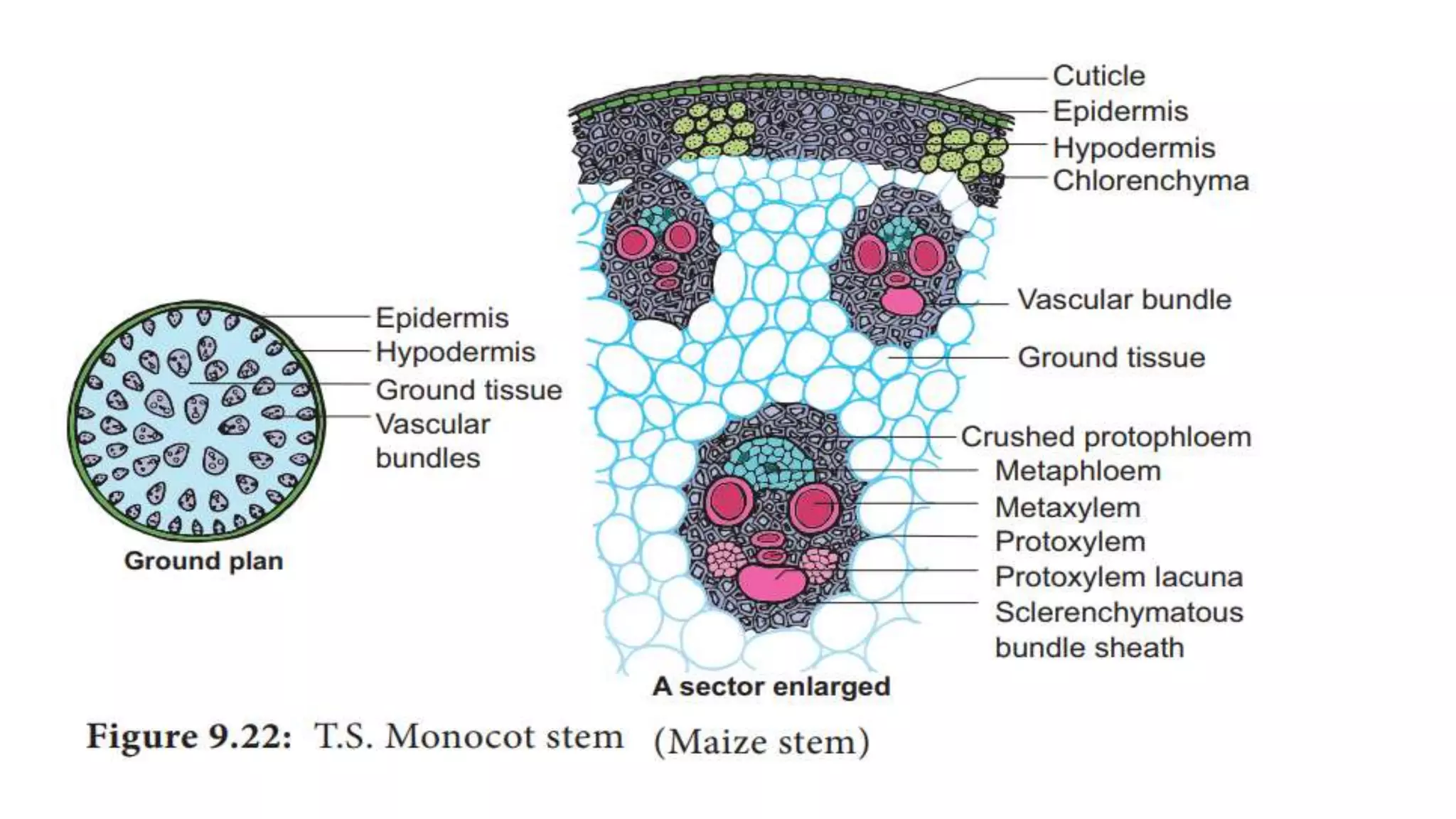
3. The internal structure of a monocot stem lacks a differentiated cortex and pith, and has scattered vascular bundles surrounded by bundle sheath, lacking medullary rays.