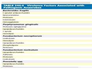
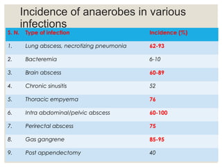
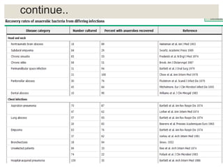
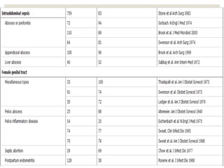
This document provides an overview of anaerobic infections. It defines anaerobes and discusses their role as commensal flora. It describes the diseases caused by anaerobic bacteria and the sites they commonly infect. The document outlines the pathogenesis of anaerobic infections and their increasing incidence. It discusses the importance of laboratory diagnosis and describes methods for specimen collection, transport, culture, and identification of anaerobic bacteria.
Tetanus is caused by Clostridium tetani bacteria entering the body through a wound. The bacteria produces a neurotoxin called tetanospasmin that causes painful muscle spasms and rigidity. Symptoms typically begin with lockjaw and stiffness of the neck muscles before becoming generalized. The disease is often fatal if untreated. Diagnosis is clinical based on symptoms, though the bacteria can sometimes be identified from wound samples. Treatment involves antitoxin administration to neutralize the toxin along with antibiotics. Prophylaxis through active immunization with tetanus toxoid vaccines or passive immunization with antitoxin is important to prevent occurrence.
This document discusses several types of hepatitis viruses. It covers hepatitis A virus, hepatitis B virus, hepatitis C virus, hepatitis D virus, and hepatitis E virus. For each virus, it describes key aspects such as transmission, pathogenesis, clinical manifestations, diagnosis, and prevention. Hepatitis viruses can cause liver inflammation and damage, and some may lead to chronic infection or liver cancer if not addressed. Vaccines exist to prevent hepatitis A and B.
Hepatitis B virus is a double-stranded DNA virus that causes hepatitis B disease. It is able to establish chronic infections and has been shown to integrate into host DNA. The virus primarily infects liver cells and establishes lifelong infections in around 5% of adults and up to 90% of infants infected. While vaccination has been effective at reducing new infections, chronic hepatitis B remains an important global public health issue.
Staphylococci are spherical bacteria that occur in grape-like clusters. Staphylococcus aureus is an important human pathogen that can cause a variety of infections, from minor skin infections to life-threatening conditions like toxic shock syndrome and endocarditis. S. aureus produces several virulence factors like toxins and enzymes that damage tissues and evade the immune system. Laboratory diagnosis involves culture, microscopy, and tests like coagulase to identify S. aureus. Antibiotics are used to treat infections, and prevention focuses on hygiene and safe food handling. Methicillin-resistant S. aureus is an antibiotic resistant form that is more difficult to treat.
Clostridium tetani is a gram-positive, rod-shaped bacterium that causes the disease tetanus. It forms spores that allow it to survive in environments without oxygen. The spores can enter the body through wounds and germinate under anaerobic conditions, producing a toxin that travels along the nervous system to the spinal cord. This toxin blocks neurotransmitters and causes painful muscle spasms, starting with the jaw and potentially leading to death if the respiratory muscles are affected. Laboratory diagnosis involves microscopy, culture studies, and biochemical tests to identify the bacterium, as well as animal inoculation experiments. Treatment involves wound cleaning, antibiotics, and vaccines to prevent further cases.
The document discusses human herpesvirus infections, focusing on herpes simplex viruses types 1 and 2. It provides details on the structure and properties of herpesviruses, describing how they cause both lytic and latent infections. The major symptoms and clinical manifestations of HSV-1 and HSV-2 infections are outlined, including oral and genital lesions as well as infections in immunocompromised individuals and newborns. Recurrent infections are also discussed.
Superficial mycoses are fungal infections that are limited to the outer layers of the skin. Common causative agents include dermatophytes, Malassezia furfur, Piedraia hortae, Trichosporon beigelii, and Exophiala werneckii. Dermatophytes such as Trichophyton, Microsporum, and Epidermophyton cause ringworm infections of the skin, hair, and nails. Clinical manifestations vary depending on the infected area but may include scaly patches with active, inflamed borders. Laboratory identification involves microscopic examination of specimens and fungal culture.
The PPT is mainly all about Mycobacterium Tuberculosis. Agents causing the disease Tuberculosis, pathogenesis, laboratory diagnosis, treatment and prophylaxis. It was made for both BSc and MSc students.
Mycetoma is a chronic subcutaneous infection caused by certain fungi or bacteria that enters through the skin via minor trauma. It results in a painless swelling, draining sinuses, and discharge containing grains. The infection commonly affects the feet, legs, and other extremities in agricultural workers and those walking barefoot in endemic areas like parts of Africa and Asia. Diagnosis involves examining biopsy samples for characteristic grains and cultures to identify the causative organism. Treatment depends on whether it is caused by bacteria (actinomycetoma), requiring prolonged antibiotics, or fungi (eumycetoma), which may require surgery and antifungal therapy. Prevention involves wearing shoes in endemic areas.
Streptococcus pyogenes is a Gram-positive bacterium that can cause a variety of infections in humans. It commonly colonizes the throat and skin. It produces toxins and enzymes that contribute to its virulence and ability to cause disease. S. pyogenes can cause suppurative infections like pharyngitis, impetigo, and necrotizing fasciitis. It can also cause non-suppurative sequelae after infection like acute rheumatic fever and glomerulonephritis. Diagnosis involves culturing samples on blood agar and testing for sensitivity to bacitracin. Treatment involves antibiotics like penicillin. Prevention focuses on proper treatment of streptococcal infections to reduce risk of
Streptococcus pyogenes is a Gram positive coccus that forms chains and causes beta hemolysis on blood agar. It is classified by Lancefield grouping based on cell wall carbohydrates and Griffith typing based on M proteins. S. pyogenes causes respiratory, skin, and genital infections and can lead to post-streptococcal sequelae like rheumatic fever and glomerulonephritis. Penicillin is usually the treatment of choice.
Laboratory diagnosis of tuberculosis pract.deepak deshkar
This document summarizes the laboratory diagnosis of tuberculosis. It describes how specimens are collected from pulmonary and extra-pulmonary sites. The specimens then undergo decontamination, concentration, and acid-fast staining for direct microscopic examination. Culture methods including solid and liquid media as well as automated systems are discussed. Biochemical tests and animal inoculation are used to identify Mycobacterium tuberculosis. Sensitivity testing evaluates resistance to anti-tubercular drugs using phenotypic and molecular methods. Molecular diagnostic techniques like PCR are also employed.
Clostridium tetani is an obligate anaerobic, gram-positive bacterium that causes the disease tetanus. It forms terminal spores that give it a distinctive drumstick appearance. Though found worldwide in soil, C. tetani enters the body through wounds and causes tetanus by producing a potent neurotoxin. It is a major cause of mortality in developing countries, with neonatal tetanus accounting for about half of worldwide cases and having a mortality rate of 85%.
Amoebiasis is an infection caused by the protozoan Entamoeba histolytica. It is common in developing parts of the world with poor sanitation. Most infections are asymptomatic, but some can cause intestinal symptoms ranging from mild diarrhea to severe dysentery. Rarely, the infection can spread beyond the intestines and cause liver abscesses or affect other organs. Diagnosis involves microscopic examination of stool samples or biopsy specimens to look for trophozoites or cysts. Treatment depends on the severity of symptoms and may involve antibiotics or antiparasitic drugs. Prevention relies on improved sanitation, water treatment, hygiene and health education.
Vibrio cholerae is the bacteria that causes cholera. It is a facultative anaerobe that grows well between 37°C and pH 7.4-9.6. It can be cultured on ordinary media like nutrient agar as well as special transport and enrichment media. Pathogenesis involves ingesting contaminated food/water, with the bacteria multiplying in the intestines and producing cholera toxin which causes hypersecretion of fluids. Diagnosis involves microscopic examination of rice water stool samples and culturing in selective media. Treatment focuses on fluid replacement therapy and oral rehydration.
Dermatophytoses, commonly known as ringworm, are fungal infections caused by dermatophytes which invade keratinized tissues like skin, hair, and nails. There are three main genera of dermatophytes - Trichophyton, Microsporum, and Epidermophyton. They are classified based on their morphology and site of infection. Dermatophytes are contracted through direct contact with infected skin, animals, or soil. Common clinical manifestations include tinea capitis, tinea corporis, tinea pedis, and tinea cruris. Laboratory diagnosis involves microscopic examination of skin or nail samples in KOH and fungal culture. Oral antifungals like terbinaf
This document discusses wound infections, including the types (exogenous and endogenous), common causative organisms, and methods for diagnosis. It notes that pus accumulation is a sign of local infection and describes redness, pain, and swelling as additional indicators. Culture-based methods are outlined for identifying bacteria and determining pathogenicity from wound specimens. The importance of discussing isolated organisms with the physician is emphasized.
Staphylococci are Gram positive cocci that commonly cause localized suppurative lesions. Staphylococcus aureus is an important pathogenic species that can cause a variety of infections like food poisoning, toxic shock syndrome, and nosocomial infections. S. aureus has developed resistance to many antibiotics like penicillin through production of beta-lactamases. Methicillin resistant S. aureus strains are a major concern as they are resistant even to methicillin and related antibiotics.
This powerpoint contains slides describing types of hepatitis viruses, pathogenesis, clinical course, laboratory diagnosis, treatment and prevention against hepatitis viruses. This presentation is intended to use by medical students, nurses, paramedics in the learning on virology. The slided could also be resource materials for the academicians.
This document provides information about Hepatitis C, including how it is transmitted, symptoms, diagnosis, treatment, and prevention. Some key points:
- Hepatitis C is a contagious liver disease caused by the hepatitis C virus that can range from mild to serious and lifelong.
- It is mostly transmitted through exposure to infectious blood, such as through contaminated medical equipment or injecting drug use.
- Most infected people do not show symptoms, though some may experience fatigue, abdominal pain, and jaundice. Chronic infection can lead to cirrhosis or liver cancer.
- Diagnosis involves antibody and RNA testing to confirm. People at high risk should be screened.
- Treatment involves antiviral therapy with
This document discusses Staphylococcus aureus and methods for its laboratory diagnosis. It notes that S. aureus is commonly found in the nose and can cause infections, while S. epidermidis lives on the skin and S. saprophyticus in the vagina. Diagnosis involves collecting samples from infections and using gram staining, culturing, and biochemical tests like catalase, coagulase and novobiocin to identify the species. Rapid PCR tests are also now used to diagnose S. aureus.
This document discusses Streptococcus pneumoniae, a common bacterium that can cause pneumonia, meningitis, and other infections. It describes the morphology and cultural characteristics of S. pneumoniae, including that it appears as paired diplococci and forms alpha-hemolytic colonies on blood agar. It also covers the antigenic properties, virulence factors, pathogenicity, epidemiology, laboratory diagnosis, prophylaxis, and treatment of infections caused by S. pneumoniae.
1. Mycobacterium is a genus of bacteria that includes M. tuberculosis and M. leprae, which are the causes of tuberculosis and leprosy, respectively.
2. These bacteria have an acid-fast staining pattern and lipid-rich cell walls that make them resistant to disinfectants and antibiotics.
3. M. tuberculosis spreads through the air and causes pneumonia and cavitary lesions in the lungs, while M. leprae spreads through skin lesions and can cause disfigurement if untreated.
This document discusses human herpes simplex virus (HSV) types 1 and 2. It covers the structure and life cycle of HSV, how it establishes lifelong latent infections in human sensory neurons, and its associated diseases. These include oral and genital lesions. HSV is commonly transmitted through oral contact and sexual activity. While incurable, antiviral medications can reduce symptoms and transmission risk.
The document discusses laboratory diagnosis of urinary tract infections, including specimen collection and transport, microscopic examination of urine to detect white blood cells, bacteria, casts, crystals and parasites, and culture of urine samples to identify causative organisms and antibiotic susceptibility testing. Appearance of urine and findings on microscopic examination can provide clues to possible urinary tract infections or other underlying conditions. Proper collection and transport of urine samples is important for accurate laboratory diagnosis of UTIs.
The document discusses the bacterium Staphylococcus aureus, including MRSA. It describes S. aureus's characteristics and how it can cause infections. MRSA was first identified in the 1940s when some S. aureus strains developed resistance to penicillin. It further discusses how MRSA is transmitted and the differences between CA-MRSA and HA-MRSA. Prevention, treatment options, and challenges like developing resistance are also covered at a high level.
This document provides an overview of anaerobic microorganisms. It discusses their typical habitats including the skin, oral cavity, gastrointestinal tract, and urogenital tract. It outlines their classification and important genera such as Bacteroides, Prevotella, and Clostridium. Virulence factors and mechanisms of pathogenesis are described. The laboratory diagnosis of anaerobic infections including specimen collection and culture techniques are explained. Common clinical infections associated with different anaerobic bacteria and their treatment conclude the document.
This document provides an overview of anaerobic microorganisms. It discusses their habitat, classification, virulence factors and pathogenesis. Key points include that anaerobes normally inhabit the skin, oral cavity, gastrointestinal tract and genitalia. They outnumber aerobic bacteria in many areas. Common pathogenic genera include Bacteroides, Prevotella, Porphyromonas, Fusobacterium, Clostridium and Peptostreptococcus. Virulence factors allow tissue invasion when mucosal barriers are breached. Laboratory diagnosis involves culture media and identification of colonies and metabolites.
COLLECTION AND TRANSPORT OF SPECIMEN LATEST.pptxSereneVarghese1
The document provides guidance on proper collection, transport, and storage of clinical specimens to ensure accurate microbiological testing and results. Key points include:
- Specimen quality directly impacts patient care, treatment decisions, and outcomes.
- Laboratories should reject poor quality specimens that could impact results.
- Proper collection, transport, and storage methods depend on specimen type to maintain organism viability until testing.
- Complete labeling and requisition forms are needed to properly identify and interpret specimen results.
Recent advances in cultivation & identification of anaerobicabhishek yadav
This document discusses recent advances in cultivating and identifying clinically significant anaerobic bacteria. It covers:
1. The challenges in detecting anaerobic infections due to their slow growth and long turnaround times for identification.
2. Methods for classifying, isolating, and identifying anaerobic bacteria including their oxygen tolerance levels, suitable specimens for culture, transport methods, and culture techniques.
3. The major infections caused by different anaerobic bacteria like Clostridium, Bacteroides, Fusobacterium species.
4. Clues that suggest anaerobic infections and approaches to the presumptive identification of isolated organisms.
Cryptosporidium parvum is a protozoan parasite that causes cryptosporidiosis. It has a life cycle involving an infective oocyst stage that is transmitted through ingestion of contaminated food or water. Infection results in watery diarrhea that can last 1-2 weeks. Diagnosis is made by identifying oocysts in stool samples microscopically or through antigen testing. Treatment focuses on rehydration though some drugs may help reduce symptoms. Prevention involves proper hygiene and water treatment.
Dr. Diwan Mahmood Khan, Assistant Professor of Microbiology,
MCDRC, Durg, Chattisgarh, India.
Topic: Opportunistic Mycoses- Candidiasis or Candidosis
For Medical Student: MBBS and BDS
This document provides guidance on proper specimen collection, transport, and aseptic technique in microbiology. It discusses the importance of collecting the right specimen from the right patient at the right time and transporting it properly to the laboratory. It provides details on proper collection and transport of various specimen types from different sites of infection, including respiratory, urinary, genital, and others. Proper labeling, containers, transport media, and aseptic technique are also covered to ensure sample quality and viability.
This document provides an overview of anaerobic bacteria. It describes the characteristics of anaerobes and how they differ from aerobic bacteria in their oxygen requirements. It also outlines methods for specimen examination, culture, identification, and the major types of clinically relevant anaerobic bacteria including Bacteroides, Prevotella, Fusobacterium, Clostridium, Actinomyces, Propionibacterium, and anaerobic cocci. Key details on morphology, habitat, diseases caused, and biochemical traits are given for important bacterial genera.
Sputum examination provides important diagnostic information by analyzing material coughed up from the lungs and respiratory tract. Key indications for sputum examination include identifying the causative organism in suspected lower respiratory infections like pneumonia or tuberculosis. Sputum samples can also be examined cytologically to detect malignant cells or investigate other infections. Proper collection and transport of sputum samples is important for microbiological culture and other tests. Staining and microscopic examination of sputum looks for bacteria, fungi, parasites and other pathogenic organisms. Molecular tests like PCR provide a rapid and sensitive method for tuberculosis diagnosis.
This document discusses Group B and D streptococci, including their taxonomy, description, epidemiology, clinical significance, and laboratory diagnosis. It focuses on Streptococcus agalactiae (Group B streptococcus) and Enterococcus (Group D streptreptococcus).
Group B streptococcus is a leading cause of neonatal infections. It commonly causes early-onset meningitis in newborns. Proper screening and treatment of colonized mothers during pregnancy can help prevent early-onset neonatal infections. Identification involves culturing vaginal/rectal swabs in selective broth followed by plating on blood agar. Phenotypic tests like CAMP and hippurate hydrolysis are used to identify S. agalactiae.
This document discusses principles of anaerobic bacteriology. It describes how anaerobic microbes differ from aerobic microbes in their ability to utilize or tolerate oxygen. There are three main categories of anaerobes - obligate anaerobes which cannot tolerate oxygen and are inhibited by it, aerotolerant anaerobes which can tolerate but not use oxygen, and facultative anaerobes which can grow with or without oxygen. Common anaerobic infections are caused by Clostridium, Bacteroides, Actinomyces, and Fusobacterium species. Proper collection and transport of specimens is important for culturing anaerobes due to their sensitivity to oxygen. Special
This document outlines the diagnostic approach and laboratory tests for evaluating a patient presenting with pyrexia of unknown origin (PUO). It describes collecting relevant clinical history and performing a physical exam. Specimens including blood, urine, sputum, CSF and tissues may be obtained for bacterial, viral, parasitic and fungal cultures and stains. Tests like blood cultures, urine cultures, sputum smears and cultures, and CSF analysis can help identify potential infectious causes. Serology, skin tests, hematology, immunology and biopsy may also provide diagnostic clues. Empiric antibiotic therapy is guided by risk factors and test results.
Neisseria is a genus of bacteria that includes the pathogenic species N. gonorrhoeae and N. meningitidis. N. gonorrhoeae causes the sexually transmitted infection gonorrhea, while N. meningitidis causes meningitis. They appear as gram-negative diplococci under microscopy. N. gonorrhoeae can be cultured on specialized media and identified through its biochemical profile and testing for oxidase production. Gonorrhea spreads through sexual contact and may disseminate throughout the body if left untreated. Diagnosis involves microscopic examination, culturing, and molecular testing of samples from infected sites. Treatment originally involved penicillin but drug resistance emerged, requiring alternative antibiotic
The document discusses the Neisseriae bacteria. It notes that Neisseria gonorrhoeae and Neisseria meningitidis are pathogenic to humans and typically found inside or associated with polymorphonuclear cells. N. gonorrhoeae causes gonorrhea infections of the genital tract, while N. meningitidis typically infects the upper respiratory tract and causes meningitis. The document provides details on the morphology, identification, culture characteristics and diseases caused by these two pathogenic Neisseriae species.
it contains how the sample is processed and how it is subjected to staiing, biochemical reactions, what are the culture medias used, and thier methods. it also includes the antimicrobial susceptibility testing
This document provides an overview of sputum examination, including indications, sample collection and transport, and various analysis methods. Key points include: sputum examination can identify causative organisms in respiratory infections or detect malignant cells; samples should be collected in the morning and transported in preservative solution; analysis includes physical examination of appearance, microbiological tests like Gram stain and culture, and examination for acid-fast bacilli via staining or molecular methods. Cytological examination of sputum can detect lung cancer but has only 65% sensitivity.
The document discusses the bacterium Bordetella bronchiseptica, which was discovered in 1906 and causes respiratory disease in various animal species. It describes the characteristics, species, virulence factors, cell structure, ecology, pathogenesis, and diagnosis and treatment of B. bronchiseptica infections. Key points include that it is a gram-negative bacterium that infects the respiratory tract of animals like dogs, pigs, and rabbits, causing diseases like kennel cough. Its virulence is regulated by temperature-sensitive gene expression and it uses adhesins and toxins to infect hosts. Diagnosis involves culturing samples and PCR techniques, while treatment involves antibiotics and vaccines.
The document discusses the processing of respiratory tract and gastrointestinal specimens to isolate and identify microorganisms. For both specimen types, collection is followed by sample preparation, culture, and identification of any pathogens present. Culture involves plating the samples on selective media and incubating to promote bacterial growth. Identification uses techniques like biochemical testing or molecular assays to determine the specific microbes. Additional tests may then characterize pathogens or assess antibiotic susceptibility as needed for clinical purposes.
Letter to MREC - application to conduct studyAzreen Aj
Application to conduct study on research title 'Awareness and knowledge of oral cancer and precancer among dental outpatient in Klinik Pergigian Merlimau, Melaka'
About this webinar: This talk will introduce what cancer rehabilitation is, where it fits into the cancer trajectory, and who can benefit from it. In addition, the current landscape of cancer rehabilitation in Canada will be discussed and the need for advocacy to increase access to this essential component of cancer care.
DECODING THE RISKS - ALCOHOL, TOBACCO & DRUGS.pdfDr Rachana Gujar
Introduction: Substance use education is crucial due to its prevalence and societal impact.
Alcohol Use: Immediate and long-term risks include impaired judgment, health issues, and social consequences.
Tobacco Use: Immediate effects include increased heart rate, while long-term risks encompass cancer and heart disease.
Drug Use: Risks vary depending on the drug type, including health and psychological implications.
Prevention Strategies: Education, healthy coping mechanisms, community support, and policies are vital in preventing substance use.
Harm Reduction Strategies: Safe use practices, medication-assisted treatment, and naloxone availability aim to reduce harm.
Seeking Help for Addiction: Recognizing signs, available treatments, support systems, and resources are essential for recovery.
Personal Stories: Real stories of recovery emphasize hope and resilience.
Interactive Q&A: Engage the audience and encourage discussion.
Conclusion: Recap key points and emphasize the importance of awareness, prevention, and seeking help.
Resources: Provide contact information and links for further support.
2024 HIPAA Compliance Training Guide to the Compliance OfficersConference Panel
Join us for a comprehensive 90-minute lesson designed specifically for Compliance Officers and Practice/Business Managers. This 2024 HIPAA Training session will guide you through the critical steps needed to ensure your practice is fully prepared for upcoming audits. Key updates and significant changes under the Omnibus Rule will be covered, along with the latest applicable updates for 2024.
Key Areas Covered:
Texting and Email Communication: Understand the compliance requirements for electronic communication.
Encryption Standards: Learn what is necessary and what is overhyped.
Medical Messaging and Voice Data: Ensure secure handling of sensitive information.
IT Risk Factors: Identify and mitigate risks related to your IT infrastructure.
Why Attend:
Expert Instructor: Brian Tuttle, with over 20 years in Health IT and Compliance Consulting, brings invaluable experience and knowledge, including insights from over 1000 risk assessments and direct dealings with Office of Civil Rights HIPAA auditors.
Actionable Insights: Receive practical advice on preparing for audits and avoiding common mistakes.
Clarity on Compliance: Clear up misconceptions and understand the reality of HIPAA regulations.
Ensure your compliance strategy is up-to-date and effective. Enroll now and be prepared for the 2024 HIPAA audits.
Enroll Now to secure your spot in this crucial training session and ensure your HIPAA compliance is robust and audit-ready.
https://conferencepanel.com/conference/hipaa-training-for-the-compliance-officer-2024-updates
PET CT beginners Guide covers some of the underrepresented topics in PET CTMiadAlsulami
This lecture briefly covers some of the underrepresented topics in Molecular imaging with cases , such as:
- Primary pleural tumors and pleural metastases.
- Distinguishing between MPM and Talc Pleurodesis.
- Urological tumors.
- The role of FDG PET in NET.
MBC Support Group for Black Women – Insights in Genetic Testing.pdfbkling
Christina Spears, breast cancer genetic counselor at the Ohio State University Comprehensive Cancer Center, joined us for the MBC Support Group for Black Women to discuss the importance of genetic testing in communities of color and answer pressing questions.
The facial nerve, also known as cranial nerve VII, is one of the 12 cranial nerves originating from the brain. It's a mixed nerve, meaning it contains both sensory and motor fibres, and it plays a crucial role in controlling various facial muscles, as well as conveying sensory information from the taste buds on the anterior two-thirds of the tongue.
Hypertension and it's role of physiotherapy in it.Vishal kr Thakur
This particular slides consist of- what is hypertension,what are it's causes and it's effect on body, risk factors, symptoms,complications, diagnosis and role of physiotherapy in it.
This slide is very helpful for physiotherapy students and also for other medical and healthcare students.
Here is summary of hypertension -
Hypertension, also known as high blood pressure, is a serious medical condition that occurs when blood pressure in the body's arteries is consistently too high. Blood pressure is the force of blood pushing against the walls of blood vessels as the heart pumps it. Hypertension can increase the risk of heart disease, brain disease, kidney disease, and premature death.
Chandrima Spa Ajman is one of the leading Massage Center in Ajman, which is open 24 hours exclusively for men. Being one of the most affordable Spa in Ajman, we offer Body to Body massage, Kerala Massage, Malayali Massage, Indian Massage, Pakistani Massage Russian massage, Thai massage, Swedish massage, Hot Stone Massage, Deep Tissue Massage, and many more. Indulge in the ultimate massage experience and book your appointment today. We are confident that you will leave our Massage spa feeling refreshed, rejuvenated, and ready to take on the world.
Visit : https://massagespaajman.com/
Call : 052 987 1315
The best massage spa Ajman is Chandrima Spa Ajman, which was founded in 2023 and is exclusively for men 24 hours a day. As of right now, our parent firm has been providing massage services to over 50,000+ clients in Ajman for the past 10 years. It has about 8+ branches. This demonstrates that Chandrima Spa Ajman is among the most reasonably priced spas in Ajman and the ideal place to unwind and rejuvenate. We provide a wide range of Spa massage treatments, including Indian, Pakistani, Kerala, Malayali, and body-to-body massages. Numerous massage techniques are available, including deep tissue, Swedish, Thai, Russian, and hot stone massages. Our massage therapists produce genuinely unique treatments that generate a revitalized sense of inner serenely by fusing modern techniques, the cleanest natural substances, and traditional holistic therapists.
2. ◦ Introduction
◦ Definition and Classification of anaerobes
◦ Anaerobic bacteria as commensal flora
◦ Diseases caused by anaerobic bacteria and site of
infection
◦ Virulence factor
◦ Pathogenesis
◦ Incidence of infection
◦ Laboratory diagnosis
◦ Antibiotic susceptibility test
◦ Antimicrobial activity
3. Introduction
◦ Anaerobes are indigenous flora of skin and mucous
membranes.
◦ Cause infections involving every organ & anatomic region of
the body.
◦ Deep seated abscesses and necrotizing lesions, are
polymicrobial, and may include obligate anaerobes,
facultative anaerobes, or microaerophiles.
◦ Within past few decades endogenous anaerobic infections
have become far more common, as Compromised host
immune response due to immunosuppressive drugs.
◦ Antimicrobial agents are used empirically due to inadequate
anaerobic culture techniques, poor quality control in vitro
susceptibility results, and difficulty in obtaining test results within
a useful time frame.
4. Why it is essential to isolate and identify anaerobic bacteria?
1)Associated with high morbidity & mortality.
2)Treatment varies with bacterial species involved.
◦ Currently > 3/4th of anaerobes isolated from different clinical
specimens are Bacteroides fragilis group, Prevotella,
Porphyromonas, Fusobacterium, anaerobic cocci, and the
anaerobic gram-positive, non-spore forming rods.
◦ Most of them are resistant to penicillin and its analogues; they
are resistant to many cephalosporins including third gen.,
tetracyclines, aminoglycosides, also emergence of resistance
to newer quinolones and clindamycins.
5. Definitions◦ Anaerobes
◦ Bacteria that require anaerobic conditions to initiate and
sustain growth
◦ Strict (obligate) anaerobes
◦ Unable to grow if > than 0.5% oxygen
◦ Moderate anaerobes
◦ Capable of growing between 2-8% oxygen
◦ Microaerophillic bacteria
◦ Grows poorly in air, but better in anaerobic conditions
◦ Facultative bacteria (facultative anaerobes)
◦ Grows both in presence and absence of air
11. PATHOGENESIS OF
ANAEROBIC INFECTIONS
◦ Infections caused by anaerobes are generally a result of
the breakdown of a mucosal barrier and the subsequent
leakage of indigenous polymicrobial flora into previously
sterile closed spaces or tissue.
◦ Three major factors are involved:
-virulence factors of the organisms,
-bacterial synergy, and
-mechanisms of abscess formation.
◦ The predominant gram-negative anaerobes in these
infections include B. fragilis, Prevotella, Fusobacterium, and
Porphyromonas spp.
12.
13. Incidence of anaerobes in various
infections
S. N. Type of infection Incidence (%)
1. Lung abscess, necrotizing pneumonia 62-93
2. Bacteremia 6-10
3. Brain abscess 60-89
4. Chronic sinusitis 52
5. Thoracic empyema 76
6. Intra abdominal/pelvic abscess 60-100
7. Perirectal abscess 75
8. Gas gangrene 85-95
9. Post appendectomy 40
17. FEATURES OF ANAEROBIC INFECTIONS
◦ Characterized by Putrid odors & foul-smelling exudates
◦ Gas in tissues and exudates
◦ Infections are always near to the site of the body which are
habitat.
◦ Necrotic tissue (gangrene), abscesses
◦ Black discoloration or black pigment containing exudates
◦ Polymicrobial.
◦ Failure to grow organism from pus if not culture anaerobically.
◦ Failure to respond to usual antibiotics like aminoglycosides,
fluoroquinolones.
◦ Infection secondary to human or animal bite.
◦ Detection of "Sulfur granules“ in discharge (due to
actinomycosis).
19. Specimen collection and Transport
Specimens suitable for anaerobic culture:
◦Material aspirated from abscesses (the best specimens are from
loculated or walled off lesions)
◦Decubitus ulcer (if obtained from base of lesion after thorough
debridement of surface debris)
◦Sulfur granules from discharging fistula
◦Tissue obtained at biopsy or autopsy
◦Fluid from normally sterile site (e.g. joints fluid)
◦Pulmonary specimens (Bronchial washings obtained with double-
lumen plugged catheter, Percutaneous lung aspirate or biopsy,
Thoracocentesis fluid, Transtracheal aspirate)
20. ◦Gastrointestinal specimens (Bile, Peritoneal fluid)
◦Genitourinary specimens (Suprapubic bladder aspirate, Uterine
contents, Biopsy of endometrial tissue obtained with an endometrial
suction curette, Culdocentesis aspirate)
◦Blood, Bone marrow, CSF
21. Specimens unsuitable for anaerobic culture:
◦Bronchial washing or brush
◦Coughed (expectorated) sputum
◦Feces (except for Clostridium difficile)
◦Gastric or small bowel contents (except in blind loop syndrome)
◦Ileostomy or colostomy drainage
◦Nasopharyngeal swab, throat swab
◦Rectal swab, urethral swab, vaginal or cervical swab
◦Secretions obtained by nasotracheal or orotracheal suction
◦Voided or catheterized urine
22. Aspiration is ideal;
Avoid Swabs
II. Collection by needle
aspiration is preferable
than swab culture
because of;
a. better survival of
pathogen
b. greater quantity of
specimen
c. less contamination
with extraneous
organism are often
achieved
23. Transport system:
◦Stuarts transport media (Na thioglycollate, Na glycerophosphate,
CaCl2, Agar, Methylene blue, Distilled water)
◦PRAS ( Pre-reduced anaerobically sterilized ) plated media (Na
thioglycollate, NaH2PO4, NaCl2)
◦Syringe and needle sealed with a sterile rubber stopper
◦CO2 vials
◦Bactec anaerobic bottle for blood culture
◦Vacutainer anaerobic transport
◦Hungate tube
25. Common Anaerobic Media
Non-selective
1.Anaerobic blood Agar: non-selective medium for
isolation of anaerobes and facultative anaerobes;
contain 5% sheep blood, hemin, L- cysine, and vit. K1
1.Supplemented Brain Heart Infusion Agar:
Enriched with addition of yeast extract, Vit. K1, Hemin
solution, L-Cystine.
26. Liquid Media
1.Robertson cooked meat broth: Ground beef,
yeast extract, Trypticase, KH2PO4; L-cystine, resazurin
solution, distilled water
2.Peptone yeast extract broth: nonselective for
cultivation of anaerobic bacteria for gas-liquid
chromatography
3.Thioglycollate broth: for cultivation of anaerobes,
as well as facultative anaerobes and aerobes.
27. •Selective
1. Bacteroides bile esculin agar (BBE)
Selective and differential for Bacteroides fragilis
group; gentamicin inhibits aerobic organisms, 20% bile
inhibits most anaerobes, esculin hydrolysis turns medium
brown.
2. Laked Kanamycin-Vancomycin blood agar
(LKV)
Selective for isolation of Prevotella and Bacteroides grp;
kanamycin inhibits facultative gram-negatives,
vancomycin inhibits gram-positives and Porphyromonas,
laked blood allows early detection (with 48 hr) of
pigmented Prevotella.
28. 3. Cycloserine cefoxitin fructose agar (CCFA)
Selective for Clostridium difficile; cycloserine and
cefoxitin acts as inhibitory of normal intestinal flora, and
differential by fructose and neutral red as pH indicator.
4. Anaerobic phenylethyl alcohol agar (PEA)
Selective for inhibition of enteric gram-negative rods
and swarming by some clostridia.
30. Anaerobic systems for cultivation:
◦Conventional anaerobic jar with evacuation and replacement of
gases with hydrogen, nitrogen and carbon dioxide.
◦GASPAK anaerobic system (sealed foil satchet with 2 tablets of: citric
acid and sodium bicarbonate & sodium borohydride and cobalt
chloride)
◦Anoxomat (uses single jar with a mixture of gases)
◦PRAS (Pre-reduced anaerobically sterilized media)
◦Anaerobic cabinet and Glove Box.
32. Anaerobic Jar Techniques- Jars are used primarily with primary plated
media or subculture plates. Oxoid jar has a metal lid, valves & a pressure
gauge.
It can be used either as an evacuation-replacement jar or, it can be
used with a disposable gas generator (Gaspak).
33. Gaspak
◦ Method of choice for preparing anaerobic jars. It is available as
disposable envelope, containing chemicals which generate H2 & CO2
on addition of water.
◦ After the inoculated plates are kept in the jar, Gaspak envelope, with
water added, is placed inside & the lid screwed tight.
◦ Presence of a cold catalyst in the envelope permits combination of H2
& O2 to produce an anaerobic environment.
◦ Gaspak is simple, effective, & eliminates the need for drawing a
vacuum & adding H2.
◦ Reduced Methylene blue is used as indicator.
39. Roll Streak System
FIG. A) Side view of roll tube, butyl rubber closure and
screw-on cap. B) Top view of closed roll tube.
40. Newer anaerobic systems
◦ Recently, anaerobic gas-generating systems have been introduced
that don’t require either catalyst or the addition of water to activate
these systems.
◦ AnaeroPack, absorbs O2 and generates CO2, but doesn’t generate
H2.
◦ Appear to be an excellent alternative to the GasPak and other
established anaerobic incubation systems.
◦ Another type of commercially available catalyst free-system i.e.,
Anaerocult (Merck, Germany), makes use of iron filings in a sachet to
which water is added, producing an O2 free, CO2-rich atmosphere.
41. Methods for diagnosis of anaerobic
infections
A) Direct examination of specimens and staining
B) Culture
C) Metabolic product detection by gas-liquid chromatography
D) Molecular methods like PCR
E) Rapid systems.
42. A) Direct examination of
clinical materials
◦ A foul odor, purulent appearance of fluid
specimens, & the presence of necrotic tissue &
gas or sulfur granules are valuable for suspicion of
anaerobes.
◦ Background & cellular characteristics of smear.
◦ Acridine orange stains are useful for detecting
bacteria in blood cultures, CSF, pleural fluid, joint
fluid, and exudates.
43. Direct Microscopic Examination
(Gram stain)
◦ Polymicrobial infection characteristic of anaerobic bacteria, and
multiple distinct morphotypes of gram-negative and gram-
positive bacteria suggestive of anaerobic infection.
◦ Bacteria seen in smears, but no growth when cultured aerobically
45. Fusobacterium nucleatum: Thin gram-negative bacteria with
tapered (pointed) ends.
Fusobacterium necrophorum: Pleomorphic, long gram-
negative rod with round ends and bizarre shapes (filaments,
coccoid forms or round bodies)
56. Growth of yellow colonies of Clostridium difficile on
cycloserine-cefoxitin-fructose agar (CCFA) with horse-
stable odor
57. Clostridium perfringens
Double zone of hemolysis on blood agar (smaller
zone of complete hemolysis due to theta-toxin, outer
zone of partial hemolysis due to alpha toxin), with
opacification of egg yolk agar (due to phospholipase
C).
58. Clostridium septicum
Gray to translucent, markedly irregular
swarming (rhizoid margins with Medusa head
pattern) over the surface of blood agar with
underlying β-hemolysis.
59. C) Gas- Liquid Chromatography
◦ Used to detect anaerobes in exudates & body fluids.
◦ A major amount of butyric acid in a specimen that contains only
thin, pointed, gram-negative rods would suggest Fusobacterium
spp.
◦ A major peak of succinate & the presence of only gram-negative
rods would suggest Bacteroides spp., Prevotella spp.
◦ A major propionate peak in a positive blood culture containing
pleomorphic, non spore forming gram-positive rods would be most
consistent with Propionibacterium spp.
◦ Direct GLC provides only presumptive clues, & should be interpreted
cautiously in polymicrobial infections.
60. D) PCR
◦ PCR amplification procedure appear promising, but are not well
commercialized.
◦ Anaerobes identified by colony PCR and sequencing of the 16S
rRNA gene using universal primers (LiPuma et al. 1999).
61. Molecular diagnosis for Clostridium
Cl. perfringens
◦ Detection of phospholipase C (lecithinase
activity) gene PCR
◦ Detection of alpha, beta, epsilon and iota
toxin gene by Multiplex PCR
62. E) Rapid methods for diagnosis of
anaerobes
◦ Two rapid systems are available for quick diagnosis of
anaerobes.
1)RapID ANA by Innovative diagnostic systems
2)AnIDENT by Analytal Products, Inc.
◦ These both systems rely on preformed enzymes and only four
hours of anaerobic incubation is required.
◦ Disadvantage is costly, and variable response.
63. Presumptive Identification of
Anaerobic Gram-Negative Bacilli1
Van Kan Col BIL IND
B. fragilis group R R R + v2
Pig Prevotella R Rs
v – v
Non-Pig Prevotella R R v – v
Pig Porphyromonas S R R – v
Fusobacterium R S S v3
+
1
BIL=growth on bile esculin agar, IND=spot indole,
R=resistant, Rs
=resistant rarely susceptible,
S=susceptible, v=variable
2
B. fragilis indole –, B. thetaiotamicron indole +
3
F. nucleatum –, F. necrophorum v
64. Anaerobic Gram-Positive Cocci:
Presumptive Identification
◦ Gram-positive, gram-variable, or gram-negative
cocci or cocci bacilli (confirm as gram-positive
cocci by susceptibility to 5-µg vancomycin disk
with inhibition zone >10 mm)
◦ Peptostreptococcus anaerobius: Growth inhibition
by sodium polyanethol sulfonate (SPS) (zone of
inhibition >12 mm around a SPS disk)
65. Definitive Species Identification of
Anaerobic Bacteria
◦Biochemical reactions in prereduced
anaerobically sterilized (PRAS) liquid media
◦Fermentation end-product analysis and/or
cell wall fatty acid profiling by gas liquid
chromatography (GLC)
◦16S rRNA gene sequencing
66. Antimicrobial susceptibility testing
TEST CONDITIONS AGAR DILUTION BROTH
MICRODILUTION AND
MACRODILUTION
Medium Brucella agar
supplemented with
hemin ( 5µg/mL),
vitamin K (1µg/mL) and
5% laked sheep blood
Brucella agar
supplemented with
hemin (5µg/mL),
vitamin K (1µg/mL)
and lysed horse
blood
Inoculum size 1X10⁵
CFU/spot 1X10⁶
CFU/spot
Incubation
conditions
Anaerobic, 350
-370
C Anaerobic, 350
-370
C
Incubation
duration
48 hrs 48 hrs