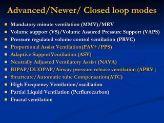
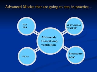
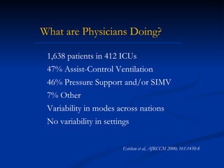
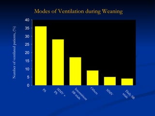
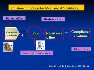
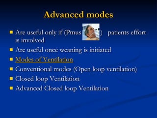
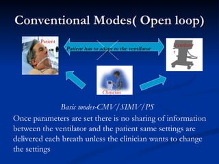
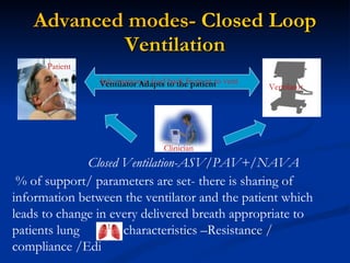
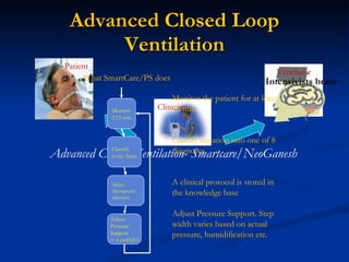
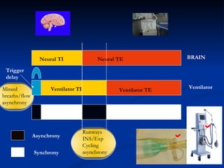
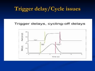
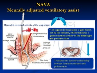
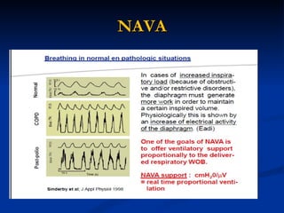
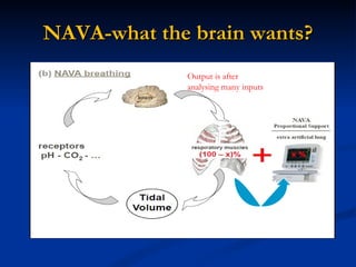
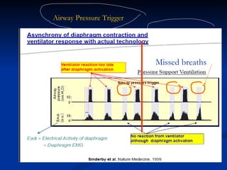
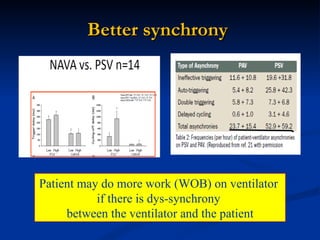
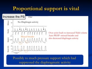
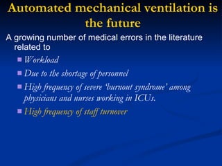
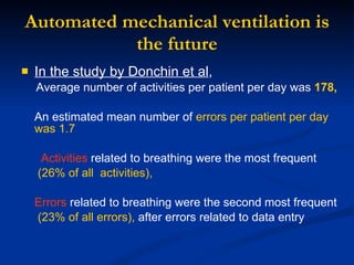
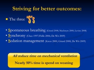
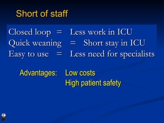
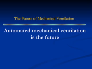
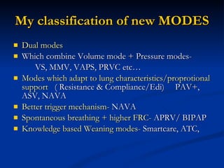
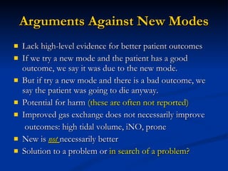
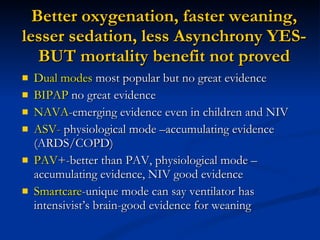
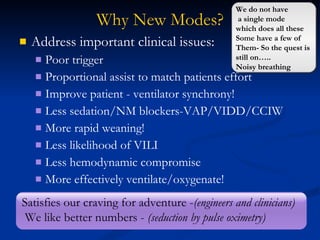
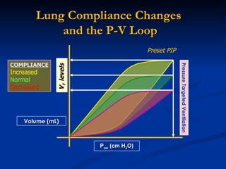
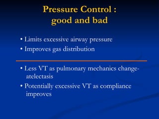
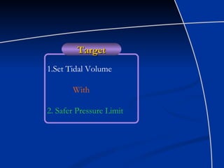
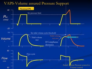
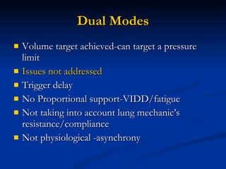
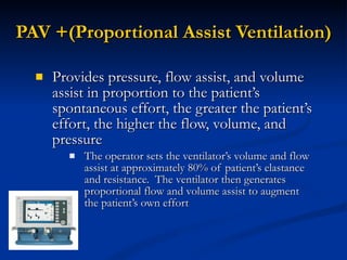
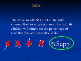
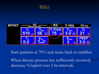
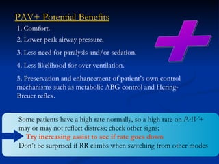
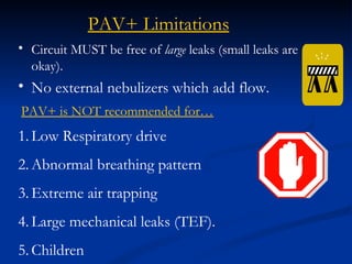
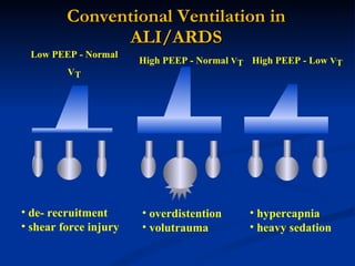
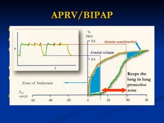
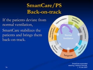
The document discusses advanced modes of mechanical ventilation. It begins by outlining newer modes such as VAPS, APRV/BIPAP, PAV+, Smartcare, and their benefits over basic modes. These advanced modes aim to improve synchrony between the patient and ventilator, reduce asynchrony issues, and make ventilation proportional to patient effort through feedback loops. The document argues that automated closed-loop ventilation is the future as it reduces workload and errors while allowing for quicker weaning and lower costs through greater ease of use and patient safety.
An excellent tool to treat refractory hypoxia. Target audience are ICU junior physicians and Respiratory Therapists. It will take away the fear of "What is APRV?" from your hearts and you will feel ready to give it a try.
Basic information on the Graphics displayed on the Ventilators. Prepared to educate about the graphics to train the professionals who work with Ventilators.
HERE IS A SEMINAR BASED ON ALL THE NEWER MODES OF MECHANICAL VENTILATION .
MY SINCERE APOLOGIES , BECAUSE I HAD TO TAKE INFORMATION FROM OTHERS SLIDES TOO , SINCE THERE IS VERY LESS INFORMATION AVAILABLE ABOUT THIS TOPIC
Presentation of Dr. Dean Hess at 10th Pulmonary Medicine Update Course, Cairo, Egypt. Pulmonary Medicine Update Course is organized by Scribe : www.scribeofegypt.com
Created by:
Rob Chatburn, RRT RRT-NPS, FAARC
Research Manager – Respiratory Therapy
Cleveland Clinic
Associate Professor
Case Western Reserve University
Presentation of Dr.Lluis Blanch at Pulmonary Critical Care Egypt 2014 , January2014, the leading critical care conference and medical exhibition in Egypt.www.pccmegypt.com
Mechanical ventilation ppt including airway, ventilator, tubings and connections, nursing management, trouble shooting common problems and issues, suctioning etc.
Non-invasive ventilation (NIV) is the use of breathing support administered through a face mask or nasal mask. Learn more about NIV in this presentation by Dr Somnath Longani, consultant Anaesthesiologist & Intensivist, Midland Healthcare & Research Center, lucknow
https://midlandhealthcare.org/
this is compiled & created to discuss the basic modes and initiation of NIV
the author is thankful to the previous authors,teachers who helped to conceptualize the NIV .
Factory Supply Best Quality Pmk Oil CAS 28578–16–7 PMK Powder in Stockrebeccabio
Factory Supply Best Quality Pmk Oil CAS 28578–16–7 PMK Powder in Stock
Telegram: bmksupplier
signal: +85264872720
threema: TUD4A6YC
You can contact me on Telegram or Threema
Communicate promptly and reply
Free of customs clearance, Double Clearance 100% pass delivery to USA, Canada, Spain, Germany, Netherland, Poland, Italy, Sweden, UK, Czech Republic, Australia, Mexico, Russia, Ukraine, Kazakhstan.Door to door service
Hot Selling Organic intermediates
These simplified slides by Dr. Sidra Arshad present an overview of the non-respiratory functions of the respiratory tract.
Learning objectives:
1. Enlist the non-respiratory functions of the respiratory tract
2. Briefly explain how these functions are carried out
3. Discuss the significance of dead space
4. Differentiate between minute ventilation and alveolar ventilation
5. Describe the cough and sneeze reflexes
Study Resources:
1. Chapter 39, Guyton and Hall Textbook of Medical Physiology, 14th edition
2. Chapter 34, Ganong’s Review of Medical Physiology, 26th edition
3. Chapter 17, Human Physiology by Lauralee Sherwood, 9th edition
4. Non-respiratory functions of the lungs https://academic.oup.com/bjaed/article/13/3/98/278874
Tom Selleck Health: A Comprehensive Look at the Iconic Actor’s Wellness Journeygreendigital
Tom Selleck, an enduring figure in Hollywood. has captivated audiences for decades with his rugged charm, iconic moustache. and memorable roles in television and film. From his breakout role as Thomas Magnum in Magnum P.I. to his current portrayal of Frank Reagan in Blue Bloods. Selleck's career has spanned over 50 years. But beyond his professional achievements. fans have often been curious about Tom Selleck Health. especially as he has aged in the public eye.
Follow us on: Pinterest
Introduction
Many have been interested in Tom Selleck health. not only because of his enduring presence on screen but also because of the challenges. and lifestyle choices he has faced and made over the years. This article delves into the various aspects of Tom Selleck health. exploring his fitness regimen, diet, mental health. and the challenges he has encountered as he ages. We'll look at how he maintains his well-being. the health issues he has faced, and his approach to ageing .
Early Life and Career
Childhood and Athletic Beginnings
Tom Selleck was born on January 29, 1945, in Detroit, Michigan, and grew up in Sherman Oaks, California. From an early age, he was involved in sports, particularly basketball. which played a significant role in his physical development. His athletic pursuits continued into college. where he attended the University of Southern California (USC) on a basketball scholarship. This early involvement in sports laid a strong foundation for his physical health and disciplined lifestyle.
Transition to Acting
Selleck's transition from an athlete to an actor came with its physical demands. His first significant role in "Magnum P.I." required him to perform various stunts and maintain a fit appearance. This role, which he played from 1980 to 1988. necessitated a rigorous fitness routine to meet the show's demands. setting the stage for his long-term commitment to health and wellness.
Fitness Regimen
Workout Routine
Tom Selleck health and fitness regimen has evolved. adapting to his changing roles and age. During his "Magnum, P.I." days. Selleck's workouts were intense and focused on building and maintaining muscle mass. His routine included weightlifting, cardiovascular exercises. and specific training for the stunts he performed on the show.
Selleck adjusted his fitness routine as he aged to suit his body's needs. Today, his workouts focus on maintaining flexibility, strength, and cardiovascular health. He incorporates low-impact exercises such as swimming, walking, and light weightlifting. This balanced approach helps him stay fit without putting undue strain on his joints and muscles.
Importance of Flexibility and Mobility
In recent years, Selleck has emphasized the importance of flexibility and mobility in his fitness regimen. Understanding the natural decline in muscle mass and joint flexibility with age. he includes stretching and yoga in his routine. These practices help prevent injuries, improve posture, and maintain mobilit
New Directions in Targeted Therapeutic Approaches for Older Adults With Mantl...i3 Health
i3 Health is pleased to make the speaker slides from this activity available for use as a non-accredited self-study or teaching resource.
This slide deck presented by Dr. Kami Maddocks, Professor-Clinical in the Division of Hematology and
Associate Division Director for Ambulatory Operations
The Ohio State University Comprehensive Cancer Center, will provide insight into new directions in targeted therapeutic approaches for older adults with mantle cell lymphoma.
STATEMENT OF NEED
Mantle cell lymphoma (MCL) is a rare, aggressive B-cell non-Hodgkin lymphoma (NHL) accounting for 5% to 7% of all lymphomas. Its prognosis ranges from indolent disease that does not require treatment for years to very aggressive disease, which is associated with poor survival (Silkenstedt et al, 2021). Typically, MCL is diagnosed at advanced stage and in older patients who cannot tolerate intensive therapy (NCCN, 2022). Although recent advances have slightly increased remission rates, recurrence and relapse remain very common, leading to a median overall survival between 3 and 6 years (LLS, 2021). Though there are several effective options, progress is still needed towards establishing an accepted frontline approach for MCL (Castellino et al, 2022). Treatment selection and management of MCL are complicated by the heterogeneity of prognosis, advanced age and comorbidities of patients, and lack of an established standard approach for treatment, making it vital that clinicians be familiar with the latest research and advances in this area. In this activity chaired by Michael Wang, MD, Professor in the Department of Lymphoma & Myeloma at MD Anderson Cancer Center, expert faculty will discuss prognostic factors informing treatment, the promising results of recent trials in new therapeutic approaches, and the implications of treatment resistance in therapeutic selection for MCL.
Target Audience
Hematology/oncology fellows, attending faculty, and other health care professionals involved in the treatment of patients with mantle cell lymphoma (MCL).
Learning Objectives
1.) Identify clinical and biological prognostic factors that can guide treatment decision making for older adults with MCL
2.) Evaluate emerging data on targeted therapeutic approaches for treatment-naive and relapsed/refractory MCL and their applicability to older adults
3.) Assess mechanisms of resistance to targeted therapies for MCL and their implications for treatment selection
Title: Sense of Taste
Presenter: Dr. Faiza, Assistant Professor of Physiology
Qualifications:
MBBS (Best Graduate, AIMC Lahore)
FCPS Physiology
ICMT, CHPE, DHPE (STMU)
MPH (GC University, Faisalabad)
MBA (Virtual University of Pakistan)
Learning Objectives:
Describe the structure and function of taste buds.
Describe the relationship between the taste threshold and taste index of common substances.
Explain the chemical basis and signal transduction of taste perception for each type of primary taste sensation.
Recognize different abnormalities of taste perception and their causes.
Key Topics:
Significance of Taste Sensation:
Differentiation between pleasant and harmful food
Influence on behavior
Selection of food based on metabolic needs
Receptors of Taste:
Taste buds on the tongue
Influence of sense of smell, texture of food, and pain stimulation (e.g., by pepper)
Primary and Secondary Taste Sensations:
Primary taste sensations: Sweet, Sour, Salty, Bitter, Umami
Chemical basis and signal transduction mechanisms for each taste
Taste Threshold and Index:
Taste threshold values for Sweet (sucrose), Salty (NaCl), Sour (HCl), and Bitter (Quinine)
Taste index relationship: Inversely proportional to taste threshold
Taste Blindness:
Inability to taste certain substances, particularly thiourea compounds
Example: Phenylthiocarbamide
Structure and Function of Taste Buds:
Composition: Epithelial cells, Sustentacular/Supporting cells, Taste cells, Basal cells
Features: Taste pores, Taste hairs/microvilli, and Taste nerve fibers
Location of Taste Buds:
Found in papillae of the tongue (Fungiform, Circumvallate, Foliate)
Also present on the palate, tonsillar pillars, epiglottis, and proximal esophagus
Mechanism of Taste Stimulation:
Interaction of taste substances with receptors on microvilli
Signal transduction pathways for Umami, Sweet, Bitter, Sour, and Salty tastes
Taste Sensitivity and Adaptation:
Decrease in sensitivity with age
Rapid adaptation of taste sensation
Role of Saliva in Taste:
Dissolution of tastants to reach receptors
Washing away the stimulus
Taste Preferences and Aversions:
Mechanisms behind taste preference and aversion
Influence of receptors and neural pathways
Impact of Sensory Nerve Damage:
Degeneration of taste buds if the sensory nerve fiber is cut
Abnormalities of Taste Detection:
Conditions: Ageusia, Hypogeusia, Dysgeusia (parageusia)
Causes: Nerve damage, neurological disorders, infections, poor oral hygiene, adverse drug effects, deficiencies, aging, tobacco use, altered neurotransmitter levels
Neurotransmitters and Taste Threshold:
Effects of serotonin (5-HT) and norepinephrine (NE) on taste sensitivity
Supertasters:
25% of the population with heightened sensitivity to taste, especially bitterness
Increased number of fungiform papillae
micro teaching on communication m.sc nursing.pdfAnurag Sharma
Microteaching is a unique model of practice teaching. It is a viable instrument for the. desired change in the teaching behavior or the behavior potential which, in specified types of real. classroom situations, tends to facilitate the achievement of specified types of objectives.
Anti ulcer drugs and their Advance pharmacology ||
Anti-ulcer drugs are medications used to prevent and treat ulcers in the stomach and upper part of the small intestine (duodenal ulcers). These ulcers are often caused by an imbalance between stomach acid and the mucosal lining, which protects the stomach lining.
||Scope: Overview of various classes of anti-ulcer drugs, their mechanisms of action, indications, side effects, and clinical considerations.
- Video recording of this lecture in English language: https://youtu.be/lK81BzxMqdo
- Video recording of this lecture in Arabic language: https://youtu.be/Ve4P0COk9OI
- Link to download the book free: https://nephrotube.blogspot.com/p/nephrotube-nephrology-books.html
- Link to NephroTube website: www.NephroTube.com
- Link to NephroTube social media accounts: https://nephrotube.blogspot.com/p/join-nephrotube-on-social-media.html
Report Back from SGO 2024: What’s the Latest in Cervical Cancer?bkling
Are you curious about what’s new in cervical cancer research or unsure what the findings mean? Join Dr. Emily Ko, a gynecologic oncologist at Penn Medicine, to learn about the latest updates from the Society of Gynecologic Oncology (SGO) 2024 Annual Meeting on Women’s Cancer. Dr. Ko will discuss what the research presented at the conference means for you and answer your questions about the new developments.
TEST BANK for Operations Management, 14th Edition by William J. Stevenson, Ve...kevinkariuki227
TEST BANK for Operations Management, 14th Edition by William J. Stevenson, Verified Chapters 1 - 19, Complete Newest Version.pdf
Lung Cancer: Artificial Intelligence, Synergetics, Complex System Analysis, S...Oleg Kshivets
RESULTS: Overall life span (LS) was 2252.1±1742.5 days and cumulative 5-year survival (5YS) reached 73.2%, 10 years – 64.8%, 20 years – 42.5%. 513 LCP lived more than 5 years (LS=3124.6±1525.6 days), 148 LCP – more than 10 years (LS=5054.4±1504.1 days).199 LCP died because of LC (LS=562.7±374.5 days). 5YS of LCP after bi/lobectomies was significantly superior in comparison with LCP after pneumonectomies (78.1% vs.63.7%, P=0.00001 by log-rank test). AT significantly improved 5YS (66.3% vs. 34.8%) (P=0.00000 by log-rank test) only for LCP with N1-2. Cox modeling displayed that 5YS of LCP significantly depended on: phase transition (PT) early-invasive LC in terms of synergetics, PT N0—N12, cell ratio factors (ratio between cancer cells- CC and blood cells subpopulations), G1-3, histology, glucose, AT, blood cell circuit, prothrombin index, heparin tolerance, recalcification time (P=0.000-0.038). Neural networks, genetic algorithm selection and bootstrap simulation revealed relationships between 5YS and PT early-invasive LC (rank=1), PT N0—N12 (rank=2), thrombocytes/CC (3), erythrocytes/CC (4), eosinophils/CC (5), healthy cells/CC (6), lymphocytes/CC (7), segmented neutrophils/CC (8), stick neutrophils/CC (9), monocytes/CC (10); leucocytes/CC (11). Correct prediction of 5YS was 100% by neural networks computing (area under ROC curve=1.0; error=0.0).
CONCLUSIONS: 5YS of LCP after radical procedures significantly depended on: 1) PT early-invasive cancer; 2) PT N0--N12; 3) cell ratio factors; 4) blood cell circuit; 5) biochemical factors; 6) hemostasis system; 7) AT; 8) LC characteristics; 9) LC cell dynamics; 10) surgery type: lobectomy/pneumonectomy; 11) anthropometric data. Optimal diagnosis and treatment strategies for LC are: 1) screening and early detection of LC; 2) availability of experienced thoracic surgeons because of complexity of radical procedures; 3) aggressive en block surgery and adequate lymph node dissection for completeness; 4) precise prediction; 5) adjuvant chemoimmunoradiotherapy for LCP with unfavorable prognosis.
7. Smartcare ASV NAVA PAV PPS Advanced/ Closed loop ventilation APRV/BIPAP DUOPAP Advanced Modes that are going to stay in practice …..
8. What are Physicians Doing? 1,638 patients in 412 ICUs 47% Assist-Control Ventilation 46% Pressure Support and/or SIMV 7% Other Variability in modes across nations No variability in settings Esteban et al, AJRCCM 2000; 161:1450-8
9. Modes of Ventilation during Weaning Esteban et al, AJRCCM 2000;161:1450 PS SIMV + PS Intermittent SB trials Others SIMV Daily SB trials Number of ventilated patients, (%)
10.
11.
12. Goals of ventilation Setting the Ventilator Ventilator-Induced Lung Injury Lung protective ventilation Gas Exchange PaO2/PaCo2 Accepting hypoxia and hypercarbia Due to low volume ventilation Patient Comfort Synchrony sedation /paralysis Early weaning Hemodynamics
13. Patient effort Ventilator assistance . Kondili et al, Br J Anesthesia 2003;91:106 Resistive load Elastic load . Pmus Paw Resistance x flow Compliance x volume + + = Equation of motion for Mechanical Ventilation Controlled ventilation Pt/vent work shared-interaction
14.
15. Conventional Modes( Open loop) Basic modes-CMV/SIMV/PS Clinician Ventilator Patient Once parameters are set there is no sharing of information between the ventilator and the patient same settings are delivered each breath unless the clinician wants to change the settings Patient has to adapt to the ventilator
16. Advanced modes- Closed Loop Ventilation Closed Ventilation-ASV/PAV+/NAVA Clinician Ventilator Patient % of support/ parameters are set- there is sharing of information between the ventilator and the patient which leads to change in every delivered breath appropriate to patients lung characteristics –Resistance / compliance /Edi Ventilator Adapts to the patient Information is feed back from pt to vent
17. Advanced Closed Loop Ventilation Advanced Closed Ventilation- Smartcare/NeoGanesh Clinician Ventilator Patient Intensivists brain What SmartCare/PS does Monitor the patient for at least 15 min Classify situation into one of 8 diagnoses A clinical protocol is stored in the knowledge base Adjust Pressure Support. Step width varies based on actual pressure, humidification etc. Monitor ≥ 15 min Select therapeutic measure Classify every 5min Adjust Pressure Support < 4 cmH2O
18. Anything close to normal physiology is Advanced What is close to physiology in positive pressure ventilation? Ventilation starts /ends / and is as much as the brain wants
19.
20. NAVA PAV+/ASV Respiratory center output Peripheral nerve transmission Muscle Electrical activation Contraction Lung distension Respiratory compliance Airway resistance Airway opening pressure Flow/volume Alveolar ventilation Gas exchange Blood gases Proportionality of support means Support stats and ends and is as much as the Brain wants Chemo receptors Lung and airway reflexes Respiratory muscle afferents If ventilator uses any of These parameter to alter Breath pattern then ventilation will Be more synchronized and Proportional to what brain wants
22. PAV+ vs. PCV /PSV example PCV 15 cmH2O PAV+ at 75% Compared to PCV, the PAV+ mode better matches patient’s effort to ventilator output. PAV+ P T P T P T P T P T P T Proportional support has synchronised inspiration to expiration cycling
23. What are the problems with conventional modes ? Trigger delay/Synchrony issues
24. Phases of ventilatory cycle Delay, Missed breaths Flow not proportionate to patients effort -dyssynchrony/overassist VIDD/ Runway Asynchrony can occur at the start of a breath (trigger asynchrony Asynchrony can occur during the breath (flow asynchrony Asynchrony can occur at the end of a breath (cycle or termination asynchrony).
26. Trigger in conventional modes Time delay We are targeting the last part of the cycle and Also add the delay from the Y piece to the machine end Trigger delay is inbuilt in the old modes
27. Ventilator TE Neural TI Neural TE Trigger delay Ventilator TI Asynchrony Synchrony BRAIN Ventilator Missed breaths/flow asynchrony Runways INS/Exp Cycling asynchrony
29. NAVA Neurally adjusted ventilatory assist Recorded electrical activity of the diaphragm % of support is based upon a gain factor, set by the clinician, which translates a given electrical activity of the diaphragm into pressure assist Translates into a positive relationship between ventilator assistance and patient effort Esophagus
32. Sinderby et al, Nature Med 1999;5:1433 Time (s) 0 1 4 3 2 0 1 4 3 2 Airway Pressure Trigger Onset of diaphragmatic electrical activity Onset of ventilator flow Neural Trigger 0 20 -5.0 0.0 0.0 0.5 -1 0 1 Flow (l/s) Volume (l) P es (cm H 2 O) P aw (cm H 2 O ) Missed breaths
33. Better synchrony Studies prove Better quality of sleep and less arousals- PAV+/NAVA Patient may do more work (WOB) on ventilator if there is dys-synchrony between the ventilator and the patient
34.
35. Proportional support is vital No Diaphragm activity Missed breaths Over assist leads to increased Tidal volume Auto PEEP –missed breaths and also decreased diaphragm activity Possibly to much pressure support which had suppressed the diaphragmatic activity Increase the PS
36.
37.
38.
39. Short of staff Closed loop = Less work in ICU Quick weaning = Short stay in ICU Easy to use = Less need for specialists Low costs High patient safety Advantages:
40. The Future of Mechanical Ventilation Automated mechanical ventilation is the future
47. Lung Compliance Changes and the P-V Loop Volume (mL) PIP levels Preset V T P aw (cm H 2 O) Volume Targeted Ventilation COMPLIANCE Increased Normal Decreased
48.
49. Lung Compliance Changes and the P-V Loop Volume (mL) Preset PIP V T levels P aw (cm H 2 O) COMPLIANCE Increased Normal Decreased Pressure Targeted Ventilation
50.
51.
52. 60 -20 60 Flow L/min Volume Switch from Pressure control to Volume control L 0 0.6 40 VAPS-Volume assured Pressure Support Normal PS If Compliance decreases P aw cmH 2 0 Set tidal volume cycle threshold Set pressure limit Tidal volume met Tidal volume not met Flow cycle
56. PAV+ uses the compliance and resistance information collected every 4-10 breaths to know what it’s fighting against . PAV+ uses the flow and volume information collected every 5 milliseconds to know what the patient wants. PAV+ combines this data with the %Supp information input by the clinician to determine how much pressure to supply to the system. PAV+
57. The clinician will NOT set a rate, tidal volume, flow or target pressure. Instead, the clinician will simply set the percentage of work that the ventilator should do. f %Supp x x x x PAV+ V . V t P i
58. PAV+ Start patients at 70% and wean back to stabilize When disease process has sufficiently reversed, decrease %Support over 2 hr intervals
59. + PAV+ Potential Benefits 1. Comfort. 2. Lower peak airway pressure. 3. Less need for paralysis and/or sedation. 4. Less likelihood for over ventilation. 5. Preservation and enhancement of patient’s own control mechanisms such as metabolic ABG control and Hering-Breuer reflex. Some patients have a high rate normally, so a high rate on PAV + may or may not reflect distress; check other signs; Try increasing assist to see if rate goes down Don’t be surprised if RR climbs when switching from other modes
64. Spontaneous ventilation in assisted breaths Diaphragm with sedation P abdominal Area of increased ventilation Area of increased perfusion Risk of over distention Risk of atelectasis Good ventilated area Area with good perfusion R ! R ! R ! Spontaneous breathing Diaphragm with low sedation1 Spontaneous ventilation in assisted breaths Controlled ventilation Better V/Q Less VILI R ! R ! R !
65. APRV settings P aw T high (4-5) Sec T low P high P low ( 1 sec) Time-triggered, Time-cycled, Pressure-limited, Spontaneous breathing is allowed at any point during the ventilatory cycle FLOW P high -This parameter is set with the goal of improving oxygenation. P low -The setting of this parameter has the goal of facilitating ventilation or CO2 clearance. It is this inverse inspiratory:expiratory (I:E) ratio that distinguishes APRV from bi-level positive airway pressure (BiPAP=1:1 or more) Inverse ratio ventilation
66.
67. 5 possible breath types in BIPAP High incidence of asynchrony issues
68.
69. Pplat = Palv; Pplat = Transpulmonary Pressure? transpulmonary pressure = 45 cm H 2 O 0 5 10 15 20 25 30 -5 -10 -15 45 cms of H2O PCV 20 cm H 2 O, PEEP 10 cm H 2 O; Pplat 30 cm H 2 O -15 cm H 2 O Active inspiratory effort
70.
71.
72.
73.
74.
75. Smartcare These therapeutic measures are based on a clinical protocol that has been tested and verified during several years of development ..
LRF -This slide shows how PAV+ software may improve ventilator synchrony. -The top three boxes represent pressure vs. time for PCV while the bottom three boxes represent the same thing for PAV+ software. -The green line represents the effort input from the patient’s diaphragm and the red line represents the pressure output from the ventilator. -In PCV the ventilator’s output is the same despite changes in the diaphragm’s input. -In PAV+ mode, the machine’s output mirrors the input of the diaphragm. -If the patient pulls a little bit, the vent pushes a little bit. If the patient pulls a lot then the vent pushes a lot.
NAVA Training Presentation 2007 NAVA Training Presentation.ppt The electrical discharge of the diaphragm is captured through the introduction of an Edi Catheter fitted with an electrode array. Since NAVA uses the Edi to control the ventilator, it is important to understand what the signal represents. All muscles (including the diaphragm and other respiratory muscles) generate electrical activity to excite muscle contraction. This electrical excitation is controlled by nerve stimulus and controlled in magnitude by adjusting the stimulation frequency (rate coding) or by adjusting the numbers of nerves that are sending the stimulus (nerve fiber recruitment). Both, the rate coding and nerve fiber recruitment will be transmitted into muscle fiber motor unit action potentials which will be summed both in time and space producing the intensity of the electrical activity measured on the muscle. To reduce the influence of external noise, the measurement of the muscle electrical activity is performed by bipolar differential recordings, where the signal difference between two single electrodes is measured. For example the resting Edi measured with electrodes in the esophagus in a healthy subject typically ranges between a few and 10 μ V. Patients with chronic respiratory insufficiency may demonstrate signals 5-7 times stronger. Due to the differential recording and low signal amplitude, measurement of Edi is sensitive to electrode filtering, external noise, and cross-talk from other muscles e.g. the heart which produces electrical amplitudes of about 10-100 times that of the diaphragm. Since, the Edi must always be present to initiate a contraction of the diaphragm it should always be possible to record the signal in healthy subjects
So: There is less work to do in the ICU Patients have shorter stays in the ICU And there is less need for specialists in the ICU All this means: Low costs: you need fewer resources to do the same job. High patient safety: the closed loop and the quick weaning always enhance safety. [Click: Next slide.]
LRF -Potential benefits as listed by Dr Younes in one of his early papers. M Younes. Proportional Assist Ventilation, A New Approach to Ventilatory Support. Theory. Am Rev Respir Dis 1992;145:114-120.
We’ve used a variety of mechanical ventilation strategies for low lung volume disorders. Ventilation with normal tidal volumes but low PEEP level requires high pressures, and the shear forces created during the inflation- derecruitment- reinflation sequence can cause severe lung injury. Using normal tidal volumes with high PEEP means even higher pressure, and can overdistend relatively healthy lung tissue. In an effort to protect the ARDS lung from further, ventilator- induced injury, some clinicians advocate a so- called “open lung approach” of high PEEP and tidal volumes of 6 mL/ kg of body weight, or less. This may result in high arterial CO2, referred to as “permissive hypercapnia”. Dr. John Luce from UCSF points out, however, that hypercapnia may be unavoidable with this strategy in patients with severe Acute Lung Injury. Really, the only permission given is to ourselves as clinicians, making high CO2 “O.K.”, which makes us feel better. But not the patient. Hypercapnia is uncomfortable, and patients usually require heavy sedation to control their ventilation. A large, prospective, multicenter trial of high versus low tidal volume use in ARDS is currently underway in the U.S.
Peak and Mean airway pressures are reduced Less invasive, less mechanical Weaning is smooth and effortless Less sedation and muscle relaxants Spontaneous breathing contributes to better gas exchange and secretion clearance. Greater comfort and less stress for patients
So, there are only four settings for APRV as seen on this graph of airway pressure and flow : • the high pressure, P- high, the CPAP level to keep the lungs open, • the duration, or time, that the CPAP pressure is held at the airway, called T- high, • the release pressure, P- low, that allows additional CO2 removal, • and the duration, or time, that pressure is released, called T- low. We see flow in and out of the lungs with spontaneous breathing during the time that the higher pressure is applied to the airway. And here we see the larger flow, or exhaled volume, from the lungs during the release. Again, it’s very important that the release time be short so that lung volume is maintained. How can we assess that? Well, I’d love to be able to actually measure FRC at the bedside in the ICU, but that really isn’t practical today. Notice that the expiratory flow tracing during the release doesn’t reach the zero line before the high pressure is re- applied. Because flow is still coming from the lungs, we know that volume remains in the lungs. In other words, we are intentionally trapping gas in the lung by limiting the release time. When we set T- high, we are really setting the frequency of releases, which is like setting the ventilator rate.
The previous case was a best case example. During weaning, patients often show signs of ventilatory instabilities such as Hyper or Hypoventilation, tachypnea, or are simply not adequately ventilated. SmartCare classifies these situations into 8 different diagnoses, and adapts the pressure support accordingly to bring the patient back-on-track. For every diagnosis a different set of therapeutic measures is incorporated into the protocol.