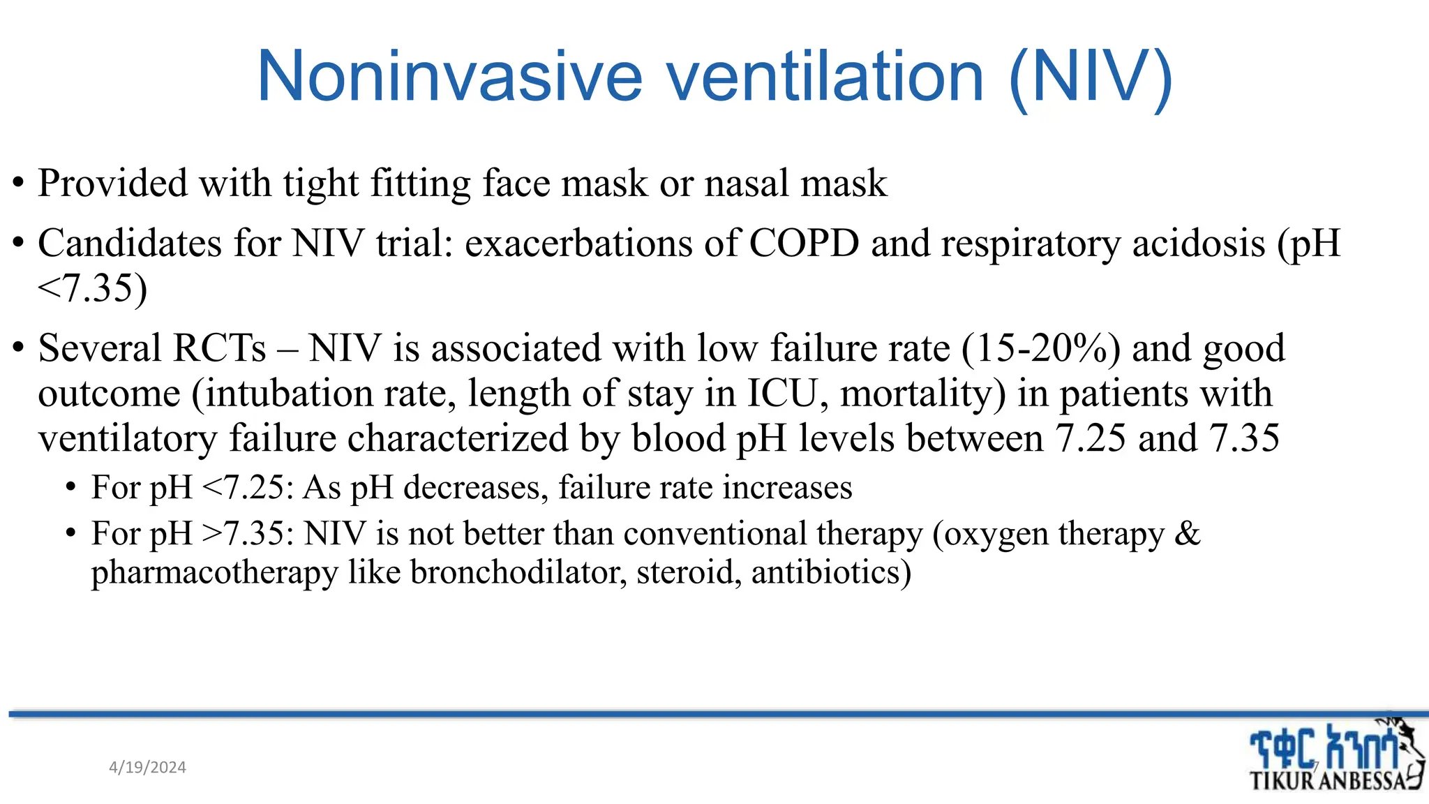
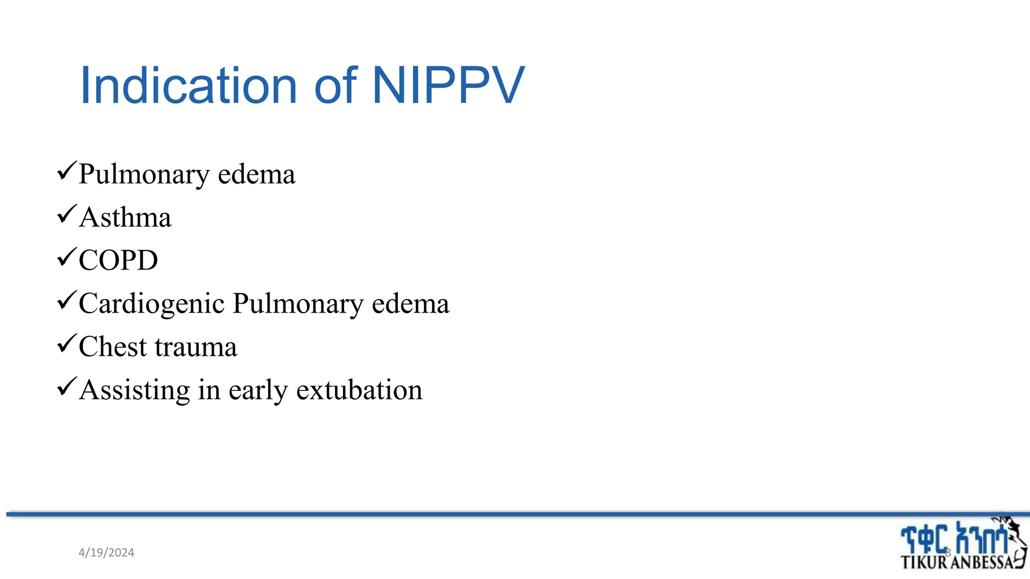
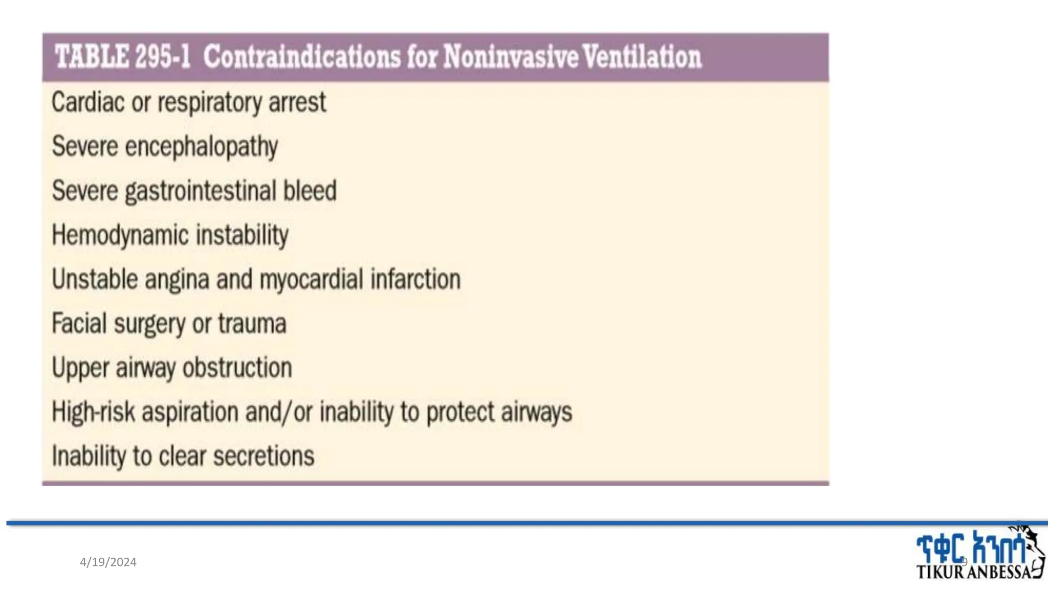
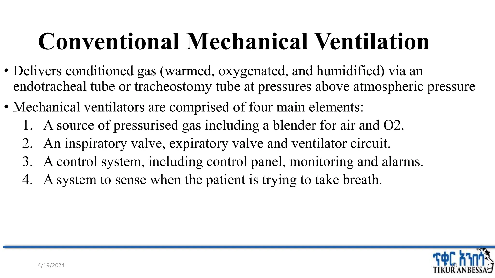
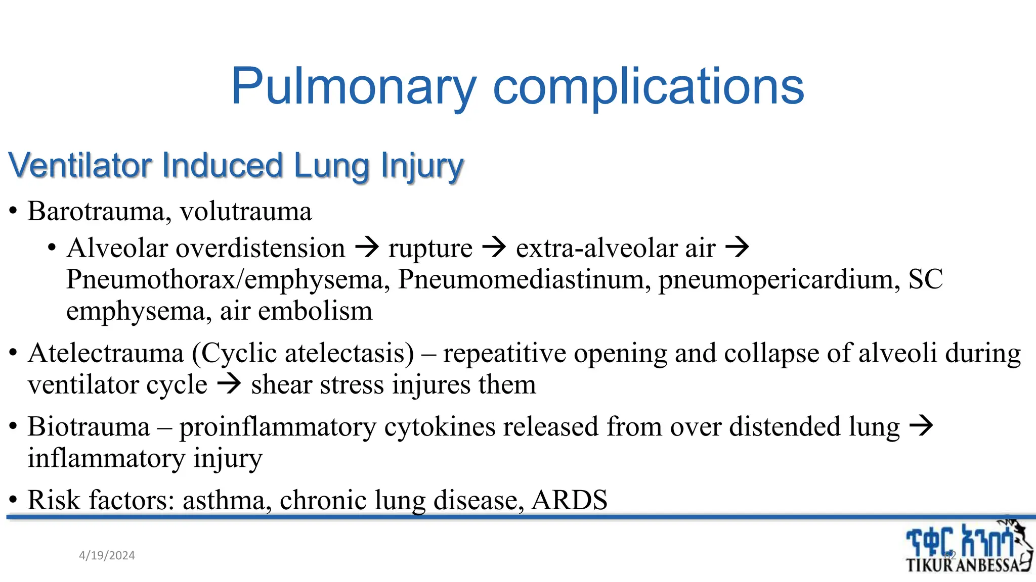
2. Outline
• Introduction
• Types of Mechanical Ventilation
• Aims of Ventilatory Support
• Indications
• Modes of ventilation
• Ventilator settings
• Monitoring
• Complications of mechanical ventilation
• Weaning
4/19/2024 2
3. Introduction
• Mechanical Ventilator: A device which assists or replaces spontaneous breathing
,Support ventilatory function and improve oxygenation.
• Inspiratory trigger predetermined air (O2 + other gases) forced into airways &
alveoli lungs inflate ↑ intraalveolar pressure termination signal – vent
stops delivery ↓airway pressure expiration follows passively due to
pressure gradiant.
4/19/2024 3
4. Several models evolving over time
• Negative pressure ventilation – past
• Positive pressure ventilation – present
4/19/2024 4
5. Negative pressure ventilators
• Developed in 1929.
• Used widely in 1940 polio
epidemic.
• The patient’s body was
encased in an iron cylinder
and negative pressure was
generated
Emerson JH, Loynes JA. The evolution of iron lungs: respirators of the body-
encasing type. Cambridge:JH Emerson & Company; 1958. Google Scholar
4/19/2024 5
6. Positive-pressure
ventilators
A positive pressure is applied to the mouth or
mask providing a positive pressure gradient
from mouth to alveoli resulting in inspiration
Invasive ventilation first used at Massachusetts
General Hospital in 1955
This can be applied with a mask (non-invasive)
or with a tracheal tube or tracheostomy
(invasive).
Current ICU ventilators are primarily positive
pressure ventilators
4/19/2024 6
7. Noninvasive ventilation (NIV)
• Provided with tight fitting face mask or nasal mask
• Candidates for NIV trial: exacerbations of COPD and respiratory acidosis (pH
<7.35)
• Several RCTs – NIV is associated with low failure rate (15-20%) and good
outcome (intubation rate, length of stay in ICU, mortality) in patients with
ventilatory failure characterized by blood pH levels between 7.25 and 7.35
• For pH <7.25: As pH decreases, failure rate increases
• For pH >7.35: NIV is not better than conventional therapy (oxygen therapy &
pharmacotherapy like bronchodilator, steroid, antibiotics)
4/19/2024 7
8. Indication of NIPPV
Pulmonary edema
Asthma
COPD
Cardiogenic Pulmonary edema
Chest trauma
Assisting in early extubation
4/19/2024 8
10. Conventional Mechanical Ventilation
• Delivers conditioned gas (warmed, oxygenated, and humidified) via an
endotracheal tube or tracheostomy tube at pressures above atmospheric pressure
• Mechanical ventilators are comprised of four main elements:
1. A source of pressurised gas including a blender for air and O2.
2. An inspiratory valve, expiratory valve and ventilator circuit.
3. A control system, including control panel, monitoring and alarms.
4. A system to sense when the patient is trying to take breath.
4/19/2024 10
12. Indications
1. Acute Respiratory Failure
• Hypoxic/Hypercapnic
2. Air way protection
• To prevent Aspiration in deeply sedated & unconscious pt
• Maintain patency in obstruction (Asthma, Anaphylaxis, Tumor)
3. Pulmonary vascular disease:
PTE, Amniotic fluid embolism, tumor emboli
4. Hyperventilation Therapy - raised ICP
5. Hypoventilation:
• Decreased central drive (General anesthesia, Drug overdose), PNS/respiratory muscle
dysfunction
6. Increased ventilatory demand
• E.g:- Severe sepsis, septic shock, severe metabolic acidosis
4/19/2024 12
13. Aims of Mechanical Ventilation
• Achieve and maintain adequate pulmonary gas exchange (ventilation and
oxygenation)
• Reduce or eliminate excessive work of breathing
• Optimize patient comfort
• Minimize the risk of complications
• Maintain and protect the airway and allow more effective clearance of secretions
4/19/2024 13
14. Modes
Mode refers to the manner in which ventilator breaths are triggered,
cycled, and limited
The trigger, either an inspiratory effort or a time based signal, defines
what the ventilator senses to initiate an assisted breath.
The limiting factors -are operator-specified values, such as airway
pressure
Cycle refers to the factors that determine the end of inspiration.
Other types of cycling include pressure cycling and time cycling.
4/19/2024 14
15. Modes of Ventilation
1. CMV
2. Assist control (AC)
3. Synchronized intermittent mandatory ventilation (SIMV)
4. Pressure support ventilation
5. CPAP
15
4/19/2024
16. Controlled mechanical ventilation (CMV)
• During CMV, the minute ventilation is determined entirely by the set
respiratory rate and tidal volume.
• This may be due to pharmacologic paralysis, heavy sedation, coma, or lack of
incentive to increase the minute ventilation because the set minute ventilation
meets or exceeds physiologic need.
16
4/19/2024
19. PRESSURE-CONTROL VENTILATION (PCV)
• Are ventilator-initiated breaths with a pressure limit.
• Limits inflation pressure
• Mandatory breath only
• Set: PIP, I:E ratio, RR, PEEP, FiO2
• Inspiration ends at delivery of set PIP.
• Tidal volume is variable, related to
• PIP, compliance, airway resistance, tubing resistance
• High VT: can be due to High PIP & low compliance
19
4/19/2024
21. PCV
Advantage
• Prevents excessive airway
pressures
• Avoid regional alveolar
over distention
• May lead to earlier
liberation from MV
Disadvantage
• Very uncomfortable and
requires deep sedation
+/- paralysis
• Unable to guarantee
minimum VE
21
Indications= patients who are at particularly
high risk from barotrauma and post thoracic
surgery.
4/19/2024
22. Assist-Control Ventilation
• ACMV is the most widely used mode of ventilation.
• In this mode, an inspiratory cycle is initiated either by the patient’s
inspiratory effort or, if none is detected within a specified time window, by
a timer signal within the ventilator.
• During AC, the clinician determines the minimal minute ventilation by
setting the respiratory rate and tidal volume.
• The patient can increase the minute ventilation by triggering additional
breaths.
22
4/19/2024
23. Cont…
• Each patient-initiated breath receives the set tidal volume from the
ventilator.
• Every breath delivered, whether patient- or timer-triggered, consists of the
operator-specified tidal volume.
• It is a mixed mode in which pts receive a mandatory breath with set tidal
volume (if volume AC) or pressure (pressure AC).
• In ACV, the mechanical breaths are limited by volume and/or flow and
cycled by volume or time.
23
4/19/2024
25. Volume pre-set assist control
• Advantages
• Relatively simple to
set
• Guaranteed minimum
minute ventilation
• Rests muscles of
respiration (if
properly set)
• Disadvantages
• Not synchronized
• Patient may “lead”
ventilator
• Inappropriate triggering
may result in excessive
minute ventilation
• lung compliance
alveolar pressure with
risk of barotrauma
• Often requires sedation to
achieve synchrony.
4/19/2024 25
26. Intermittent Mandatory
Ventilation
• IMV allows the patient to breathe spontaneously between machine-cycled
or mandatory breaths.
• In the most frequently used synchronized mode (SIMV), mandatory breaths
are delivered in synchrony with the patient’s inspiratory efforts at a
frequency determined by the operator.
• SIMV differs from ACMV in that only a preset number of breaths are
ventilator-assisted.
26
4/19/2024
28. Cont…
• IMV is similar to AC in two ways:
• The clinician determines the minimal minute ventilation (by setting the
respiratory rate and tidal volume) and
• The patient is able to increase the minute ventilation.
• However, IMV differs from AC in the way that the minute ventilation is
increased.
• SIMV allows patients with an intact respiratory drive to exercise
inspiratory muscles between assisted breaths; thus it is useful for both
supporting and weaning intubated patients.
28
4/19/2024
29. Pressure support (PSV)
• The ventilator applies a pre-determined amount of positive pressure to the
airways upon inspiration.
• This form of ventilation is patient-triggered, flow-cycled, and pressure-
limited.
• The patient breathes spontaneously.
• With PSV, patients receive ventilator assistance only when the ventilator
detects an inspiratory effort.
• PSV is well tolerated by most patients who are being weaned from MV.
• Comfortable mode for awake and conscious patient
• Can be combined with SIMV mode.
29
4/19/2024
31. Continuous positive airway pressure (CPAP)
• Is the way of delivering PEEP
but also maintains the set
pressure through out the
respiratory cycle.
• Allows spontaneous
breathing at elevated
baseline pressure.
• Set P = 5-15cmH2O
• CPAP is most commonly
used in the management of
sleep related breathing
disorders, cardiogenic
pulmonary edema, and 31
4/19/2024
34. Trigger
• Trigger sensitivity
• sensitivity preferable
• Flow triggering generally more sensitive than pressure triggering
• flow or less negative pressure sensitivity
• Trigger types
• pressure triggering or
• A trigger sensitivity of -1 to -3 cmH2O is typically set.
• flow triggering
• initiated when the return flow is less than the delivered flow.
4/19/2024 34
35. Tidal volume
• Optimal tidal volume for mechanically ventilated patients:-
• without existing lung disease 6-8ml/kg of IBW.
• For ARDS-4-6 ml/kg of IBW
• Female PBW= 45.5 + 0.91 (Ht – 152.4)
• Male PBW = 50 + 0.91 (Ht – 152.4)
• As Tv inc., peak airway pressure also inc.(>45cmH20 barotrauma)
4/19/2024 35
36. Respiratory rate
• Initial respiratory rate between 12 and 16 breaths per minute is reasonable.
• RR adjustment-
• Modification according to mode
• Desired PH & paco2
• autoPEEP(if >5cmH20)
• For ALI/ARDS-up to 35’
• MV= TV X RR
4/19/2024 36
37. PEEP
• PEEP is used to mitigate end-expiratory alveolar collapse.
• An initial PEEP is 5 cmH2O.
• 20 cmH2O may be used in patients with ARDS.
• Elevated levels of PEEP-
• reduced preload(CO)
• increases risk of barotrauma and
• impaired cerebral venous outflow (increases ICP)
4/19/2024 37
38. Fraction of inspired oxygen(Fio2)
• % of oxygen in each breath.
• Titrated to meet patient needs.
• Goal-
• PaO2 above 60 mmHg and
• SpO2 above 90 percent.
4/19/2024 38
39. Inspiratory time
• Set as:
• % of respiratory cycle
• I:E ratio
• Indirectly, by setting flow
• Expiratory time not set
• Remaining time after inspiration before next breath
4/19/2024 39
40. Inspiratory Time
• Longer inspiratory time
• Improved oxygenation
• Higher mean airway pressure
• Re-distribution
• Lower peak airway pressure
• More time available to deliver set tidal volume
• Shorter inspiratory time
• Less risk of gas trapping and PEEPi
• Less effect on cardiovascular system
4/19/2024 40
41. Inspiratory to expiratory (I:E) ratio
• Inspiratory time is equal to tidal volume divided by flow rate.
• Normal I:E ratio: 1:2 to 1:3.
• Can be reduced to 1:4 or 1:5 in COPD (longer expiratory time)
• inverse I:E ratio may be necessary in states of low compliance, such as ARDS.
42. Flow rate
• The peak flow rate is the maximum flow delivered by the ventilator during
inspiration.
• Peak flow rates of 60 L per minute may be sufficient, although higher rates are
frequently necessary.
• Increased peak flow rates –
increase the peak airway pressure.
decreased inspiratory time lowers the mean airway pressure, which can
decrease oxygenation.
4/19/2024 42
43. Inspiratory flow Pattern
• Different flow rates are associated with different airflow
waveforms.
1. Square waveforms
flow accelerates very quickly and then reaches a set rate,
which is maintained throughout inspiration. Flow rate is
constant
This waveform allows for an adequate I:E ratio with a normal
flow rate.
Inspiratory
arm
44. Inspiratory flow Pattern
2. Ramp waveforms
flow rate rapidly accelerates but is followed by a
gentle tapering.
This waveform may require much higher flow
rates to obtain an adequate I:E ratio.
This waveform can be used if abnormally high
PIP is encountered.
Inspiratory
arm
Expiratory
arm
45. Pressures
• Peak inspiratory pressure- the pressure required to administer a set tidal volume
• As the tidal volume increases, the pressure required to administer that volume also
increases..
• Pawp above 45 cmH2O -the risk of barotrauma is increased.
• plateau pressure - is the pressure after the delivery of the tidal volume but before
the patient is allowed to exhale.
• is maintained < 30 cmH2O.
• Mean airway pressure- refers to the mean across the entire respiratory cycle,
both inspiration and expiration
4/19/2024 45
46. Mean airway pressure
Mean airway pressure
Time
Pressure
Time
Pressure
Tidal volume
4/19/2024 46
47. Mean airway pressure
Mean airway pressure
Time
Pressure
Time
Pressure
Inspiratory time
4/19/2024 47
53. • Incomplete expiration leading
to air trapping
• Causes include
• High minute ventilation
• Expiratory flow limitation
• Expiratory resistance
Auto PEEP
54.
55. Patient-ventilator dys-synchrony
• State when the respiratory cycle of the patient does not match
that of the ventilator.
• It can happen during triggering process, flow delivery or breath
cycling.
• Clinical features: ↑HR, RR; ↓SO2; ↑expiratory mm activity,
coughing, anxiety, visible inspiratory effort without triggering the
ventilator; Ventilator waveforms
• Consequences include: ↑Work Of Breath, prolongation of MV,
perceived need for deeper sedation
56. Vent triggers to start inspiration does not always correlate with the patient attempt to start inspiration
Airflow what the patient is demanding does not match what the vent is providing
Vent and patient went transitions from inspiration to expiration at different times
57. Principles of monitoring
• Follow the patient clinical condition
• Assess ventilation and oxygenation – ABGs
• Follow vent parameters
• Flow and pressure time curves
• Pressure and volume alarms
• Always evaluate for weaning readiness
57
4/19/2024
58. Alarms
• Never ignore an alarm
• Common causes of alarms
• Dislodgement: check ETT length of insertion (av. 22) at incisor
• Obstruction: secretion, blood clot, fighting patient biting the ETT
• Suction, sedation
• Pneumothorax: barotrauma
• Equipment failure: air leak, disconnected corrugator tube, ↓ humidifier
water, uninflated ETT cuff
58
4/19/2024
59. Complications of MV
• During Act of intubation
• Artificial air way related complication
• Pulmonary complications
• Non pulmonary complications
4/19/2024 59
60. During Act of intubation
• Dental trauma
• Aspiration – bag ventilation for preoxygenation can cause gastric distension +
irritation during ETT insertion vomiting
• Laryngeal trauma – hoarseness, URTO
• Laryngeal edema, ulceration, granuloma, vocal cord paralysis
• Bronchospasm – mechanical irritation
• Esophageal intubation
• Right main bronchus intubation – it goes too far
4/19/2024 60
61. Artificial air way related complication
• Failure of alarms or ventilator, Alarm “turned off”
• Disconnection, circuit leaks
• Obstruction by secretions
• Inadequate nebulization or humidification
• Overheated inspired air, resulting in hyperthermia
• Dyssynchronous breathing pattern
• Tube kinked, plugged
• Cuff failure
• Migration – ETT migrates with suctioning, coughing, movement
• Tracheal stenosis – pressure necrosis ulceration, scarring
4/19/2024 61
62. Pulmonary complications
Ventilator Induced Lung Injury
• Barotrauma, volutrauma
• Alveolar overdistension rupture extra-alveolar air
Pneumothorax/emphysema, Pneumomediastinum, pneumopericardium, SC
emphysema, air embolism
• Atelectrauma (Cyclic atelectasis) – repeatitive opening and collapse of alveoli during
ventilator cycle shear stress injures them
• Biotrauma – proinflammatory cytokines released from over distended lung
inflammatory injury
• Risk factors: asthma, chronic lung disease, ARDS
4/19/2024 62
63. VILI – management
• Protective lung ventilation principle
• The plateau airway pressure be immediately lowered to ≤35 cm H 2 O
• lowering the tidal volume, positive end-expiratory pressure, or inspiratory flow
• increasing sedation, administering neuromuscular blockade,
• advancing treatment of the underlying medical condition (eg, bronchodilators
for asthma)
4/19/2024 63
64. Oxygen toxicity
• Inappropriate O2 supplementation is deleterious
• Hyperoxia ↑reactive oxygen species (superoxide anion, hydroxyl radical,
H2O2) [+depleted antioxidant system] cellular injury
• There is no single threshold of FiO2 defining a safe upper limit for prevention of
oxygen toxicity.
• Reducing FiO2 to lowest tolerable limit is a good principle for all patients
4/19/2024 64
65. Infectious Complications
Risk factors
• ETT bypasses the glottis closure protective mechanism allowing
seepage of oropharyngeal content into airways.
• ETT impairs cough reflex
• Airway and parenchymal injury (primary illness + positive
pressure ventilation)
• ICU environment itself – heavy antibiotics use, sick patients in
close proximity
4/19/2024 65
66. Non pulmonary complications
• Hemodynamics
Decreased venous return
Reduced right ventricular output
Reduced left ventricular output
• Gastrointestinal
Erosive esophagitis, stress ulceration, diarrhea, acalculous cholecystitis, and
hypomotility
• Renal – acute renal failure ( decrease CO, release of inflammatory Mediators), acid
base disturbance.
• MSS- bed sore, contracture
• CNS: Critical illness polyneuropathy, myopathy,
• ICU psychosis etc.
66
4/19/2024
67. Weaning from mechanical ventilation
• The process of decreasing ventilator support & Discontinuation
of mechanical ventilation or liberation from the mechanical
ventilator
• three step process that consists of Readiness Testing ,Weaning &
extubation
67
4/19/2024
68. Readiness testing
• Readiness Testing – 2 goals
• Identify patients who are ready to wean
• Protect against premature weaning
• Objective clinical criteria and weaning predictors used
68
4/19/2024
69. Patients with uncertainty
• For patients in whom uncertainty exists as to whether the readiness criteria will predict a
successful weaning trial, we sometimes use a weaning predictor to identify potential
candidates suitable for weaning or to confirm lack of readiness to wean (eg, patients with
borderline readiness criteria or suspected respiratory muscle weakness).
• Use of weaning predictors is most pertinent among patients in whom the risk associated
with a failed spontaneous trial is significantly elevated (eg, patients with prolonged
mechanical ventilation, patients with critical care neuromyopathy).
• Among the predictors, the rapid shallow breathing index (RSBI) is our preferred
weaning predictor because it is well studied, easy to measure, and no alternative
predictor has been shown to be superior.
• For patients who have an RSBI <105 breaths/minute/L (measured without ventilatory
support), we initiate a weaning trial.
• For patients who have an RSBI ≥105 breaths/minute/L (measured without ventilatory
support), we maintain full ventilatory support.
4/19/2024 69
70. Choosing a weaning method
• For patients who have been intubated for more than 24 hours and have been
deemed as ready to wean ,we suggest a weaning trial.
• Daily spontaneous breathing trials (SBTs) with inspiratory pressure support is our
preferred method of weaning based upon randomized trials that have shown it is
efficient, safe, and effective
• Approximately 50 to 75 percent of patients pass the initial SBT and are
able to be successfully extubated
4/19/2024 70
71. • Spontaneous breathing Trial
• with minimal ventilator
support a low level of
pressure support(5-8mmH2O),
or CPAP (5mmH2O)
• Breathing through the ETT
without any ventilator support
(T-piece using 1–5 cmH2O
• Duration of a weaning trial
• 30 min to 120 min
71
4/19/2024
72. Extubation
• Extubation refers to removal of the endotracheal tube. It is the final step in
liberating a patient from mechanical ventilation.
• Extubation should not be performed until it has been determined that the patient's
medical condition is stable, a weaning trial has been successful, the airway is
patent, and any potential difficulties in reintubation have been identified. Most
patients are extubated during daytime hours
• Prior to Extubation Evaluate for Airway protection and Airway patency
4/19/2024 72
73. Airway protection
• Is the ability to guard against aspiration during spontaneous breathing It requires sufficient
cough strength and an adequate level of consciousness
• Universally accepted threshold levels of cough strength, level of consciousness, and suctioning
frequency that prohibit extubation have not been established. For many patients, it is reasonable
to delay extubation if the cough strength is weak, the GCS is <8 , or suctioning is required more
frequently than every two to three hours.
1. Cough strength (peak expiratory flow rate (PEF) >60 L/min)
2. Secretions (< 2.5 ml/hr) no more suctioning than once 2-3 hrs
3. Level of consciousness (able to obey command) 73
4/19/2024
74. Airway patency
• Cuff leak test: (assessing the presence of air movement around a deflated endotracheal
tube cuff)
• To identify pts at risk to develop post extubation laryngeal edema.
• A "cuff leak" refers to normal airflow around the ETT after the cuff of the ETT is
deflated. Its absence suggests there is reduced space between the ETT and the larynx.
• The cuff leak predicts post-extubation stridor with a sensitivity of 15 to 85 % and a
specificity of 70 to 99 %.
74
4/19/2024
75. Cont’d
The cuff leak can be detected qualitatively or quantitatively:
• Qualitative assessment by deflating the cuff and then listening for air movement
around the ETT using a stethoscope.
• Quantitatively is by deflating the ETT cuff and measuring the difference
between the inspired and expired TV of ventilator-delivered breaths during
volume-cycled MV (<110 ml or 12-24% of the delivered tidal volume) =
diminished patency
75
4/19/2024
76. How do we prevent post-extubation complications?
• Post extubation complications
Post-extubation laryngeal edema
Hypoxia and re-intubation
• Short course of glucocorticoid therapy before extubation.
Methylprednisolone (20 mg) administered every four hours for a total of four
doses.
Alternatively, a single dose of methylprednisolone 40 mg four hours prior to
extubation may be used 76
4/19/2024
77. Extubation procedure
• The patient is placed into an upright position and both the oral cavity and the
endotracheal tube (ETT) are suctioned.
• Instructions are given for the patient to take a deep breath and then exhale.
• During exhalation, the cuff is deflated and the ETT is removed in a single, smooth
motion. Orogastric tubes are typically removed simultaneously.
77
4/19/2024
78. References
• Up-to-date, online
• Hand book of Mechanical Ventilation, User’s guide
• Practical Guide for Mechanical Ventilation,4th edition
• Harrison’s 21st edition
4/19/2024 78
Editor's Notes
The principal benefits of MV during respiratory failure are improved gas exchange (by improving ventilation-perfusion (V/Q) matching) and decreased work of breathing.
Invasive ventilation first used at Massachusetts General Hospital in 1955
Candidates for NIV trial: exacerbations of COPD and respiratory acidosis (pH <7.35)
Several RCTs – NIV is associated with low failure rate (15-20%) and good outcome (intubation rate, length of stay in ICU, mortality) in patients with ventilatory failure characterized by blood pH levels between 7.25 and 7.35
For pH <7.25: As pH decreases, failure rate increases
For pH >7.35: NIV is not better than conventional therapy (oxygen therapy & pharmacotherapy like bronchodilator, steroid, antibiotics)
Hyperventilation — Use of mechanical ventilation to lower PaCO2 to 26 to 30 mmHg has been shown to rapidly reduce ICP through vasoconstriction and a decrease in the volume of intracranial blood; a 1 mmHg change in PaCO2 is associated with a 3 percent change in CBF. Hyperventilation also results in respiratory alkalosis, which may buffer post-injury acidosis. The effect of hyperventilation on ICP is short-lived (1 to 24 hours). Following therapeutic hyperventilation, the patient's respiratory rate should be tapered back to normal over several hours to avoid a rebound effect.
The most common reasons for instituting MV are
acute respiratory failure with hypoxemia which accounts for ~65% of all ventilated cases, and
hypercarbic ventilatory failure—e.g., due to coma (15%), exacerbations of chronic obstructive pulmonary disease (COPD; 13%), and neuromuscular diseases (5%).
Time cycling – lasts for a fixed period of time
Volume cycline – lasts until a preset volume is delivered before switching to expiratory phase
Pressure cycle – lasts until a preset pressure is reached
Flow cycle – inspiratory phasts occurs till when the gas flow falls below acertain level
The patient does not initiate additional minute ventilation above that set on the ventilator.
CMV does not require any patient work.
Or by the patient
Time triggered
time cycled
pressure limited
Good for VILI
Very uncomfortable and requires deep sadation +/- paralysis
Unable to guarantee minimum VE
In ACV, mechanical breaths can be triggered by the ventilator or the patient. With the former, triggering occurs when a certain time has elapsed after the previous inspiration if the patient fails to make a new inspiratory muscle effort
The inspiratory flow-shape delivery is usually a square (constant) during ACV, although some ventilators also permit sinusoidal and/or ramp (ascending or descending) gas flows.
The advantages are its relative simplicity, the tidal volume and minimum ventilatory rate are setting, thus guaranteeing a minimum minute ventilation and, if properly set, it rests the muscles of respiration.
The disadvantages:
Not synchronized with patient’s breathing - ventilator initiated breath may come on top of a patient initiated breath.
Patient may “lead” ventilator (ie try to suck gas out of the ventilator) if inspiratory flow not high enough
Inappropriate triggering (eg as a result of hiccoughs) may result in excessive minute ventilation
Fall in lung compliance results in high alveolar pressure with risk of barotrauma
Often requires sedation to achieve synchrony.
With this mode, the operator sets the number of mandatory breaths of fixed volume to be delivered by the ventilator; between those breaths, the patient can breathe spontaneously.
If the patient fails to initiate a breath, the ventilator delivers a fixed-tidal-volume breath and resets the internal timer for the next inspiratory cycle
Specifically, patients increase the minute ventilation by spontaneous breathing, rather than patient-initiated ventilator breaths.
High insp presure-large tidal volume-decreased RR
Combined with SIMV
Indicated for patients with small spontaneous tidal volume and difficult to wean patients.
It is a mode used primarily for weaning from mechanical ventilation.
The purpose of adding PSV for pt initiated breath is to overcome the resistance of ET tube and Vent circuit
Flow cycle threshold
Refers to the delivery of a continuous level of positive airway pressure.
Patient controls rate and tidal volume
No breath delivered by machine
The trigger sensitivity determines how easy it is for the patient to trigger the ventilator to deliver a breath. In general increased sensitivity is preferable in order to improve patient-ventilator synchrony (ie to stop the patient "fighting" the ventilator) but excessively high sensitivity may result in false or auto-triggering (ie ventilator detects what it "thinks" is an attempt by the patient to breath although the patient is apnoeic). Triggering may be flow-triggered or pressure triggered. Flow triggering is generally more sensitive. The smaller the flow or the smaller the negative pressure the more sensitive the trigger
During volume-limited ventilation, the tidal volume is set by the clinician and remains constant. During pressure-limited ventilation, the tidal volume is variable. It is directly related to the inspiratory pressure level and compliance, but indirectly related to the resistance of the ventilator tubing. The clinician typically changes the tidal volume by adjusting the inspiratory pressure level.
Although normalization of pH through elimination of CO2 is desirable, the risk of lung damage associated with the large volume and high pressures needed to achieve this goal has led to the acceptance of permissive hypercapnia.
Protective ventilatory strategy
For patients receiving AC-the RR is typically set four breaths per minute below the patient's native rate
For patients receiving SIMV the rate is set to ensure that at least 80 percent of the patient's total minute ventilation is delivered by the ventilator.
maintain FRC,
treat shunt,
dec. work of breathing
A typical initial applied PEEP is 5 cmH2O. However, up to 20 cmH2O may be used in patients with ARDS.
Recruitment maneuvers
It can be set as a percentage of the respiratory cycle or as a ratio of inspiration to expiration, the so called I:E ratio. Note that on most ventilators the expiratory time is not set directly but is merely the remaining time after inspiration before the next breath
Longer inspiratory times result in improved oxygenation due to a higher mean airway pressure and the longer time available for re-distribution of gas from more to less compliant alveoli. At the same time peak airway pressures are reduced because more time is available to deliver the set tidal volume. On the other hand shorter inspiratory times reduce the risk of gas trapping and intrinsic PEEP by leaving more time for expiration. The cardiovascular effects of mechanical ventilation may also be lessened by the lower mean intrathoracic pressure.
COPD
Plateau pressure
Static pressure in the alveoli at the end of inspiration
Should be <30 cmH2O to prevent volutrauma
Airway pressure can be increased in a number of ways. It is easier to understand why these methods increase mean pressure if one remembers that the mean airway pressure refers to the mean across the entire respiratory cycle, both inspiration and expiration.
The most obvious method of increasing the pressure is to increase the tidal volume but this will also increase the peak and plateau airway pressures and therefore increase the chance of ventilator induced lung injury.
Prolonging the inspiratory time increases the mean pressure without increasing the peak pressure
Waveform oscillations indicate secretions in the larger air ways, ET Tube or ventilator circuit
Large oscillations throughout the breath phase are more likely in proximal airways and fine oscillationsrepresent more distal airway
High minute ventilation can be high RR or Vt, giving less time for expiration
Expiratory flow limitation – airway collapse, bronchospasm, inflammation
Expiratory resistance – narrow or kinked tube, inspissated secretions, asynchrony
Auto PEEP is worse when pressure triggering is used
Auto PEEP = End expiratory alveolar pressure-applied PEEP , EEAP measured by end expiratory breath hold
Rx – Correct underlying cause
Adjust I:E ratio
Applied PEEP – can improve expiratory limitation, 50-80 % of measured Auto-PEEP
Ideally, the patient’s and the ventilator respiratory cycle should be synchronous (there is less than 200 millisecond dyssynchrony as there is delay of the machine recognizing the patients inspiratory effort)
• Alarms must never be ignored or disarmed
Auscultate/chest x-ray or U/S
Correct accordingly
Communicate biomedical engineer
Change machine if possible
Oxidant injury to airways and lung paranchyma. The safe oxygen concentration or duration of exposure remains unclear.
Monitoring how well weaning is going well? V/S, ABG, work of breathing
Objective clinical criteria : OPTIONAL
Hgb > 8-10
Core T˚ < 38-38.5 ˚C
Mentation: awake and alert or easily arousable
Pressure support to overcome ETT resistance
This may be due to laryngeal edema, another laryngeal injury, secretions, or a large ETT within a relatively small larynx.
Pts without a cuff leak are at increased risk for post-extubation stridor.
Despite all precautions, ~10–15% of extubated patients require reintubation.
Following extubation, the oral cavity is again suctioned and supplemental oxygen is administered by facemask.
The oxyhemoglobin saturation, HR, RR, and BP are monitored throughout the extubation process.
Patients with increased secretions may require more frequent or nasotracheal suctioning.