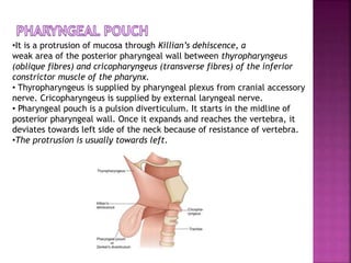
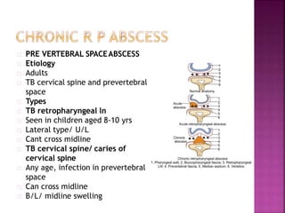
This document discusses various non-thyroid neck swellings including:
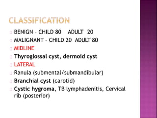
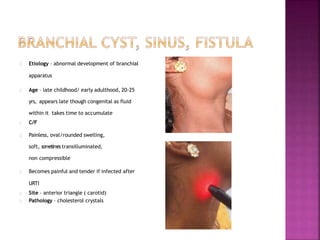
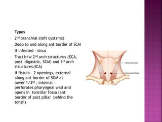
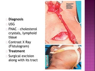
1. Benign cysts such as branchial cysts, dermoid cysts, and ranulas.
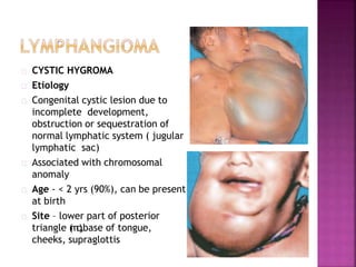
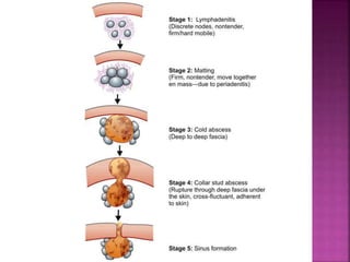
2. Inflammatory conditions including cystic hygroma and tuberculosis lymphadenitis.
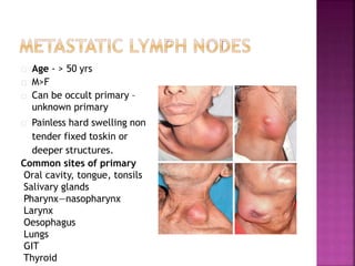
3. Malignancies including lymphomas presenting as neck node enlargement and metastases from unknown primary cancers.
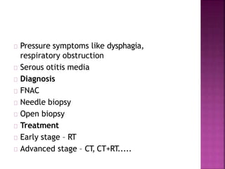
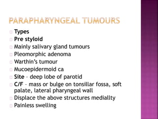
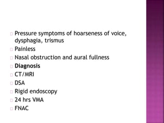
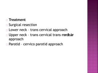
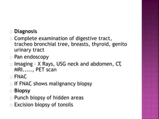
Diagnosis and treatment options are provided for each condition in brief.
THYROIDECTOMY- Operative Surgery
Dear viewers,
Greetings from “Surgical Educator”
Because of the popular demand by viewers of the YouTube channel “Surgical Educator”, I have decided to create and upload videos on common surgeries. I have already uploaded two videos on open and Laparoscopic Appendicectomy. In this video today, I have discussed Thyroidectomy Surgery. However, these videos are not real surgeries but the theoretical aspect of operative surgery like going through an atlas of operative surgery. Along with these videos, I recommend you to watch real operative surgery videos as well and I will give a link for each surgery in the end of the video as end-cards, which I think will be very useful. This will give a very good opportunity for the surgical trainees to mentally rehearse various surgical steps in a sequential manner prior to actual surgery. You can watch the video in the following links:
Surgicaleducator.blogspot.com
Youtube.com/c/surgicaleducator
Thank you for watching the videos.
Which operation for pilonidal sinus? Open or closed? Midline or eccentric closure? An algorithm for the operative treatment of symptomatic pilonidal sinus disease is proposed. In addition to intra-operative photos of the operation of Rhomboid excision and Limberg flap.
Title: Sense of Smell
Presenter: Dr. Faiza, Assistant Professor of Physiology
Qualifications:
MBBS (Best Graduate, AIMC Lahore)
FCPS Physiology
ICMT, CHPE, DHPE (STMU)
MPH (GC University, Faisalabad)
MBA (Virtual University of Pakistan)
Learning Objectives:
Describe the primary categories of smells and the concept of odor blindness.
Explain the structure and location of the olfactory membrane and mucosa, including the types and roles of cells involved in olfaction.
Describe the pathway and mechanisms of olfactory signal transmission from the olfactory receptors to the brain.
Illustrate the biochemical cascade triggered by odorant binding to olfactory receptors, including the role of G-proteins and second messengers in generating an action potential.
Identify different types of olfactory disorders such as anosmia, hyposmia, hyperosmia, and dysosmia, including their potential causes.
Key Topics:
Olfactory Genes:
3% of the human genome accounts for olfactory genes.
400 genes for odorant receptors.
Olfactory Membrane:
Located in the superior part of the nasal cavity.
Medially: Folds downward along the superior septum.
Laterally: Folds over the superior turbinate and upper surface of the middle turbinate.
Total surface area: 5-10 square centimeters.
Olfactory Mucosa:
Olfactory Cells: Bipolar nerve cells derived from the CNS (100 million), with 4-25 olfactory cilia per cell.
Sustentacular Cells: Produce mucus and maintain ionic and molecular environment.
Basal Cells: Replace worn-out olfactory cells with an average lifespan of 1-2 months.
Bowman’s Gland: Secretes mucus.
Stimulation of Olfactory Cells:
Odorant dissolves in mucus and attaches to receptors on olfactory cilia.
Involves a cascade effect through G-proteins and second messengers, leading to depolarization and action potential generation in the olfactory nerve.
Quality of a Good Odorant:
Small (3-20 Carbon atoms), volatile, water-soluble, and lipid-soluble.
Facilitated by odorant-binding proteins in mucus.
Membrane Potential and Action Potential:
Resting membrane potential: -55mV.
Action potential frequency in the olfactory nerve increases with odorant strength.
Adaptation Towards the Sense of Smell:
Rapid adaptation within the first second, with further slow adaptation.
Psychological adaptation greater than receptor adaptation, involving feedback inhibition from the central nervous system.
Primary Sensations of Smell:
Camphoraceous, Musky, Floral, Pepperminty, Ethereal, Pungent, Putrid.
Odor Detection Threshold:
Examples: Hydrogen sulfide (0.0005 ppm), Methyl-mercaptan (0.002 ppm).
Some toxic substances are odorless at lethal concentrations.
Characteristics of Smell:
Odor blindness for single substances due to lack of appropriate receptor protein.
Behavioral and emotional influences of smell.
Transmission of Olfactory Signals:
From olfactory cells to glomeruli in the olfactory bulb, involving lateral inhibition.
Primitive, less old, and new olfactory systems with different path
Local Advanced Lung Cancer: Artificial Intelligence, Synergetics, Complex Sys...Oleg Kshivets
Overall life span (LS) was 1671.7±1721.6 days and cumulative 5YS reached 62.4%, 10 years – 50.4%, 20 years – 44.6%. 94 LCP lived more than 5 years without cancer (LS=2958.6±1723.6 days), 22 – more than 10 years (LS=5571±1841.8 days). 67 LCP died because of LC (LS=471.9±344 days). AT significantly improved 5YS (68% vs. 53.7%) (P=0.028 by log-rank test). Cox modeling displayed that 5YS of LCP significantly depended on: N0-N12, T3-4, blood cell circuit, cell ratio factors (ratio between cancer cells-CC and blood cells subpopulations), LC cell dynamics, recalcification time, heparin tolerance, prothrombin index, protein, AT, procedure type (P=0.000-0.031). Neural networks, genetic algorithm selection and bootstrap simulation revealed relationships between 5YS and N0-12 (rank=1), thrombocytes/CC (rank=2), segmented neutrophils/CC (3), eosinophils/CC (4), erythrocytes/CC (5), healthy cells/CC (6), lymphocytes/CC (7), stick neutrophils/CC (8), leucocytes/CC (9), monocytes/CC (10). Correct prediction of 5YS was 100% by neural networks computing (error=0.000; area under ROC curve=1.0).
Adv. biopharm. APPLICATION OF PHARMACOKINETICS : TARGETED DRUG DELIVERY SYSTEMSAkankshaAshtankar
MIP 201T & MPH 202T
ADVANCED BIOPHARMACEUTICS & PHARMACOKINETICS : UNIT 5
APPLICATION OF PHARMACOKINETICS : TARGETED DRUG DELIVERY SYSTEMS By - AKANKSHA ASHTANKAR
263778731218 Abortion Clinic /Pills In Harare ,sisternakatoto
263778731218 Abortion Clinic /Pills In Harare ,ABORTION WOMEN’S CLINIC +27730423979 IN women clinic we believe that every woman should be able to make choices in her pregnancy. Our job is to provide compassionate care, safety,affordable and confidential services. That’s why we have won the trust from all generations of women all over the world. we use non surgical method(Abortion pills) to terminate…Dr.LISA +27730423979women Clinic is committed to providing the highest quality of obstetrical and gynecological care to women of all ages. Our dedicated staff aim to treat each patient and her health concerns with compassion and respect.Our dedicated group ABORTION WOMEN’S CLINIC +27730423979 IN women clinic we believe that every woman should be able to make choices in her pregnancy. Our job is to provide compassionate care, safety,affordable and confidential services. That’s why we have won the trust from all generations of women all over the world. we use non surgical method(Abortion pills) to terminate…Dr.LISA +27730423979women Clinic is committed to providing the highest quality of obstetrical and gynecological care to women of all ages. Our dedicated staff aim to treat each patient and her health concerns with compassion and respect.Our dedicated group of receptionists, nurses, and physicians have worked together as a teamof receptionists, nurses, and physicians have worked together as a team wwww.lisywomensclinic.co.za/
micro teaching on communication m.sc nursing.pdfAnurag Sharma
Microteaching is a unique model of practice teaching. It is a viable instrument for the. desired change in the teaching behavior or the behavior potential which, in specified types of real. classroom situations, tends to facilitate the achievement of specified types of objectives.
Recomendações da OMS sobre cuidados maternos e neonatais para uma experiência pós-natal positiva.
Em consonância com os ODS – Objetivos do Desenvolvimento Sustentável e a Estratégia Global para a Saúde das Mulheres, Crianças e Adolescentes, e aplicando uma abordagem baseada nos direitos humanos, os esforços de cuidados pós-natais devem expandir-se para além da cobertura e da simples sobrevivência, de modo a incluir cuidados de qualidade.
Estas diretrizes visam melhorar a qualidade dos cuidados pós-natais essenciais e de rotina prestados às mulheres e aos recém-nascidos, com o objetivo final de melhorar a saúde e o bem-estar materno e neonatal.
Uma “experiência pós-natal positiva” é um resultado importante para todas as mulheres que dão à luz e para os seus recém-nascidos, estabelecendo as bases para a melhoria da saúde e do bem-estar a curto e longo prazo. Uma experiência pós-natal positiva é definida como aquela em que as mulheres, pessoas que gestam, os recém-nascidos, os casais, os pais, os cuidadores e as famílias recebem informação consistente, garantia e apoio de profissionais de saúde motivados; e onde um sistema de saúde flexível e com recursos reconheça as necessidades das mulheres e dos bebês e respeite o seu contexto cultural.
Estas diretrizes consolidadas apresentam algumas recomendações novas e já bem fundamentadas sobre cuidados pós-natais de rotina para mulheres e neonatos que recebem cuidados no pós-parto em unidades de saúde ou na comunidade, independentemente dos recursos disponíveis.
É fornecido um conjunto abrangente de recomendações para cuidados durante o período puerperal, com ênfase nos cuidados essenciais que todas as mulheres e recém-nascidos devem receber, e com a devida atenção à qualidade dos cuidados; isto é, a entrega e a experiência do cuidado recebido. Estas diretrizes atualizam e ampliam as recomendações da OMS de 2014 sobre cuidados pós-natais da mãe e do recém-nascido e complementam as atuais diretrizes da OMS sobre a gestão de complicações pós-natais.
O estabelecimento da amamentação e o manejo das principais intercorrências é contemplada.
Recomendamos muito.
Vamos discutir essas recomendações no nosso curso de pós-graduação em Aleitamento no Instituto Ciclos.
Esta publicação só está disponível em inglês até o momento.
Prof. Marcus Renato de Carvalho
www.agostodourado.com
Knee anatomy and clinical tests 2024.pdfvimalpl1234
This includes all relevant anatomy and clinical tests compiled from standard textbooks, Campbell,netter etc..It is comprehensive and best suited for orthopaedicians and orthopaedic residents.
These lecture slides, by Dr Sidra Arshad, offer a quick overview of the physiological basis of a normal electrocardiogram.
Learning objectives:
1. Define an electrocardiogram (ECG) and electrocardiography
2. Describe how dipoles generated by the heart produce the waveforms of the ECG
3. Describe the components of a normal electrocardiogram of a typical bipolar lead (limb II)
4. Differentiate between intervals and segments
5. Enlist some common indications for obtaining an ECG
6. Describe the flow of current around the heart during the cardiac cycle
7. Discuss the placement and polarity of the leads of electrocardiograph
8. Describe the normal electrocardiograms recorded from the limb leads and explain the physiological basis of the different records that are obtained
9. Define mean electrical vector (axis) of the heart and give the normal range
10. Define the mean QRS vector
11. Describe the axes of leads (hexagonal reference system)
12. Comprehend the vectorial analysis of the normal ECG
13. Determine the mean electrical axis of the ventricular QRS and appreciate the mean axis deviation
14. Explain the concepts of current of injury, J point, and their significance
Study Resources:
1. Chapter 11, Guyton and Hall Textbook of Medical Physiology, 14th edition
2. Chapter 9, Human Physiology - From Cells to Systems, Lauralee Sherwood, 9th edition
3. Chapter 29, Ganong’s Review of Medical Physiology, 26th edition
4. Electrocardiogram, StatPearls - https://www.ncbi.nlm.nih.gov/books/NBK549803/
5. ECG in Medical Practice by ABM Abdullah, 4th edition
6. Chapter 3, Cardiology Explained, https://www.ncbi.nlm.nih.gov/books/NBK2214/
7. ECG Basics, http://www.nataliescasebook.com/tag/e-c-g-basics
3. Etiology – abnormal development of branchial
apparatus
Age – late childhood/ early adulthood, 20-25
yrs, appears late though congenital as fluid
within it takes time to accumulate
C/F
Painless, oval/rounded swelling,
soft, sometimes transilluminated,
non compressible
Becomes painful and tender if infected after
URTI
Site – anterior triangle ( carotid)
Pathology – cholesterol crystals
4. Types
2nd branchial cleft cyst(mc)
Deep to and along ant border of SCM
If infected – sinus
Tract b/w 2nd arch structures (ECA,
post digastric, SCM) and 3rd arch
structures(ICA)
If fistula – 2 openings, external
along ant border of SCM at
lower 1/3rd , internal –
perforates pharyngeal wall and
opens in tonsillar fossa (ant
border of post pillar behind the
tonsil)
5. 3rd branchial cleft cyst
Uncommon, deep to both ECA and ICA, sup to
hypoglossal nerve and vagus nerve
Opens into pyriform fossa
1st branchial cleft cyst
Less common
Along ant border of mandible, angle of
mandible, below ear lobe
Opens into EAC
7. CYSTIC HYGROMA
Etiology
Congenital cystic lesion due to
incomplete development,
obstruction or sequestration of
normal lymphatic system ( jugular
lymphatic sac)
Associated with chromosomal
anomaly
Age - < 2 yrs (90%), can be present
at birth
Site – lower part of posterior
triangle (mc),base of tongue,
cheeks, supraglottis
8. C/F
Painless, slow growing, fluctuant, soft
swelling, with indiscrete margins, partially
reducible, varies in size, transilluminated,
increase in size on coughing or crying
If infected – painful and increase in size
Pathology – contains multiple loculi of clear
lymph
9. Complications
Stridor – if involve larynx, pharynx
Respiratory difficulty
Feeding problem
Difficult labour
Diagnosis
Antenatal USG
CT, MRI
11. Head and neck – 7% of dermoid cyst
MC site – floor of mouth post or lateral to
frenulum, midline (submental)
C/F
Slow growing, painless cystic swelling, non
transilluminated, can lead to difficulty in
swallowing, speech and respiration
Children and young adults, 10-15 yrs
Pathology – contains epidermoid appendages
like hair, hair follicles, sweat glands,
sebaceous glands
12. Types
Sublingual – MC
Floor of mouth, above myelohyoid
Cervical
At submental triangle, below myelohyoid,
double chin appearance
Diagnosis – USG Neck
D/D – sebaceous cyst – skin mobile in dermoid
cyst over swelling
Treatment – complete surgical excision
13. Etiology
Mucous retention cyst of
sublingual salivary glands due to
obstruction of their secretory
ducts
Types
Intra oral
Cystic translucent bluish mass in
lateral partof floor of mouth, pushes
tongue up
Plunging ranula – neck swelling in
submental/ submandibular
region, painless, transilluminated
14. Complications
Difficulty in swallowing
Difficulty in chewing
Difficulty in speaking
Treatment
Excision along with sublingual gland
Marsupialization if large (as thin walled so
grows into various structures
If ruptures – recurrence
Plunging ranula – trans cervical approach
15. •It is a protrusion of mucosa through Killian’s dehiscence, a
weak area of the posterior pharyngeal wall between thyropharyngeus
(oblique fibres) and cricopharyngeus (transverse fibres) of the inferior
constrictor muscle of the pharynx.
• Thyropharyngeus is supplied by pharyngeal plexus from cranial accessory
nerve. Cricopharyngeus is supplied by external laryngeal nerve.
• Pharyngeal pouch is a pulsion diverticulum. It starts in the midline of
posterior pharyngeal wall. Once it expands and reaches the vertebra, it
deviates towards left side of the neck because of resistance of vertebra.
•The protrusion is usually towards left.
16. Stages
1. Small diverticulum pointing towards vertebra. It is asymptomatic
and incidentally diagnosed by barium meal X-ray. Foreign body
sensation in pharynx may be present.
2. Large, globular diverticulum with vertical mouth/opening causing
regurgitation, violent cough, dysphagia, respiratory infection.
Regurgitation is more after meals and while turning the neck.
Problems in pharyngeal pouch
• Progressive dysphagia
• Respiratory problems like pneumonia, lung abscess
• Abscess in the neck due to infection in the pouch
• Weight loss and cachexia
17. Differential Diagnosis
Branchial cyst.
Cold abscess in the neck.
Lymph cyst.
Haemangioma neck.
Clinical Features
• Pain, dysphagia, recurrent respiratory infection, swelling in the neck on
the left side which is smooth, soft and tender.
• Regurgitation during night while turning neck, smooth, soft, tender
swelling in the posterior triangle of the left side of the neck; typical
gurgling noise while swallowing—are typical features. It is common in
males.
• Swelling is deep to sternocleidomastoid muscle below the level of
thyroid cartilage; initially soft and emptying; impulse on coughing may
be evident unless opening of the pouch is blocked due to recurrent
inflammation.
19. Treatment
• Antibiotic is started to control infection.
• Pharyngeal pouch is excised by an oblique neck incision (approach from
neck). As there is cricopharyngeal spasm, cricopharyngeal myotomy (i.e.
cutting of cricopharyngeal circular muscle fibres without opening
mucosa) is done to
prevent the recurrence.
Indications for surgery
• Progressive symptoms.
• Recurrent respiratory complications.
• Dysphagia.
Complications of surgery
• Infection, either mediastinitis or lung infection (Pneumonia or lung
abscess)
• Pharyngeal fistula
• Abscess in the neck
• Oesophageal stenosis and recurrence
20. • It is a unilateral narrow necked, air containing diverticulum resulting
from herniation of laryngeal mucosa.
• It occurs in professional trumpet players, glass blowers and in people
with chronic cough.
Types
a. External: It is situated in the anterior third of the laryngeal
ventricle, between the false cords and thyroid cartilage, herniates
through the thyrohyoid membrane where it is pierced by superior
laryngeal nerve.
b. Internal: Confi ned within the larynx, presents as a distention
of false cords.
c. Combined.
21. Clinical Features
• Swelling in the neck in relation to larynx, adjacent to thyrohyoid
membrane which is smooth, soft, resonant and is more prominent while
blowing, coughing and Valsalva manoeuvre.
• It moves upwards during swallowing with expansile impulse on
coughing.
• Infection is quite common in the sac of laryngocele, leading to the
blockade of opening of the sac causing an abscess.
• Pus often may be discharged into the pharynx repeatedly.
• Hoarseness and cough.
• If large, causes obstruction to larynx.
22. Diagnosis
Clinical features, X-ray neck, laryngoscopy, CT scan.
Treatment
•External laryngocele: Excision through neck incision. Neck of the sac
should be ligated. Thyrohyoid membrane is repaired using 3 zero
nonabsorbable polypropylenes sutures.
•Internal laryngocele: Marsupialisation, with the help of laryngoscope.
Typical laryngocele in the neck which becomes
prominent after blowing. X-ray lateral show
radiolucent
air in the neck.
23. U/L
MC – young children (1-8 yrs)
Etiology – due to focus of infection in tonsils,
adenoids, dental, oral cavity
JD lymph nodes
C/F – fever, malaise, ln enlarged and tender
Diagnosis – WBC count, USG
Treatment – antibiotic therapy, surgical
drainage of abscess
24. Chronic infection of
lymph nodes due to
Mycobacterium
tuberculosis
Route of infection – I/L
tonsil, secondary to
pulmonary TB,
hematogenous
C/F
Painless, unilateral, gradual
increase in size mostcommon seen
in posterior triangle
Evening rise of temp, night sweats,
weight loss
Stages
Adenitis – enlarged ln
Periadenitis – matted ln (2-3 ln)
25.
26. Cold abscess – central caseation within ln
Collar stud abscess (dumb bell shaped) –
rupture of cold abscess, pus enters sup fascia
below the skin
Discharging sinus – pus ruptures through skin
Diagnosis
Mantoux test/ tuberculin skin test – positive(>
10 mm)
USG – matted ln with central necrosis
Chest X Ray PAview – pulmonary TB
27. FNAC – granulomas, acid fast bacilli
Excision biopsy
C/S
CBC
Treatment
ATT
Complete excision along with surrounding
fibrous capsule – if residual ln after ATT
If active pulmonary TB – excision not done
28. M avium complex (avium and intercellulare)
M fortuitum
M kansassi
M scrofulaceum
Age – children < 6 yrs
Site – pre auricular, submental, upper jugular
Diagnosis – tuberculin test positive (10-15
mm)
Treatment – coplete surgical excision
29. Extra rib arising from C7 vertebra
attached to1st rib
Right side is most common, but can be left side
or bilateral
Types
1. Complete bony: Cervical rib is radio-opaque,
anteriorly ends
over the first rib or manubrium.
2. Complete fibrous: Cannot be demonstrated
radio logically.
3. Combined: Partly bony partly fibrous.
4. Partial bony: With free end expanding as bony
mass, which
is felt in the neck.
30. C/F
Bony hard lump in supra clavicular region
Compression of branchial plexus and subclavian artery
Branchial plexus compression – tingling, numbness, pain along
upper forearm and fingers
Loss of power of hand
31. Subclavian artery compression –
excessive sweating of hands, cold
and numb hands, pale and blue
hands due to cyanosis, pain in
forearm worsens on exercise
Diagnosis
Adson’s test – positive – weak pulse
on turning neck on same side
X Ray
Treatment
Asymptomatic – no treatment
Symptomatic – excision by
supraclavicular, transaxillary
approach
32. Carotid bodies – chemoreceptor organs
containing cells situated at bifurcation of
CCA contain acetylcholine and
catecholamine stimulated by increase
pco2, decrease po2, increase H+ (higher
altitudes)
Site – carotid triangle at CCAbifurcation
Age – mc 5th decade
Region – high altitude areas like Tibet,
Peru
Etiology – chronic hyperplasia in
high altitude areas -> carotid body
hyperplasia
Familial – 10% autosomal
dominant
33. C/F
Painless slow growing swelling of many years
duration in carotid triangle
Pulastile
Compressible – size decreases with carotid
compression and increases on release of
pressure
Mobility from side to side and not up and
down
Bruit, thrill +
Can extend to parapharyngeal space and
oropharynx pushing the tonsil medially
34. If large can cause pressure symptoms like
dysphagia, change in voice
Pressure on swelling can lead to faintness
(carotid body syncope)
Rare regional and distant metastasis
Diagnosis
Serum catecholamines
24 hrs urine vanellyl mandelic acid
CECT
MRI with gadolinum
MRI angiography/ DSA
35. Lyre’s sign – widening of
angle/ splaying between ICA
and ECA onangiography
Avoid FNAC, open biopsy as highly
vascular
Treatment
Younger age/ no metastasis/ fit
– surgical resection by trans
cervical approach
Large tumours – do arterial
embolization firstto decrease
bleeding
Elderly > 50 yrs/ metastasis/ unfit -
RT
36. Children and young adults
55% of paediatric ca
Hodgkin’s/ non hodgkin’s
C/F
Painless, mobile, non tender, discrete, rubbery,
progressively enlarging lymph nodes in the neck
Other sites of ln enlargement – axilla, groin and
abdomen
Hypertrophy of spleen and liver
Hypertrophy of waldeyer’s ring including tonsils
Fever
37. Pressure symptoms like dysphagia,
respiratory obstruction
Serous otitis media
Diagnosis
FNAC
Needle biopsy
Open biopsy
Treatment
Early stage – RT
Advanced stage – CT, CT+RT.....
38. Types
Pre styloid
Mainly salivary gland tumours
Pleomorphic adenoma
Warthin’s tumour
Mucoepidermoid ca
Site – deep lobe of parotid
C/F – mass or bulge on tonsillar fossa, soft
palate, lateral pharyngeal wall
Displace the above structures mediallty
Painless swelling
39. Post styloid
Neurogenic tumours
Schwannomas/ neurilemmomas
Neurofibroma
Paraganglioma
Malignant schwannoma
C/F
Firm neck mass showing bulge in lateral
pharyngeal wall
Can displace the lateral pharyngeal wall
medially
40. Pressure symptoms of hoarseness of voice,
dysphagia, trismus
Painless
Nasal obstruction and aural fullness
Diagnosis
CT/MRI
DSA
Rigid endoscopy
24 hrs VMA
FNAC
42. Congenital torticolis
Age – at birth
Etiology
Birth trauma – venous obstruction
or haematoma formation
during..... Labour..... Leads to
infarction of central portion of SCM
which leads to fibrosis
Fibrosis causes contraction or
shortening ofSCM
Swelling in the SCM
43. C/F
Circumscribed firm mass palpable in middle
1/3rd of SCM
Torticolis – face turned to opposite side,
head fixed on shoulder on same side
Asymmetry of head and face
Treatment
Conservative – regular active and passive
neck movements to avoid contraction
Surgery – division of SCM at its lower end
44. Age - > 50 yrs
M>F
Can be occult primary –
unknown primary
Painless hard swelling non
tender fixed toskin or
deeper structures.
Common sites of primary
Oral cavity, tongue, tonsils
Salivary glands
Pharynx—nasopharynx
Larynx
Oesophagus
Lungs
GIT
Thyroid
45. Diagnosis
Complete examination of digestive tract,
tracheo bronchial tree, breasts, thyroid, genito
urinary tract
Pan endoscopy
Imaging – X Rays, USG neck and abdomen, CT,
MRI....., PET scan
FNAC
If FNAC shows malignancy biopsy
Biopsy
Punch biopsy of hidden areas
Excision biopsy of tonsils
46. Treatment
Depends on primary site
Occult primary – RND
Post op RT to nasopharynx, I/L tonsil, C/L
neck....., base of tongue
Need to do regular follow up
47. Spreading cellulitis (mainly B/L)
involving submandibular,
submental and sublingual
spaces
Myelohyoid divides the
submandibular space into lower
submaxillary and upper sublingual
space
Etiology
Age 20-50 yrs
Organisms – streptococci,
staphylococci, H.influenza, E
coli, pseudomonas
MC – dental infections, lower
premolar andmolar
48. Dental extraction
Tonsillar infection
Fracture mandible
Injury to oral mucosa – tongue, floor of
mouth
Submandibular sialadenitis
Post radiotherapy osteoradionecrosis of
mandible
ONLY LOCAL SPREAD NO LYMPHATIC SPREAD
49. C/F
Marked progressively painful odynophagia
Trismus
Tongue pushed upwards and backwards
Swollen tender woody hard swelling in
submandibular and submental region
Marked rapidly increasing cellulitis
Drooling of saliva
Diagnosis
Clinical features, increased leucocyte count
X Ray/ CT/ MRI
50. Complications
Spread to retropharyngeal space,
parapharyngeal space and mediastinum
Airway obstruction due to laryngeal oedema,
tongue push up, swelling
Septicaemia
Tongue necrosis
Aspiration leading to pneumonia and lung
abscess
Treatment
Medical – antibiotics, fluids, analgesics
51. Surgical
Tracheostomy if airway compromised
I&D of abscess
Intra oral – if localised to sublingual space
External/cervical – if involves submandibular
region
Steps
Transverse incision between angles of
mandible two finger breaths below margin of
mandible
Vertical incision in midline
52. Serous fluid drained
Incision not closed. Antibiotic soaked ribbon
gauze placed and dressing done daily
Wound allowed to heal by secondary
intention
Extraction of infected teeth
53. ACUTE R P ABSCESS
Etiology
Age
Mc children < 3-4 yrs
Boys
Adults
Suppuration of RP ln due to infections of
adenoids, nasopharynx, PNS, nasal cavity and
tonsils
Petrositis due to acute mastoiditis
Penetrating injury to post pharyngeal wall due to
trauma or iatrogenic
54. FB impaction at cricopharynx and upper
oesophagus
Organisms – streptococci, staphylococci
C/F
Dysphagia and odynophagia
Airway obstruction leading to stridor/stertor
Croupy cough
Torticolis – stiff rigid neck
Hot potato voice
Rapidly increasing sore throat
Drooling of saliva
55. Fever, malaise
Lymphadenopathy
U/L bulge in post pharyngeal wall, cant cross
midline due to median raphe
Diagnosis
X Ray soft tissue neck lateral view
Air shadow in prevertebral space/ widening
of prevertebral space (normal width 3.5 mm,
> 50% width)/ presence of gas
CT Scan/ MRI
56. Complications
Spread to mediastinum and danger space
(most dangerous)
Septicaemia
Meningitis
Airway obstruction
Treatment
Hospitalization
IV antibiotics
IV fluids
steroids
57. Tracheostomy – if stridor
I&D of abscess
Intra oral
No GA – chance of rupture
Position – supine with head low/ rose
position
Vertical incision at most fluctuant area on lat
part of post pharyngeal wall
Do suction to prevent aspiration
58. PRE VERTEBRAL SPACEABSCESS
Etiology
Adults
TB cervical spine and prevertebral
space
Types
TB retropharyngeal ln
Seen in children aged 8-10 yrs
Lateral type/ U/L
Cant cross midline
TB cervical spine/ caries of
cervical spine
Any age, infection in prevertebral
space
Can cross midline
B/L/ midline swelling
59. C/F
Slow in onset/ insidious
Less severe symptoms
Dysphagia
Throat discomfort
Fluctuant swelling in midline or lateral
Non tender enlarged JD ln
Painless lump in throat
Dyspnoea
Chronic cough, evening rise of temp, night
sweats, loss of appetite, loss of weight
60. Diagnosis
X Ray cervical spine
Caries
Loss of normal curvature/ straightening of
cervical spine
Bony destruction of vertebra
X Ray Neck – prevertebral widening
X Ray Chest – TB, mediastinitis
CT/MRI
FNAC
Mantoux test
61. Complications
Can extend to danger space, mediatinum and
parapharyngeal space
Airway obstruction and laryngeal oedema
Pus can extend to coccyx
Spontaneous rupture leading to pneumonia, lung
abscess
Septicaemia
Treatment
ATT
IV fluids
Tracheostomy
62. I&D of abscess
Transcervical approach
Vertical incision at anterior or posterior
border of SCM
Orthopaedics treatment for caries spine
63. PHARYNGO MAXILLARY ABSCESS/ LATERAL
PHARYNGEALABSCESS
Etiology
Any age but common in young adults
Organisms – staphylococci, streptococci, bacteroides,
E coli
Infection from peritonsillar space (mc),
retropharyngeal space, parotid space
Tonsillitis, adenoiditis, pharyngitis,sialadenitis
Dental infections – last molar, infected cysts, fistulas
CSOM/ASOM – bezold’s abscess
Penetrating injuries to neck
Iatrogenic – during procedures, inj
64. C/F
High fever, odynophagia, sore throat, torticolis
Anterior compartment
Prolapse of tonsils and tonsillar fossa
Trismus due to spasm of pterygoid muscles
Swelling at angle of mandible
Odynophagia and dysphagia
Bulging of tonsil, soft palate
Posterior compartment
Pharyngeal bulging behind posterior pillar
Swelling in parotid region
65. CN palsy – IX, X, XI, XII CN
I/L palsy of palate, larynx, tongue
Horner’s syndrome – involvement of sympathetic
chain – I/L anhidrosis, ptosis, enophthalmos,
constricted pupil
Diagnosis
CT/ FNAC/ USG/ X Ray
Complications
Airway obstruction/ laryngeal oedema
Thrombophlebitis of jugular vein
Carotid artery rupture
Mediastinitis/ RP abscess
67. I&D of abscess
Transcervical approach
GA
Horizontal incision 2-3 cm below angle of
mandible (level of hyoid)
Abscess is aspirated
Drain placed for 2-3 days
AVOID TRANS ORAL APPROACH – chance of
damage to greater vessels