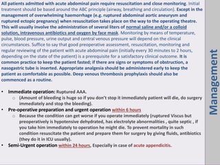
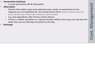
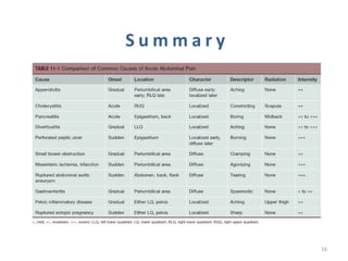
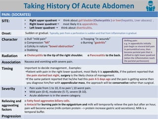
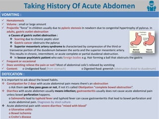
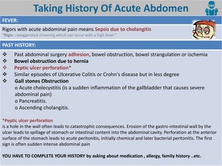
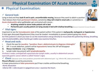
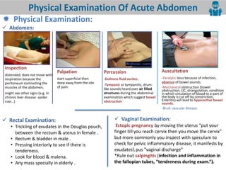
The document provides an overview of the approach to acute abdomen. It defines acute abdomen and outlines the general approach using the SOAP method - taking a history, performing a physical exam, ordering investigations, and creating a treatment plan. Common causes of acute abdomen are then discussed through various case scenarios involving factors like age, location of pain, onset, character, and associated symptoms. A detailed guide is given for examining the abdomen and evaluating vital signs, jugular venous pressure, lymph nodes, and potential referrals from other organ systems. Key blood tests are also outlined to check for indicators of issues like infection, hemorrhage, or electrolyte imbalances.

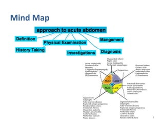
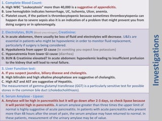
![5. Lactate:
A. Product of anaerobic metabolism: if there is bowel ischemia.
6. Arterial blood gases [ABGs]: (remember.. Diarrhea : Acidosis, Vomiting : Alkalosis)
A. Reflex the respiratory and metabolic states.
B. Do it if ischemia is suspected, severe sepsis, metabolic acidosis and before anesthesia.
Serum calcium: Patients with hypercalcemia may complain of abdominal pain as a result of
abnormal gastrointestinal motility, nephrolithiasis, peptic ulcer disease, pancreatitis or malignancy.
A low calcium level is one of the poor prognostic factors in patients with severe acute pancreatitis.
Blood glucose: Measurement of blood glucose is important, as diabetic ketoacidosis may present
with acute abdominal pain, and also because any serious illness can result in poor glycemic control,
particularly in diabetic patients.
Urinalysis:
Dipstick testing: Haematuria may result from a wide range of conditions but in the context of acute
abdominal pain may indicate a urinary tract tumour, infection or nephrolithiasis. Glucose or ketones
in the urine indicate recent starvation or possible diabetic ketoacidosis. Protein, bilirubin or casts in
the urine suggest renal or liver disease.
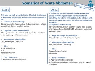
7. Chest x-ray:
A. Perforation of hollow viscous (commonly duodenal ulcer perforation), see air under the
diaphragm (pneumoperitoneum) . Ask for upright chest x ray.
The erect chest X-ray (CXR) is the most appropriate investigation for the detection of free intra-
peritoneal gas and should be carried out in any patient who might have a perforation.
A visceral perforation is the most common cause of free intra-peritoneal gas.
Investigations](https://image.slidesharecdn.com/l20-acuteabdomen-230914083232-47f6321a/85/L20-Acute-Abdomen-pdf-11-320.jpg)
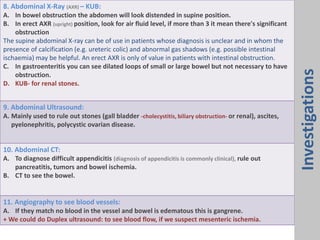
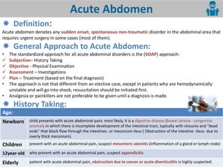
![Diagnosis
Contrast radiology:
A. The contrast used is usually water-soluble, the main issue that requires the use of contrast X-
rays is determining the presence or absence of obstruction or perforation (In up to 50% of
patients with a perforated peptic ulcer, no free gas can be identified on plain radiography)
B. CT with rectal contrast is now more commonly performed for the assessment of patients with
large bowel obstruction.
C. CT without contrast is now more commonly used to detect renal tract calculi.
Endoscopic investigations:
Flexible sigmoidoscopy is commonly performed on patients who present with an acute abdomen
associated with rectal bleeding and in those patients with large bowel obstruction to evaluate the
anorectum. Additional information can be obtained from a colonoscopy. Furthermore, a sigmoid
volvulus can often be deflated by careful sigmoidoscopy. Upper gastrointestinal endoscopy is used
to investigate patients with acute upper abdominal pain in whom a perforated peptic ulcer has
been excluded.
• Acute Abdomen + Shock – Acute Pancreatitis/ Ruptured AAA (abdominal aortic aneurysm)
resuscitate & immediate surgery otherwise patient may die in minutes. Category A
• Generalized Peritonitis – Ruptured Viscus. Category B
• Localized Peritonitis, e.g. RLQ rebound tenderness means Acute
Appendicitis. Category C
• Bowel Obstruction (distention of the abdomen with no movement
during respiration). Category D
• Medical Causes: [Lobar Pneumonia, Acute Inferior MI "if the patient have epigastric pain and
you think of MI you can rule it out by doing ECG or Cardiac enzyme (troponin)"].
Investigations
Category A :
The most immediate
intervention
Category D :
is the least](https://image.slidesharecdn.com/l20-acuteabdomen-230914083232-47f6321a/85/L20-Acute-Abdomen-pdf-13-320.jpg)