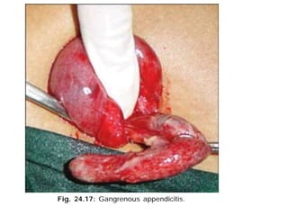
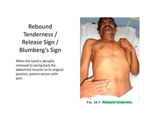
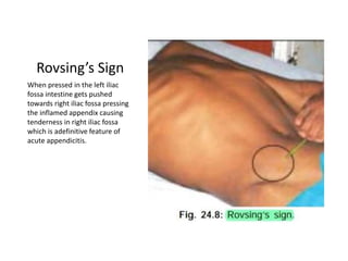
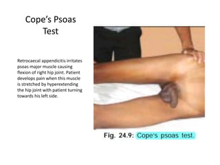
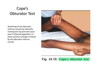
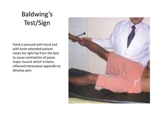
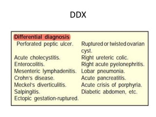
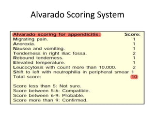
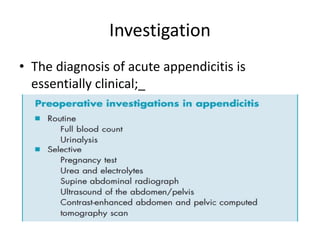
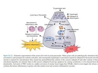
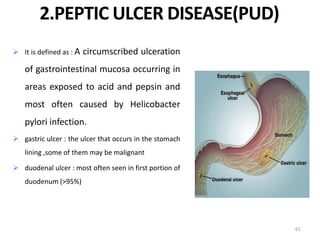
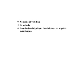
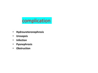
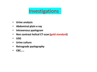
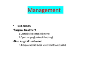
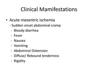
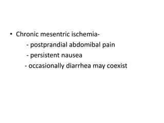
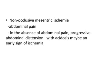
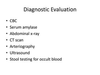
Acute abdomen and appendicitis are medical emergencies that require prompt diagnosis and treatment. The document discusses the clinical presentation of acute abdomen, with a focus on acute appendicitis. Key signs of acute appendicitis include abdominal pain that starts around the umbilicus and later localized to the right lower quadrant, as well as nausea, vomiting, fever and rebound tenderness on physical exam. A diagnosis is typically made clinically based on history and exam findings, supported by blood tests and imaging as needed. Treatment involves surgical removal of the appendix (appendectomy), most often via laparoscopy. Delays in diagnosis and treatment can lead to complications from appendicitis.
Bladder injuries may result from blunt,Penetrating and Iatrogenic trauma.
Full bladder is more susceptible to injury than empty bladder.
Management varies from conservative to surgical aiming to directly repair the injury.
Bladder injuries may result from blunt,Penetrating and Iatrogenic trauma.
Full bladder is more susceptible to injury than empty bladder.
Management varies from conservative to surgical aiming to directly repair the injury.
Describes the concept of a surgical abdomen, acute abdominal pain, emergency intervention and approach to management, including the controversial use of analgesic and antibiotics in emergency room.
This presentation explains in detail the definition, pathophysiology, signs & symptoms, management, and prognosis of intestinal obstruction, ileus, and volvulus.
These simplified slides by Dr. Sidra Arshad present an overview of the non-respiratory functions of the respiratory tract.
Learning objectives:
1. Enlist the non-respiratory functions of the respiratory tract
2. Briefly explain how these functions are carried out
3. Discuss the significance of dead space
4. Differentiate between minute ventilation and alveolar ventilation
5. Describe the cough and sneeze reflexes
Study Resources:
1. Chapter 39, Guyton and Hall Textbook of Medical Physiology, 14th edition
2. Chapter 34, Ganong’s Review of Medical Physiology, 26th edition
3. Chapter 17, Human Physiology by Lauralee Sherwood, 9th edition
4. Non-respiratory functions of the lungs https://academic.oup.com/bjaed/article/13/3/98/278874
MANAGEMENT OF ATRIOVENTRICULAR CONDUCTION BLOCK.pdfJim Jacob Roy
Cardiac conduction defects can occur due to various causes.
Atrioventricular conduction blocks ( AV blocks ) are classified into 3 types.
This document describes the acute management of AV block.
Lung Cancer: Artificial Intelligence, Synergetics, Complex System Analysis, S...Oleg Kshivets
RESULTS: Overall life span (LS) was 2252.1±1742.5 days and cumulative 5-year survival (5YS) reached 73.2%, 10 years – 64.8%, 20 years – 42.5%. 513 LCP lived more than 5 years (LS=3124.6±1525.6 days), 148 LCP – more than 10 years (LS=5054.4±1504.1 days).199 LCP died because of LC (LS=562.7±374.5 days). 5YS of LCP after bi/lobectomies was significantly superior in comparison with LCP after pneumonectomies (78.1% vs.63.7%, P=0.00001 by log-rank test). AT significantly improved 5YS (66.3% vs. 34.8%) (P=0.00000 by log-rank test) only for LCP with N1-2. Cox modeling displayed that 5YS of LCP significantly depended on: phase transition (PT) early-invasive LC in terms of synergetics, PT N0—N12, cell ratio factors (ratio between cancer cells- CC and blood cells subpopulations), G1-3, histology, glucose, AT, blood cell circuit, prothrombin index, heparin tolerance, recalcification time (P=0.000-0.038). Neural networks, genetic algorithm selection and bootstrap simulation revealed relationships between 5YS and PT early-invasive LC (rank=1), PT N0—N12 (rank=2), thrombocytes/CC (3), erythrocytes/CC (4), eosinophils/CC (5), healthy cells/CC (6), lymphocytes/CC (7), segmented neutrophils/CC (8), stick neutrophils/CC (9), monocytes/CC (10); leucocytes/CC (11). Correct prediction of 5YS was 100% by neural networks computing (area under ROC curve=1.0; error=0.0).
CONCLUSIONS: 5YS of LCP after radical procedures significantly depended on: 1) PT early-invasive cancer; 2) PT N0--N12; 3) cell ratio factors; 4) blood cell circuit; 5) biochemical factors; 6) hemostasis system; 7) AT; 8) LC characteristics; 9) LC cell dynamics; 10) surgery type: lobectomy/pneumonectomy; 11) anthropometric data. Optimal diagnosis and treatment strategies for LC are: 1) screening and early detection of LC; 2) availability of experienced thoracic surgeons because of complexity of radical procedures; 3) aggressive en block surgery and adequate lymph node dissection for completeness; 4) precise prediction; 5) adjuvant chemoimmunoradiotherapy for LCP with unfavorable prognosis.
Tom Selleck Health: A Comprehensive Look at the Iconic Actor’s Wellness Journeygreendigital
Tom Selleck, an enduring figure in Hollywood. has captivated audiences for decades with his rugged charm, iconic moustache. and memorable roles in television and film. From his breakout role as Thomas Magnum in Magnum P.I. to his current portrayal of Frank Reagan in Blue Bloods. Selleck's career has spanned over 50 years. But beyond his professional achievements. fans have often been curious about Tom Selleck Health. especially as he has aged in the public eye.
Follow us on: Pinterest
Introduction
Many have been interested in Tom Selleck health. not only because of his enduring presence on screen but also because of the challenges. and lifestyle choices he has faced and made over the years. This article delves into the various aspects of Tom Selleck health. exploring his fitness regimen, diet, mental health. and the challenges he has encountered as he ages. We'll look at how he maintains his well-being. the health issues he has faced, and his approach to ageing .
Early Life and Career
Childhood and Athletic Beginnings
Tom Selleck was born on January 29, 1945, in Detroit, Michigan, and grew up in Sherman Oaks, California. From an early age, he was involved in sports, particularly basketball. which played a significant role in his physical development. His athletic pursuits continued into college. where he attended the University of Southern California (USC) on a basketball scholarship. This early involvement in sports laid a strong foundation for his physical health and disciplined lifestyle.
Transition to Acting
Selleck's transition from an athlete to an actor came with its physical demands. His first significant role in "Magnum P.I." required him to perform various stunts and maintain a fit appearance. This role, which he played from 1980 to 1988. necessitated a rigorous fitness routine to meet the show's demands. setting the stage for his long-term commitment to health and wellness.
Fitness Regimen
Workout Routine
Tom Selleck health and fitness regimen has evolved. adapting to his changing roles and age. During his "Magnum, P.I." days. Selleck's workouts were intense and focused on building and maintaining muscle mass. His routine included weightlifting, cardiovascular exercises. and specific training for the stunts he performed on the show.
Selleck adjusted his fitness routine as he aged to suit his body's needs. Today, his workouts focus on maintaining flexibility, strength, and cardiovascular health. He incorporates low-impact exercises such as swimming, walking, and light weightlifting. This balanced approach helps him stay fit without putting undue strain on his joints and muscles.
Importance of Flexibility and Mobility
In recent years, Selleck has emphasized the importance of flexibility and mobility in his fitness regimen. Understanding the natural decline in muscle mass and joint flexibility with age. he includes stretching and yoga in his routine. These practices help prevent injuries, improve posture, and maintain mobilit
Couples presenting to the infertility clinic- Do they really have infertility...Sujoy Dasgupta
Dr Sujoy Dasgupta presented the study on "Couples presenting to the infertility clinic- Do they really have infertility? – The unexplored stories of non-consummation" in the 13th Congress of the Asia Pacific Initiative on Reproduction (ASPIRE 2024) at Manila on 24 May, 2024.
Prix Galien International 2024 Forum ProgramLevi Shapiro
June 20, 2024, Prix Galien International and Jerusalem Ethics Forum in ROME. Detailed agenda including panels:
- ADVANCES IN CARDIOLOGY: A NEW PARADIGM IS COMING
- WOMEN’S HEALTH: FERTILITY PRESERVATION
- WHAT’S NEW IN THE TREATMENT OF INFECTIOUS,
ONCOLOGICAL AND INFLAMMATORY SKIN DISEASES?
- ARTIFICIAL INTELLIGENCE AND ETHICS
- GENE THERAPY
- BEYOND BORDERS: GLOBAL INITIATIVES FOR DEMOCRATIZING LIFE SCIENCE TECHNOLOGIES AND PROMOTING ACCESS TO HEALTHCARE
- ETHICAL CHALLENGES IN LIFE SCIENCES
- Prix Galien International Awards Ceremony
Explore natural remedies for syphilis treatment in Singapore. Discover alternative therapies, herbal remedies, and lifestyle changes that may complement conventional treatments. Learn about holistic approaches to managing syphilis symptoms and supporting overall health.
New Directions in Targeted Therapeutic Approaches for Older Adults With Mantl...i3 Health
i3 Health is pleased to make the speaker slides from this activity available for use as a non-accredited self-study or teaching resource.
This slide deck presented by Dr. Kami Maddocks, Professor-Clinical in the Division of Hematology and
Associate Division Director for Ambulatory Operations
The Ohio State University Comprehensive Cancer Center, will provide insight into new directions in targeted therapeutic approaches for older adults with mantle cell lymphoma.
STATEMENT OF NEED
Mantle cell lymphoma (MCL) is a rare, aggressive B-cell non-Hodgkin lymphoma (NHL) accounting for 5% to 7% of all lymphomas. Its prognosis ranges from indolent disease that does not require treatment for years to very aggressive disease, which is associated with poor survival (Silkenstedt et al, 2021). Typically, MCL is diagnosed at advanced stage and in older patients who cannot tolerate intensive therapy (NCCN, 2022). Although recent advances have slightly increased remission rates, recurrence and relapse remain very common, leading to a median overall survival between 3 and 6 years (LLS, 2021). Though there are several effective options, progress is still needed towards establishing an accepted frontline approach for MCL (Castellino et al, 2022). Treatment selection and management of MCL are complicated by the heterogeneity of prognosis, advanced age and comorbidities of patients, and lack of an established standard approach for treatment, making it vital that clinicians be familiar with the latest research and advances in this area. In this activity chaired by Michael Wang, MD, Professor in the Department of Lymphoma & Myeloma at MD Anderson Cancer Center, expert faculty will discuss prognostic factors informing treatment, the promising results of recent trials in new therapeutic approaches, and the implications of treatment resistance in therapeutic selection for MCL.
Target Audience
Hematology/oncology fellows, attending faculty, and other health care professionals involved in the treatment of patients with mantle cell lymphoma (MCL).
Learning Objectives
1.) Identify clinical and biological prognostic factors that can guide treatment decision making for older adults with MCL
2.) Evaluate emerging data on targeted therapeutic approaches for treatment-naive and relapsed/refractory MCL and their applicability to older adults
3.) Assess mechanisms of resistance to targeted therapies for MCL and their implications for treatment selection
These lecture slides, by Dr Sidra Arshad, offer a quick overview of physiological basis of a normal electrocardiogram.
Learning objectives:
1. Define an electrocardiogram (ECG) and electrocardiography
2. Describe how dipoles generated by the heart produce the waveforms of the ECG
3. Describe the components of a normal electrocardiogram of a typical bipolar leads (limb II)
4. Differentiate between intervals and segments
5. Enlist some common indications for obtaining an ECG
Study Resources:
1. Chapter 11, Guyton and Hall Textbook of Medical Physiology, 14th edition
2. Chapter 9, Human Physiology - From Cells to Systems, Lauralee Sherwood, 9th edition
3. Chapter 29, Ganong’s Review of Medical Physiology, 26th edition
4. Electrocardiogram, StatPearls - https://www.ncbi.nlm.nih.gov/books/NBK549803/
5. ECG in Medical Practice by ABM Abdullah, 4th edition
6. ECG Basics, http://www.nataliescasebook.com/tag/e-c-g-basics
Pulmonary Thromboembolism - etilogy, types, medical- Surgical and nursing man...VarunMahajani
Disruption of blood supply to lung alveoli due to blockage of one or more pulmonary blood vessels is called as Pulmonary thromboembolism. In this presentation we will discuss its causes, types and its management in depth.
Title: Sense of Smell
Presenter: Dr. Faiza, Assistant Professor of Physiology
Qualifications:
MBBS (Best Graduate, AIMC Lahore)
FCPS Physiology
ICMT, CHPE, DHPE (STMU)
MPH (GC University, Faisalabad)
MBA (Virtual University of Pakistan)
Learning Objectives:
Describe the primary categories of smells and the concept of odor blindness.
Explain the structure and location of the olfactory membrane and mucosa, including the types and roles of cells involved in olfaction.
Describe the pathway and mechanisms of olfactory signal transmission from the olfactory receptors to the brain.
Illustrate the biochemical cascade triggered by odorant binding to olfactory receptors, including the role of G-proteins and second messengers in generating an action potential.
Identify different types of olfactory disorders such as anosmia, hyposmia, hyperosmia, and dysosmia, including their potential causes.
Key Topics:
Olfactory Genes:
3% of the human genome accounts for olfactory genes.
400 genes for odorant receptors.
Olfactory Membrane:
Located in the superior part of the nasal cavity.
Medially: Folds downward along the superior septum.
Laterally: Folds over the superior turbinate and upper surface of the middle turbinate.
Total surface area: 5-10 square centimeters.
Olfactory Mucosa:
Olfactory Cells: Bipolar nerve cells derived from the CNS (100 million), with 4-25 olfactory cilia per cell.
Sustentacular Cells: Produce mucus and maintain ionic and molecular environment.
Basal Cells: Replace worn-out olfactory cells with an average lifespan of 1-2 months.
Bowman’s Gland: Secretes mucus.
Stimulation of Olfactory Cells:
Odorant dissolves in mucus and attaches to receptors on olfactory cilia.
Involves a cascade effect through G-proteins and second messengers, leading to depolarization and action potential generation in the olfactory nerve.
Quality of a Good Odorant:
Small (3-20 Carbon atoms), volatile, water-soluble, and lipid-soluble.
Facilitated by odorant-binding proteins in mucus.
Membrane Potential and Action Potential:
Resting membrane potential: -55mV.
Action potential frequency in the olfactory nerve increases with odorant strength.
Adaptation Towards the Sense of Smell:
Rapid adaptation within the first second, with further slow adaptation.
Psychological adaptation greater than receptor adaptation, involving feedback inhibition from the central nervous system.
Primary Sensations of Smell:
Camphoraceous, Musky, Floral, Pepperminty, Ethereal, Pungent, Putrid.
Odor Detection Threshold:
Examples: Hydrogen sulfide (0.0005 ppm), Methyl-mercaptan (0.002 ppm).
Some toxic substances are odorless at lethal concentrations.
Characteristics of Smell:
Odor blindness for single substances due to lack of appropriate receptor protein.
Behavioral and emotional influences of smell.
Transmission of Olfactory Signals:
From olfactory cells to glomeruli in the olfactory bulb, involving lateral inhibition.
Primitive, less old, and new olfactory systems with different path
3. What is Acute Abdomen?
• It is sudden severe attack of abdominal pain to
such an extent that patient is in severe agony
and often in shock.
• It is in many cases a medical emergency,
requiring urgent and specific diagnosis and
surgery.
• Because there is progressive underlying intra-
abdominal disorder, undue delay in diagnosis
and treatment adversely affect outcome.
4. Causes of acute Abdomen?
INTRA-ABDOMINAL CAUSES
Inflammation—Acute appendicitis, acute cholecystitis,
acute salpingitis, acute diverticulitis, acute Crohn’s, acute
mesenteric adenitis, primary acute peritonitis.
Perforation of bowel
Acute intestinal obstruction – In the lumen (roundworm,
gallstones),
in the wall (stricture, intussusception, tumour),
outside wall (hernia, bands, adhesions, volvulus).
6. Con.t
EXTRA-ABDOMINAL CAUSES
• In the abdominal wall—Abdominal wall abscess,
Meleney’s spreading gangrene, rupture of
abdominal wall muscles, inferior epigastric artery
tear and haematoma foformation
• In thorax – Lobar pneumonia, diaphragmatic
pleurisy, pericarditis, angina pectoris, coronary
disease.
•
•
7. Con.t
• Retroperitoneal causes—Acute pyelonephritis,
retroperitoneal lymphadenitis and lymphangitis,
ruptured aortic aneurysm.
• Diseases of spine, spinal cord and intercostal nerves
•
• – Pott’s tuberculous spine, gastric crisis of Tabes dorsalis,
herpes zoster of intercostal nerves with neuralgia.
• Other causes – Malaria, typhoid, porphyria, diabetic
crisis, sickle cell disease, purpura, haemophilia, etc.
8. Approach to Acute Abdomen
• The first step is to obtain a history from the
patient. But even before that, initial observations
provide clues to the direction which the history
should take; general appearance, gait, position in
bed, facial expression, tone of the speech.
• History of pain, Fever, chills and rigors are
important in acute abdomen.
• History of jaundice may be significant in acute
chole cystitis, pancreatitis.
9. Con.t
History taking should be in this order:
Presenting complaint
Duration of the presenting complaint
History of the presenting complaint
Past history
Drug history and allergies
Social history,Family history,Review of the
systems.
10. Con.t
Physical Examination
• Observation of the patient’s general
condition is useful even before starting to
take the history, especially in the emergency
situation.
• If a patient resorts to the knee–elbow
position to get relief from pain, it indicates
that the pain is of pancreatic origin.
11.
12. Con.t
Inspection
In a patient with acute abdominal pain, always
check to see if the abdominal wall moves with
respiration. If only the thorax moves then
peritonitis should be suspected.
• Grey Turner’s sign
• Cullen’s sign
13.
14. Con.t
Plapation
Signs of parietal peritoneal irritation (tenderness,
guarding, rebound tenderness, rigidity)
Abdominal masses
Percussion
Percussion is a very sensitive and refined method of testing
for rebound tenderness. If the patient winces with pain
on ababdoial percussion it denotes underlying peritonitis.
15. Con.t
Auscultation
Not heared during generalized peritonitis
High-pitched bowel sounds are heard during early stages of
mechanical intestinal obstruction.
A succussion splash is found most often in patients with gastric
stasis due to gastric outlet obstruction.
16. Clinical presentation of Acute
Abdomen
Pain
Pain is the most common presentation of acute abdomen.
• Mode of onset of pain
• Change in position of pain or spread of pain
• aggravating and relieving factors:
• Type of pain;
• Visceral Pain: dull and aching in character, poorly localized and
arises from distention or spasm of fibers innervating the wall
of hollow or solid organs.
• Parietal Pain: sharp, very well localized and arises from
irritation of somatically innervated parietal peritoneal.
• Referred Pain: aching and perceived to be near the surface of
the body.
18. Con.t
Bowel Habits
diarrhoea
constipation
Bloody putrid stool
Abdominal Distention
often presents with fullness of abdomen which is gradually
progressive and is associated with constipation and vomiting.
19. Acute appendicitis
One of the most common surgical emergencies
encountered by general surgeons.
It is inflammation of the Appendix.
20. Eitology
• 1, Altered diet (Decreased dietary fibre and increased
consumption of refined carbohydrates)
• 2, Familial susceptibility (individuals with long
retrocecal appendix)
3, Obstruction of the lumen of appendix (Faecoliths,
stricture, foreign body, roundworm or threadworm.)
(4) Distal colonic obstruction.
(5) Abuse of purgatives
*out of all this "faecoliths" is the comonest cause.
21. Types
• 1, Acute non-obstructive
appendicitis:(Inflammation of the mucus
membrane)
• 2, Acute obstructive appendicitis: pus collects and
it become blackish, gangrenous, oedematous and
rapidly progresses leading to perforation either at
the tip or at the base of the appendix.
• 3, Recurrent appendicitis
• 4, Subacute appendicitis
22.
23. Clinical features
• It is rare before the age of two, common in
children and other age groups.
• Pain: Visceral pain starts around the umbilicus
due to distension of the appendix, later after few
hours somatic pain occurs in right iliac fossa due
to irritation of parietal peritoneum by the
inflamed apappendix.
Pain eventually becomes severe and diffuse
which signifies spread of infection into the general
peritoneal cavity.
24. Con.t
• Vomiting: Due to reflex pylorospasm
• Constipation is the usual feature but diarrhoea
can occur if appendix is in post-ileal or pelvic
positions.
• Urinary frequency: Inflamed appendix may
come in contact with bladder and can cause
bladder irritation.
• _x0000_Fever, tachycardia, foetor oris are
other features_x0000_
26. Rebound
Tenderness /
Release Sign /
Blumberg’s Sign
When the hand is abruptly
removed to spring back the
abdominal muscles to its original
position, patient winces with
pain.
27. Rovsing’s Sign
When pressed in the left iliac
fossa intestine gets pushed
towards right iliac fossa pressing
the inflamed appendix causing
tenderness in right iliac fossa
which is adefinitive feature of
acute appendicitis.
28. Cope’s Psoas
Test
Retrocaecal appendicitis irritates
psoas major muscle causing
flexion of right hip joint. Patient
develops pain when this muscle
is stretched by hyperextending
the hip joint with patient turning
towards his left side.
29. Cope’s
Obturator Test
Stretching of the obturator
internus muscle by internally
rotating the hip joint will cause
pain if inflamed appendix is in
pelvic position and gets irritated
by the obturator internus
muscle.
30. Baldwing’s
Test/Sign
Flank is pressed with hand and
with knee extended patient
raises his right hip from the bed
to cause contraction of psoas
major muscle which irritates
inflamed retrocaecal appendix to
develop pain.
34. Management
Surgery
The surgical procedure for the removal of the
appendix is called an appendectomy.
Open appendectomy: This procedure consists of
the removal of the infected appendix through a
single large incision in the lower right area of the
abdomen. The incision in a laparotomy is usually
2 to 3 inches (51 to 76 mm) long. Under general
Anesthesia
35. Con.t
• Laparoscopic Appendoctomy
• This surgical procedure consists of making
three to four incisions in the abdomen, each
0.25 to 0.5 inches (6.4 to 12.7 mm) long. This
type of appendectomy is made by inserting a
special surgical tool called a laparoscope into
one of the incisions
38. • Definition
– It is an inflammatory disorder of the pancreas that is
characterized by edema and when severe necrosis.
– Ranges from mild self limiting inflammation to sever
critical disease
• Etiology
– Gallstones(present in the 6th decade) and alcohol(present
in the 3rd or 4th decade) accounting for up to 80% of cases
– Gender difference is related with etiology
39. Gallstones
• Transient incompetence caused by the
passage of a stone through the shinicter of
oddi might allow duodenal fluid and bile to
reflux into the pancreatic duct
• Gallstone obstructing the pancreatic duct
leading to ductal hypertension-> minor ductal
disruption->extravasation of pancreatic juice
40. Alcohol
• Ethanol acts on acinar and stellate cells
• Ethanol induced spasm of sphincter of oddi
• Ethanol also induce ductal permeability
• Ethanol increases the protein content of
pancreatic juice and decreases bicarbonate
levels and trypsin inhibitor concentration.
42. Pathophysiology
• Begins with the activation of digestive
zymogens inside acinar cells -> cell injury
• Severity determined by events that occur after
acinar cell injury.
• Physiologic protective mxs
– Inactive precursors
– Separation of site of production and activation of
enzymes
– Trypsin inhibitors in pancreas
43.
44. Clinical Presentation and Diagnosis
• Acute onset of severe constant epigastric pain
that often radiates through to the mid back
• Nausea, repeated vomiting
• elevation of serum amylase/lipase(>3x upper
limit of normal)
• In some cases urinary clearance of pancreatic
enzymes
• Imaging(usually CT scan) only required when the
above diagnostic criteria are not met
48. Management
• Initial assessment (first 4 hours)
– IV Analgesia
• For mild pain- NSAIDS
• For severe pain – Opoids ; avoid morphine
– Fluid resuscitation
• Reassessment (4-6 hrs)
– Assess response to fluid resuscitation
– Determine etiology
– ERCP if concomitant cholangitis is present
– Commence enteral nutrition
49. Cont.
• Managing local complication
– Percutaneous catheter drainage in patients with
suspected infected complications
• Indication for laparotomy
– Failed step up approach
– Acute abdomen(ischemia or perforation)
– Sever abdominal compartment syndrome
• Cholecystectomy to prevent gallstone
pancreatitis recurrence
50. Cholelithiasis
• Often asymptomatic.
• Most common symptom is biliary colic which
usually develops 1-2 hours after fatty meal
• Gallbladder contraction forces the stone against
the orifice of the cystic duct->rise in intraluminal
pressure and pain
• Dull pressure like discomfort in the RUQ or
epigastrium that may radiate to the back and
right shoulder
51. Acute Cholecystitis
• Inflammation of the gallbladder.
• Calculous cholecystitis 90-95% of cases
• Can occur without gallstones :- Acalculous
cholecystitis
52.
53. Risk Factors
• Calculous cholecystitis
– Risk factor for gallstones
• Female
• Obesity
• >age, px, crohn’s disease …
– Previous gallbladder attacks
• Acalculous cholecystitis
– Mostly seen in ICU patients
– Usually associated with major illness such as
polytrauma, burns, major surgery, elderly with DM
55. Clinical manifestations
• In contrast to biliary colic, the pain of acute
cholecystitis doesn’t subside(may persist for
several days)
• Patient is often febrile, complains of anorexia,
nausea or vomiting and maybe reluctant to
move as the inflammatory process creates
focal peritonitis.
56. Clinical signs
• Tenderness and guarding often present in the
RUQ
• A mass, gallbladder and adherant omentum is
occasionally palpable
• Positive murphy’s sign
• Boa’s sign:- an area ofof hyperesthesia between
the 9th and 11th ribs posteriorly on the right
side
• Jaundice- indicates Mirizzi’s syndrome
57. Investigation
• WBC count
• Blood and urine sugar estimation to rule out DM
• Bilirubin level
• Abdominal ultrasound- most commonly used
• Pericholecystic fluid
• GB wall thickening
• Dilation of bile duct
• Sonographic murphy’s sign
• HIDA
• CT scan
58. Management
• IV fluids, broad spectrum antibiotics and analgesia
• Cholecystectomy is the definitive treatment.
• Early cholecystectomy performed with in 72 hours of
illness is preferred over delayed one that is
performed 6-10 weeks after onset of the illness
• Laparoscopic cholecystectomy is preferred
• Unfit for surgery patients are treated with antibiotics
with cholecystectomy scheduled for 2 months later
60. Outline
• Introduction to PUD
• Etiology and Pathogenesis of PUD
• Clinical manifestaions of PUD
• Investigations
• Complication
• Rx of Peptic Ulcer Disease
60
61. It is defined as : A circumscribed ulceration
of gastrointestinal mucosa occurring in
areas exposed to acid and pepsin and
most often caused by Helicobacter
pylori infection.
gastric ulcer : the ulcer that occurs in the stomach
lining ,some of them may be malignant
duodenal ulcer : most often seen in first portion of
duodenum (>95%)
61
2.PEPTIC ULCER DISEASE(PUD)
68. Perforated Peptic Ulcer
• Focal defects in the gastric or duodenal
mucosa that extend into the submucosa or
deeper
69. Perforated Peptic Ulcer
Stages of PUD Perforation
1st phase - Stage of chemical peritonitis
• within 2 hours of onset
2nd phase - Stage of reaction (illusion)
• Between 2-6 hours
3rd phase - Stage of diffuse bacterial peritonitis
• After 6 hours
70. Clinical feature of perforated PUD
• Abdominal pain
– Duodenal ulcer
– Gastric ulcer
• Anemia, stool color change
• Vomiting with/without hematemesis
• Active ulcer can perforate
– Severe stabbing/penetrating agonizing
epigasric pain that even the patient
recognize the exact time of the incident
later generalized
71. Management
Rx GOALS
Medical
• PPI
• high dose H2RAs and
sucralfate
• Antacids
ƚ stop smoking alcohol
and NSAIDs (aspirin).
71
72. Indications for
surgery in PUD
Management
Surgical
• bleeding,
• perforation,
• obstruction, and
• intractability
72
73. The goals of operation
To prevent gastric acid secretion
• Achieved by:
• l.anterctomy- removal of the gastrin-secretion
portion of stomach
• 2.vagotomy alone decrease the acid secretion
by 50%
• 3.Combination of vagotomy and anterectomy
decrease acid secretion by 85%
79. definition
• Urolithiasis(urethralstone)-a small ,hard deposit that forms
in the kidneys and is often painful when passed.
• Ureteric stone-stones come down from pelvis of the kidney and
may get impacted at any site of anatomical narrowing of ureter.
80. • Most stones pass
spontaneously from the
ureter but there are five
sites of narrowing where
the stones may be
arrested.
• Obstruction of urethral
lumen result in severe and
spasmodic pain which is
known as urethral colic.
NORMAL ANATOMICAL NARROWINGS OF URETER
Uretral orifice
83. Clinical feature
Ureteric colic is an intermittent attack of pain which occurs when a
stone passes through the ureter.
• Pain in the loin radiating to groin: which is sever, colicky , intolerable
and lasts for a few hours. the pain may be aggravated by exercise
and relieved by rest.
• when stone descends in to lower ureter, pain radiates to testicles ,
labia majora , and to the upper portion of thigh due to the irritation
of genitofemoral nerve.
84. • Upper urinary tract obstruction-induce renal pain(renal colic) which
radiates from the flank anterioly toward umblicus.
• Right mid uretral obstruction-the pain is reffered to the RLQ of the
abdomen and may be misinterpreted as appendicitis.
• Left mid-ureteral obstruction-pain is referred to the LLQ and can
present in a similar way to diverticulitis.
• Stones lodged in the lower ureter, especially at the vesicoureteric
junction, may cause irritative bladder symptoms.
85. Nausea and vomiting
Hematuria
Guardind and rigidity of the abdomen on physical
examination
89. Indications for surgical removal of
ureteric calculus
• Repeated attacks of pain and the stone is not moving
• Stone is enlarging
• Complete obstruction of the kidney
• Urine is infected
• Stone is too large to pass
• Stone is obstructing solitary kidney or there is bilateral
obstruction.
90. Prevention of renal stone disease
•Fluid management: 1.5L/day
• Dietary adjustments: red meat to be avoided (rich in uric acid)
• Ultrasound to be done once in 6 months
96. • Non-occlusive mesentric ischemia
-abdominal pain
- in the absence of abdominal pain, progressive
abdominal distension. with acidosis maybe an
early sign of ischemia
99. • In chronic mesentric ischemia the goal is to
revascularize mesentric circulation and
prevent development of bowel infarction
• Mesentric occlusive disease can be treated by
transaortic endarterectomy or mesentric
artery bypass.
100. • Ischemic Colitis
-Most commonly from non-occlusive causes
-splenic flexure and cecum has poor tolerance to
hypoperfusion
- May occur in healthy young individuals from
vasospasm due to extreme exercise or illicit drug
use
101. - Less abdominal pain than small bowel
ischemia
- Pt’s recover with conseravtive management,
normalization of the collateral flow and
resolution of the precipitating factors.