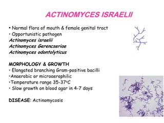
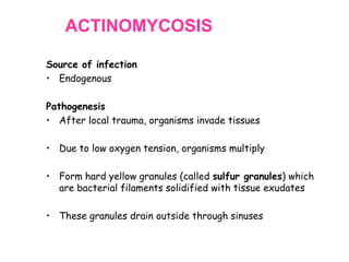
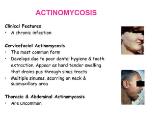
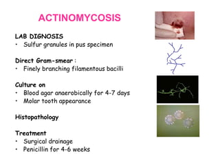
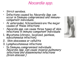
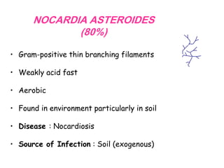
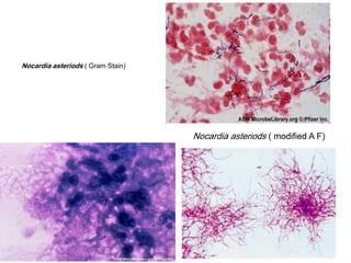
Actinomyces and Nocardia
Objectives
To be familiar with
• The clinical infections associated with

Actinomycetes

• The clinical infection caused by Nocardia spp.
• The microscopic morphology and colonial
morphology of Nocardia and Actinomycetes
• Antimicrobial therapy
ACTINOMYCES ISRAELII
• Normal flora of mouth & female genital tract
• Opportunistic pathogen

Actinomyces israelii
Actinomyces Gerencseriae
Actinomyces odontolyticus
MORPHOLOGY & GROWTH
• Elongated branching Gram-positive bacilli
•Anaerobic or microaerophilic
•Temperature range 35-37oC
• Slow growth on blood agar in 4-7 days
DISEASE: Actinomycosis
ACTINOMYCOSIS
Source of infection
• Endogenous

Pathogenesis
• After local trauma, organisms invade tissues
• Due to low oxygen tension, organisms multiply
• Form hard yellow granules (called sulfur granules) which
are bacterial filaments solidified with tissue exudates
• These granules drain outside through sinuses
ACTINOMYCOSIS
Clinical Features
• A chronic infection
Cervicofacial Actinomycosis
• The most common form
• Develops due to poor dental hygiene & tooth
extraction. Appear as hard tender swelling
that drains pus through sinus tracts
• Multiple sinuses, scarring on neck &
submaxillary area
Thoracic & Abdominal Actinomycosis
• Are uncommon
ACTINOMYCOSIS
LAB DIGNOSIS
• Sulfur granules in pus specimen
Direct Gram-smear :
• Finely branching filamentous bacilli
Culture on
• Blood agar anaerobically for 4-7 days
• Molar tooth appearance
Histopathology
Treatment
• Surgical drainage
• Penicillin for 4-6 weeks
Nocardia spp.
• Strict aerobes.
• Infections caused by Nocardia Spp. can
occur in Immuno-compromised and immunocompetent individuals.
• N. asteriodes, N.brasiliensis are the major
causes of these infections
• Nocardia spp. can cause three types of skin
infections in immuno-competent individuals
1. Mycetoma (chronic, localized, painless,
subcutaneous infection)
2. Skin abscesses or cellulitis
3. Lymphocutaneous infections
• In Immuno-compromised individuals

Nocardia Spp. Can cause invasive pulmonary
infections and disseminated infections
(brain abscess )
NOCARDIA ASTEROIDES
(80%)
• Gram-positive thin branching filaments
• Weakly acid fast
• Aerobic
• Found in environment particularly in soil
• Disease : Nocardiosis
• Source of Infection : Soil (exogenous)
Nocardia asteriods ( Gram Stain)

Nocardia asteriods ( modified A F)
NOCARDIOSIS : CLINICAL FEATURES
Pulmonary Nocardiosis
• Due to inhalation of organism
• Pneumonia-like abscesses
• Usually in immunocompromised patients
Skin & Subcutaneous Tissue Infection
• Usually after trauma like thorne prick
• May present as sinus tract like
actinomycosis
NOCARDIOSIS
LAB DIAGNOSIS
Specimen : Sputum or pus
Staining with :
• Gram-stain or Weak Acid-Fast
Culture on:
• Blood agar
• LJ agar
TREATMENT
• Surgical drainage
• Trimethoprim-sulphamethoxazole

Actinomyces & nocardia

  • 1.
  • 2.
    Objectives To be familiarwith • The clinical infections associated with Actinomycetes • The clinical infection caused by Nocardia spp. • The microscopic morphology and colonial morphology of Nocardia and Actinomycetes • Antimicrobial therapy
  • 3.
    ACTINOMYCES ISRAELII • Normalflora of mouth & female genital tract • Opportunistic pathogen Actinomyces israelii Actinomyces Gerencseriae Actinomyces odontolyticus MORPHOLOGY & GROWTH • Elongated branching Gram-positive bacilli •Anaerobic or microaerophilic •Temperature range 35-37oC • Slow growth on blood agar in 4-7 days DISEASE: Actinomycosis
  • 4.
    ACTINOMYCOSIS Source of infection •Endogenous Pathogenesis • After local trauma, organisms invade tissues • Due to low oxygen tension, organisms multiply • Form hard yellow granules (called sulfur granules) which are bacterial filaments solidified with tissue exudates • These granules drain outside through sinuses
  • 5.
    ACTINOMYCOSIS Clinical Features • Achronic infection Cervicofacial Actinomycosis • The most common form • Develops due to poor dental hygiene & tooth extraction. Appear as hard tender swelling that drains pus through sinus tracts • Multiple sinuses, scarring on neck & submaxillary area Thoracic & Abdominal Actinomycosis • Are uncommon
  • 6.
    ACTINOMYCOSIS LAB DIGNOSIS • Sulfurgranules in pus specimen Direct Gram-smear : • Finely branching filamentous bacilli Culture on • Blood agar anaerobically for 4-7 days • Molar tooth appearance Histopathology Treatment • Surgical drainage • Penicillin for 4-6 weeks
  • 7.
    Nocardia spp. • Strictaerobes. • Infections caused by Nocardia Spp. can occur in Immuno-compromised and immunocompetent individuals. • N. asteriodes, N.brasiliensis are the major causes of these infections • Nocardia spp. can cause three types of skin infections in immuno-competent individuals 1. Mycetoma (chronic, localized, painless, subcutaneous infection) 2. Skin abscesses or cellulitis 3. Lymphocutaneous infections • In Immuno-compromised individuals Nocardia Spp. Can cause invasive pulmonary infections and disseminated infections (brain abscess )
  • 8.
    NOCARDIA ASTEROIDES (80%) • Gram-positivethin branching filaments • Weakly acid fast • Aerobic • Found in environment particularly in soil • Disease : Nocardiosis • Source of Infection : Soil (exogenous)
  • 9.
    Nocardia asteriods (Gram Stain) Nocardia asteriods ( modified A F)
  • 10.
    NOCARDIOSIS : CLINICALFEATURES Pulmonary Nocardiosis • Due to inhalation of organism • Pneumonia-like abscesses • Usually in immunocompromised patients Skin & Subcutaneous Tissue Infection • Usually after trauma like thorne prick • May present as sinus tract like actinomycosis
  • 11.
    NOCARDIOSIS LAB DIAGNOSIS Specimen :Sputum or pus Staining with : • Gram-stain or Weak Acid-Fast Culture on: • Blood agar • LJ agar TREATMENT • Surgical drainage • Trimethoprim-sulphamethoxazole