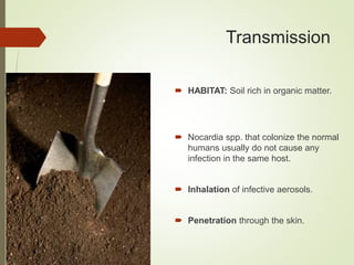
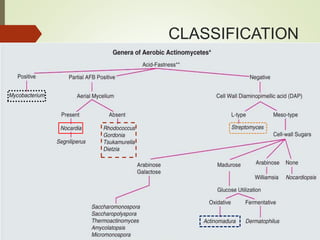
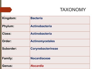
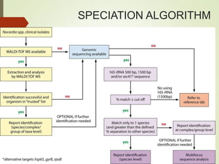
AEROBIC
ACTINOMYCETES
PRESENTED BY:DR.AAMIR
ACTINOMYCETES
 Better known for the production of a wide range of
 Antibiotics
 Anticancer drugs
 Industrial enzymes
 Only a few species of this order are human pathogens
 Mostly oppurtunistic
ACTINOMYCETES
 Thin, Gram +ve, non-motile, non-sporing, non- capsulated filaments
containing muramic acid.
 Morphological resemblance to fungi, cellular organization typical of
bacteria
 Related to Mycobacteria and Corynebacteria
 Mostly free living, mainly soil
 Slow growers
AEROBIC ACTINOMYCETES
CLASSIFICATION
NOCARDIA
History
 Named after Edmond Nocard
 Described & isolated the organism in
cattle with bovine farcy(1888).
 In 1889 given the name Nocardia
farcinica by Trevisan
 First human case of Nocardiosis was
reported in 1890 by Eppinger.
 From brain abscess
 Cladothrix asteroides
 Later renamed Nocardia asteroides
Edmond Nocard
TAXONOMY
Kingdom: Bacteria
Phylum: Actinobacteria
Class: Actinobacteria
Order: Actinomycetales
Suborder: Corynebacterineae
Family: Nocardiaceae
Genus: Nocardia
INTRODUCTION
 Most important genus among aerobic
actinomycetes.
 Saprophytic
 Nearly 100 species isolated
 Reside in soil
 Contribute to decay of organic matter
 Responsible for localized or
disseminated infections in animals
and humans
Characteristics
 Gram-positive bacilli showing a
branching, beaded, and filamentous
form.
 Stain poorly with Gram stain
 Appear to be Gram negative with
intracellular Gram-positive granules
 Usually weakly acid fast
 Acid fastness differentiates it from
other similar bacteria, such as
Actinomyces.
 Catalase positive
 CELL WALL COMPOSITION:
 Short chain mycolic acids
 Peptidoglycan
 Meso-DAP
 Arabinose
 Galactose
Nocardia species
 Of 100 species , 40 species are known to be human
pathogens.
 Most commonly reported species from clinical sources:
 Nocardia asteroides complex
 Nocardia nova (most commonly isolated)
 Nocardia farcinica (most resistant & likely to disseminate)
 Nocardia cyriacigeorgica
 Nocardia abscessus
 Nocardia brasiliensis (skin,sub-cutenous,lymphocutaneous)
Nocardia vs Fungi
Characteristic Nocardia Fungi
Filament/hyphae 0.5–1.0μm in diameter 1.5 to ≥15μm
Reproduction Binary Fission, Hyphae
Fragmentation
Sexually/Asexually
Cell wall composition Mycolic acid Chitins+glucans+mannans+
other fungal specific
proteins
Treatment Anti-bacterials Anti-fungals
Epidemiology
 Nocardia is everywhere in the environment:
 Soil,
 Organic matter, and
 Water.
 Common animal infection
 Outbreaks in oncology and transplant wards and surgical wounds have
occured from :
 Fomites,
 Hospital construction with resultant contaminated dust,
 Health care worker hands.
 United States: 500-1,000 new cases of Nocardiosis occur every
year.
 60% of Nocardiosis cases are associated with pre-existing immune
compromise.
 Men greater risk than women; 3:1
Transmission
 HABITAT: Soil rich in organic matter.
 Nocardia spp. that colonize the normal
humans usually do not cause any
infection in the same host.
 Inhalation of infective aerosols.
 Penetration through the skin.
People at risk
 Pathogenic bacteria have low virulence in Immuno-competent
 Nocardiosis is increasingly found in the immunocompromised
individuals.
 More common in patients with HIV
 Received organ, bone marrow, or stem cell transplantation
 Cirrhosis, lymphoreticular malignancies, and SLE.
 Chronic pulmonary disease, such as emphysema, bronchitis,
bronchiectasis, etc.
 Small children
 Elderly
PATHOGENESIS
 Disease manifestations of Nocardiosis are determined by
 Strain characteristics
 Inoculation site
 Tissue tropism
 Ability to survive initial neutrophilic leukocyte phagocytic attack
 Nature of the immune response.
 T-cell–mediated immunity is the principal protective immune
response to Nocardiosis
 Most problematic in individuals with impaired T-cell–mediated immunity.
 Chronic granulomatous disease patients-more vulnerable to this
infection
Pathogenesis(contd.)
Outer lipids-cytokines IL-1β & IL-6(MACROPHAGES)
=>powerful granulomatous reaction
Catalase & Superoxide Dismutase inactivate reactive
oxygen species that would otherwise prove toxic to the bacteria
Cord Factor-
interferes with phagocytosis by macrophages by preventing the
fusion of phagosome with lysosome.
Gives rise to cell wall–deficient forms (L-forms) that
can be isolated from within macrophages many days later
CLINICAL MANIFESTATIONS
Pulmonary
Manifestation
Pneumonia
Disseminated
Disease
Brain
abscess
Skin
Manifestations
Superficial
infection
Lympho-
cutaneous
syndrome
Actino-
mycetoma
Eye Infection
Keratitis
Endopthalmatitis
Pulmonary Manifestations
 Most common spp –
 N. cyriaci-georgica,
 N. nova,
 N. farcinica.
 Suppurative in nature, but
granulomatous or mixed responses
occur
 Endobronchial inflammatory
masses,
 Pneumonia,
 Lung abscess,
 Cavitary disease
Skin Manifestations
Cellulitis,
Lympho-
cutaneous
syndrome,
Actino-
mycetoma .
Cellulitis
 N. brasiliensis and N. otitidis caviarum complex
 Begins 1–3 weeks after a recognized breach of the skin, (often with soil
contamination)
 Sub-acute cellulitis, with pain, swelling, erythema, and warmth, develops
over days to weeks
 Lesions are usually firm and not fluctuant.
 May progress to involve underlying muscles, tendons, bones, or joints
 Dissemination is rare.
Lympho-Cutaneous Syndrome
(Sporotrichoid Nocardiosis)
 Associated with N. brasiliensis
 Similar disease occurs with other pathogens, most notably Sporothrix
schenckii and Mycobacterium marinum
 Begins as a pyodermatous nodule at the site of inoculation with
 Central ulceration
 Purulent or honey-colored drainage
 appear along lymphatics that drain the primary lesion.
ACTINOMYCETOMA(Madura Foot)
 Nocardia asteroides causes infections
worldwide,
 N. brasiliensis is limited to the southern United
States and Central and South America
 Organism enters the body through breaks in the
skin
 Causes a localized infection involving skin ,
cutaneous , and subcutaneous tissue
 Chronic, indurated, granulomatous masses,
mostly found on the lower extremities
 Characteristic features in Mycetoma
 Swelling (tumifaction)
 Draining sinuses
 Granules
 Tends to invade underlying connective tissue,
muscle, bone

 Also caused by fungi (eumycetoma)
Disseminated Disease
 Disseminated infection- characterized by
widespread abscess formation.
 Most common site - brain.
 Eye (particularly the retina), skin and
subcutaneous tissues, kidneys, joints, bone,
and heart
 Brain abscesses are usually supra-tentorial ,
multiloculated , single or multiple
Nocardial keratitis
 Most commonly identified agents -“N.
asteroides complex” and N. brasiliensis
 Well described in Asia and has been reported in
travelers returning from Asia
 Aggressive ocular infection, typically following
corneal trauma or minor surgical procedures to
the eye
 Appropriate therapy - keratitis resolves with
good visual outcomes
 Nocardial endophthalmitis can develop :
 after eye surgery.
 during disseminated disease.
DIAGNOSIS
 IMAGING STUDIES
 Plain chest radiography
 CT chest scanning
 CT or MRI Brain
 CSF analysis(meningitis)
 Skin ,lung or brain biopsies
LAB DIAGNOSIS
 SPECIMEN:
 Sputum is a frequently received specimen.
 Other specimen:
 Respiratory secretions(BAL, bronchial washings)
 Biopsies-skin ,lung
 Pus from abscesses.
MICROSCOPY
GRAM
STAIN
ACID
FAST
STAIN
SILVER
STAIN
GRAM STAIN
 Routine Gram staining
 Examination of sputum or pus
for crooked, branching,
beaded, gram-positive
filaments 1 μm wide and up to
50 μm long
ACID FAST STAIN
 Most Nocardiae are acid-fast in
direct smears if a weak acid is used
for de-colorization
 Modified Kinyoun,
 Ziehl-Neelsen,
 Fite-Faraco methods
Modified Acid Fast stain
SILVER STAIN
 Gomori methanemine
silver stain of tissue
specimen-biopsy
CULTURE
 Grow on most non-selective media
used routinely for culture of bacteria
e.g
 Sheep’s blood agar,
 Brain–heart infusion agar,
 SAB without antibiotics
 LJ medium
 Middlebrook 7H10 agar
 Specimen containing mixed flora
(e.g., respiratory secretions) use of
selective media e.g
 Thayer-Martin agar with antibiotics
 Paraffin agar.
 Buffered charcoal-yeast
extract(BCYE) medium
CULTURE
 N. asteroides grows well at 25°C,
35°C to 37°C, and at 42°C to 45°C
 Growth is enhanced by incubation
in 10% CO2.
 Growth of Nocardia species may
take 48 hours to 7 to 14 days,
 But typical colonies are usually
seen after 3 to 5 days
 Nocardia colonies vary from white,
to tan, orange and red in color.
CULTURE
 Appear either buff or pigmented,
 Waxy cerebriform colonies
 Dry, chalky-white appearance if aerial
hyphae are produced.
 Characteristic earthy odor
CULTURE
 Lowenstein–Jensen media:
 Whitesh chalky adherent colonies
of Nocardia species
 Tap-water agar:
 Growth shows aerIal hyphae
Orange colony of Nocardia
Aerial hyphae of Nocardia
Other etiologies
Molecular Methods
 Nocardia isolates were previously classified on the basis of
phenotypic tests( hydrolysis of casein, tyrosine, xanthine, hypoxanthine, and
testosterone and by sugar utilization tests.)
 Phenotypic methods are relatively expensive, slow, and limited by
their inability to differentiate between members :largely replaced by
molecular identification tests.
 Gene targets
 65-kda heat shock protein (hsp) gene
 16S rRNA gene
 secA1 gene
Methods
 PCR-RFLP analysis:
 Nocardia 16S rRNA gene and the hsp65 gene (441 bp)
 Good, although incomplete, agreement for species identification
 Cant identify the more uncommon Nocardia species
 RIBOTYPING:
 identify a small number of Nocardia species
 differentiate between N. asteroides sensu stricto and N. farcinica
 limited by the need for multiple probes to identify different species
 GENE SEQUENCING
 Rapid and identifies most isolates reliably
 Sequencing of the first 500 to 606 base pairs (bp) of the 5′ end of the 16S rRNA gene
is currently the most informative approach
 Accuracy of identification is dependent on the quality of the gene repositories
MALDI-TOF
 Recent studies- analysis of bacterial cell wall proteins by MALDI-
TOF MS is a reliable, cheap, and rapid method for identification of
Nocardia
 Has become the method of choice in many laboratories.
SPECIATION ALGORITHM
TREATMENT
Mild/Moderate disease TMP-SMX in Two Divided doses
Severe disease TMP-SMX, Amikacin , and Ceftriaxone or
Imipenem.
Keratitis Topical sulfonamide or Amikacin drops plus a
sulfonamide or an alternative oral drug
Nocardial infections tend to relapse (particularly in patients with
chronic granulomatous disease), and long courses of antimicrobial
therapy are necessary
Treatment Duration
 N
Surgical treatment
 Brain abscesses should be aspirated, drained, or excised if:
 the diagnosis is unclear
 abscess is large and accessible,
 abscess fails to respond to chemotherapy
 In deep or extensive mycetoma cases, drainage or excision
of heavily involved tissue may facilitate healing
Prophylaxis
 Use of SMX and TMP in high-risk populations to prevent
Pneumocystis disease or urinary tract infections appears to reduce
but not eliminate the risk of Nocardiosis.
 Incidence of Nocardiosis is low enough and prophylaxis solely to
prevent this disease is not recommended.
Clinical Outcomes
 Dependent on the site and extent of disease and underlying
host factors.
 Cure rates:
 almost 100% - skin or soft tissue involvement,
 90% in - pleuropulmonary disease,
 63% in disseminated infection, and
 50% in brain abscess.
PREVENTION
 No specific ways to prevent infection.
 People(weakened immune systems )should
wear shoes as well as clothing covering the
skin, open wounds, and cuts when they are
working with the soil.
 Hospitals should maintain strong infection
control practices to avoid outbreaks of
Nocardiosis.
 Organ transplant recepients might be given
antibiotics to prevent bacterial infections.
OTHER AEROBIC ACTINOMYCETES
RHODOCOCCUS SPP.
 Closely related to the Nocardia
 Lack aerial “hyphae”
 Gram-positive , partially acid-fast.
 Non-motile , non-sporulating
 Coccoid or rod-like
 Rhodococcus equi -most important human
pathogen in the genus.
Rhodococcosis
 Zoonotic disease, presumably by a
respiratory route
 Immunocompromised individuals(defects in
cell-mediated immunity)
 Primarily pulmonary disease-clinically mimic
tuberculosis
 Also causes bacteremia, endophthalmitis,
osteomyelitis, pleurisy with effusion and
wound infections
 Mortality
 Immunocompetent = 11%
 Immunocompromised (HIV)= 50 to 55%
 Non hiv = 20 to 25 %
ACTINOMADURA SPP.
 Actinomadura spp. are soil organisms that are introduced
through the skin by trauma.
 primarily in tropical and subtropical countries
 India and Tunisia (A. madurae) or
 Senegal, Chad, and Somalia (A. pelletieri)
 Etilogic agent of Actinomycetoma (Madura foot)
 A. madurae infections are superficial, and can be found on
any part of the host ; Most commonly the foot
STREPTOMYCES SPP.
 Soil organisms- Saprophytes
 Production of two-thirds of the world’s naturally occurring
antibiotics
 S. somaliensis causes actinomycetoma
 worldwide distribution
 Recovered from patients with mycetoma in Saudi Arabia,
Nigeria, Niger, Sudan, Somalia, South Africa, Venezuela, India,
and Mexico
 Madura skull-infections involving head & neck

Aerobic actinomycetes

  • 1.
  • 2.
    ACTINOMYCETES  Better knownfor the production of a wide range of  Antibiotics  Anticancer drugs  Industrial enzymes  Only a few species of this order are human pathogens  Mostly oppurtunistic
  • 3.
    ACTINOMYCETES  Thin, Gram+ve, non-motile, non-sporing, non- capsulated filaments containing muramic acid.  Morphological resemblance to fungi, cellular organization typical of bacteria  Related to Mycobacteria and Corynebacteria  Mostly free living, mainly soil  Slow growers
  • 5.
  • 6.
  • 7.
  • 8.
    History  Named afterEdmond Nocard  Described & isolated the organism in cattle with bovine farcy(1888).  In 1889 given the name Nocardia farcinica by Trevisan  First human case of Nocardiosis was reported in 1890 by Eppinger.  From brain abscess  Cladothrix asteroides  Later renamed Nocardia asteroides Edmond Nocard
  • 9.
    TAXONOMY Kingdom: Bacteria Phylum: Actinobacteria Class:Actinobacteria Order: Actinomycetales Suborder: Corynebacterineae Family: Nocardiaceae Genus: Nocardia
  • 10.
    INTRODUCTION  Most importantgenus among aerobic actinomycetes.  Saprophytic  Nearly 100 species isolated  Reside in soil  Contribute to decay of organic matter  Responsible for localized or disseminated infections in animals and humans
  • 11.
    Characteristics  Gram-positive bacillishowing a branching, beaded, and filamentous form.  Stain poorly with Gram stain  Appear to be Gram negative with intracellular Gram-positive granules  Usually weakly acid fast  Acid fastness differentiates it from other similar bacteria, such as Actinomyces.
  • 12.
     Catalase positive CELL WALL COMPOSITION:  Short chain mycolic acids  Peptidoglycan  Meso-DAP  Arabinose  Galactose
  • 13.
    Nocardia species  Of100 species , 40 species are known to be human pathogens.  Most commonly reported species from clinical sources:  Nocardia asteroides complex  Nocardia nova (most commonly isolated)  Nocardia farcinica (most resistant & likely to disseminate)  Nocardia cyriacigeorgica  Nocardia abscessus  Nocardia brasiliensis (skin,sub-cutenous,lymphocutaneous)
  • 14.
    Nocardia vs Fungi CharacteristicNocardia Fungi Filament/hyphae 0.5–1.0μm in diameter 1.5 to ≥15μm Reproduction Binary Fission, Hyphae Fragmentation Sexually/Asexually Cell wall composition Mycolic acid Chitins+glucans+mannans+ other fungal specific proteins Treatment Anti-bacterials Anti-fungals
  • 15.
    Epidemiology  Nocardia iseverywhere in the environment:  Soil,  Organic matter, and  Water.  Common animal infection  Outbreaks in oncology and transplant wards and surgical wounds have occured from :  Fomites,  Hospital construction with resultant contaminated dust,  Health care worker hands.
  • 16.
     United States:500-1,000 new cases of Nocardiosis occur every year.  60% of Nocardiosis cases are associated with pre-existing immune compromise.  Men greater risk than women; 3:1
  • 17.
    Transmission  HABITAT: Soilrich in organic matter.  Nocardia spp. that colonize the normal humans usually do not cause any infection in the same host.  Inhalation of infective aerosols.  Penetration through the skin.
  • 18.
    People at risk Pathogenic bacteria have low virulence in Immuno-competent  Nocardiosis is increasingly found in the immunocompromised individuals.  More common in patients with HIV  Received organ, bone marrow, or stem cell transplantation  Cirrhosis, lymphoreticular malignancies, and SLE.  Chronic pulmonary disease, such as emphysema, bronchitis, bronchiectasis, etc.  Small children  Elderly
  • 19.
    PATHOGENESIS  Disease manifestationsof Nocardiosis are determined by  Strain characteristics  Inoculation site  Tissue tropism  Ability to survive initial neutrophilic leukocyte phagocytic attack  Nature of the immune response.  T-cell–mediated immunity is the principal protective immune response to Nocardiosis  Most problematic in individuals with impaired T-cell–mediated immunity.  Chronic granulomatous disease patients-more vulnerable to this infection
  • 20.
    Pathogenesis(contd.) Outer lipids-cytokines IL-1β& IL-6(MACROPHAGES) =>powerful granulomatous reaction Catalase & Superoxide Dismutase inactivate reactive oxygen species that would otherwise prove toxic to the bacteria Cord Factor- interferes with phagocytosis by macrophages by preventing the fusion of phagosome with lysosome. Gives rise to cell wall–deficient forms (L-forms) that can be isolated from within macrophages many days later
  • 21.
  • 22.
    Pulmonary Manifestations  Mostcommon spp –  N. cyriaci-georgica,  N. nova,  N. farcinica.  Suppurative in nature, but granulomatous or mixed responses occur  Endobronchial inflammatory masses,  Pneumonia,  Lung abscess,  Cavitary disease
  • 23.
  • 24.
    Cellulitis  N. brasiliensisand N. otitidis caviarum complex  Begins 1–3 weeks after a recognized breach of the skin, (often with soil contamination)  Sub-acute cellulitis, with pain, swelling, erythema, and warmth, develops over days to weeks  Lesions are usually firm and not fluctuant.  May progress to involve underlying muscles, tendons, bones, or joints  Dissemination is rare.
  • 25.
    Lympho-Cutaneous Syndrome (Sporotrichoid Nocardiosis) Associated with N. brasiliensis  Similar disease occurs with other pathogens, most notably Sporothrix schenckii and Mycobacterium marinum  Begins as a pyodermatous nodule at the site of inoculation with  Central ulceration  Purulent or honey-colored drainage  appear along lymphatics that drain the primary lesion.
  • 26.
    ACTINOMYCETOMA(Madura Foot)  Nocardiaasteroides causes infections worldwide,  N. brasiliensis is limited to the southern United States and Central and South America  Organism enters the body through breaks in the skin  Causes a localized infection involving skin , cutaneous , and subcutaneous tissue  Chronic, indurated, granulomatous masses, mostly found on the lower extremities
  • 27.
     Characteristic featuresin Mycetoma  Swelling (tumifaction)  Draining sinuses  Granules  Tends to invade underlying connective tissue, muscle, bone   Also caused by fungi (eumycetoma)
  • 28.
    Disseminated Disease  Disseminatedinfection- characterized by widespread abscess formation.  Most common site - brain.  Eye (particularly the retina), skin and subcutaneous tissues, kidneys, joints, bone, and heart  Brain abscesses are usually supra-tentorial , multiloculated , single or multiple
  • 29.
    Nocardial keratitis  Mostcommonly identified agents -“N. asteroides complex” and N. brasiliensis  Well described in Asia and has been reported in travelers returning from Asia  Aggressive ocular infection, typically following corneal trauma or minor surgical procedures to the eye  Appropriate therapy - keratitis resolves with good visual outcomes  Nocardial endophthalmitis can develop :  after eye surgery.  during disseminated disease.
  • 30.
    DIAGNOSIS  IMAGING STUDIES Plain chest radiography  CT chest scanning  CT or MRI Brain  CSF analysis(meningitis)  Skin ,lung or brain biopsies
  • 31.
    LAB DIAGNOSIS  SPECIMEN: Sputum is a frequently received specimen.  Other specimen:  Respiratory secretions(BAL, bronchial washings)  Biopsies-skin ,lung  Pus from abscesses.
  • 32.
  • 33.
    GRAM STAIN  RoutineGram staining  Examination of sputum or pus for crooked, branching, beaded, gram-positive filaments 1 μm wide and up to 50 μm long
  • 34.
    ACID FAST STAIN Most Nocardiae are acid-fast in direct smears if a weak acid is used for de-colorization  Modified Kinyoun,  Ziehl-Neelsen,  Fite-Faraco methods
  • 35.
  • 36.
    SILVER STAIN  Gomorimethanemine silver stain of tissue specimen-biopsy
  • 37.
    CULTURE  Grow onmost non-selective media used routinely for culture of bacteria e.g  Sheep’s blood agar,  Brain–heart infusion agar,  SAB without antibiotics  LJ medium  Middlebrook 7H10 agar  Specimen containing mixed flora (e.g., respiratory secretions) use of selective media e.g  Thayer-Martin agar with antibiotics  Paraffin agar.  Buffered charcoal-yeast extract(BCYE) medium
  • 38.
    CULTURE  N. asteroidesgrows well at 25°C, 35°C to 37°C, and at 42°C to 45°C  Growth is enhanced by incubation in 10% CO2.  Growth of Nocardia species may take 48 hours to 7 to 14 days,  But typical colonies are usually seen after 3 to 5 days  Nocardia colonies vary from white, to tan, orange and red in color.
  • 39.
    CULTURE  Appear eitherbuff or pigmented,  Waxy cerebriform colonies  Dry, chalky-white appearance if aerial hyphae are produced.  Characteristic earthy odor
  • 40.
    CULTURE  Lowenstein–Jensen media: Whitesh chalky adherent colonies of Nocardia species  Tap-water agar:  Growth shows aerIal hyphae
  • 42.
  • 43.
  • 44.
  • 46.
    Molecular Methods  Nocardiaisolates were previously classified on the basis of phenotypic tests( hydrolysis of casein, tyrosine, xanthine, hypoxanthine, and testosterone and by sugar utilization tests.)  Phenotypic methods are relatively expensive, slow, and limited by their inability to differentiate between members :largely replaced by molecular identification tests.  Gene targets  65-kda heat shock protein (hsp) gene  16S rRNA gene  secA1 gene
  • 47.
    Methods  PCR-RFLP analysis: Nocardia 16S rRNA gene and the hsp65 gene (441 bp)  Good, although incomplete, agreement for species identification  Cant identify the more uncommon Nocardia species  RIBOTYPING:  identify a small number of Nocardia species  differentiate between N. asteroides sensu stricto and N. farcinica  limited by the need for multiple probes to identify different species  GENE SEQUENCING  Rapid and identifies most isolates reliably  Sequencing of the first 500 to 606 base pairs (bp) of the 5′ end of the 16S rRNA gene is currently the most informative approach  Accuracy of identification is dependent on the quality of the gene repositories
  • 48.
    MALDI-TOF  Recent studies-analysis of bacterial cell wall proteins by MALDI- TOF MS is a reliable, cheap, and rapid method for identification of Nocardia  Has become the method of choice in many laboratories.
  • 49.
  • 50.
    TREATMENT Mild/Moderate disease TMP-SMXin Two Divided doses Severe disease TMP-SMX, Amikacin , and Ceftriaxone or Imipenem. Keratitis Topical sulfonamide or Amikacin drops plus a sulfonamide or an alternative oral drug Nocardial infections tend to relapse (particularly in patients with chronic granulomatous disease), and long courses of antimicrobial therapy are necessary
  • 51.
  • 52.
    Surgical treatment  Brainabscesses should be aspirated, drained, or excised if:  the diagnosis is unclear  abscess is large and accessible,  abscess fails to respond to chemotherapy  In deep or extensive mycetoma cases, drainage or excision of heavily involved tissue may facilitate healing
  • 53.
    Prophylaxis  Use ofSMX and TMP in high-risk populations to prevent Pneumocystis disease or urinary tract infections appears to reduce but not eliminate the risk of Nocardiosis.  Incidence of Nocardiosis is low enough and prophylaxis solely to prevent this disease is not recommended.
  • 54.
    Clinical Outcomes  Dependenton the site and extent of disease and underlying host factors.  Cure rates:  almost 100% - skin or soft tissue involvement,  90% in - pleuropulmonary disease,  63% in disseminated infection, and  50% in brain abscess.
  • 55.
    PREVENTION  No specificways to prevent infection.  People(weakened immune systems )should wear shoes as well as clothing covering the skin, open wounds, and cuts when they are working with the soil.  Hospitals should maintain strong infection control practices to avoid outbreaks of Nocardiosis.  Organ transplant recepients might be given antibiotics to prevent bacterial infections.
  • 56.
  • 57.
    RHODOCOCCUS SPP.  Closelyrelated to the Nocardia  Lack aerial “hyphae”  Gram-positive , partially acid-fast.  Non-motile , non-sporulating  Coccoid or rod-like  Rhodococcus equi -most important human pathogen in the genus.
  • 58.
    Rhodococcosis  Zoonotic disease,presumably by a respiratory route  Immunocompromised individuals(defects in cell-mediated immunity)  Primarily pulmonary disease-clinically mimic tuberculosis  Also causes bacteremia, endophthalmitis, osteomyelitis, pleurisy with effusion and wound infections  Mortality  Immunocompetent = 11%  Immunocompromised (HIV)= 50 to 55%  Non hiv = 20 to 25 %
  • 59.
    ACTINOMADURA SPP.  Actinomaduraspp. are soil organisms that are introduced through the skin by trauma.  primarily in tropical and subtropical countries  India and Tunisia (A. madurae) or  Senegal, Chad, and Somalia (A. pelletieri)  Etilogic agent of Actinomycetoma (Madura foot)  A. madurae infections are superficial, and can be found on any part of the host ; Most commonly the foot
  • 60.
    STREPTOMYCES SPP.  Soilorganisms- Saprophytes  Production of two-thirds of the world’s naturally occurring antibiotics  S. somaliensis causes actinomycetoma  worldwide distribution  Recovered from patients with mycetoma in Saudi Arabia, Nigeria, Niger, Sudan, Somalia, South Africa, Venezuela, India, and Mexico  Madura skull-infections involving head & neck

Editor's Notes

  • #15 Nocardia and other aerobic actinomycetes reproduce by fission (like other bacteria) as compared to the way in which fungi reproduce asexually by mitosis-derived conidia, as well as in some instances, sexually by means of more elaborate reproductive structures and meiotically-derived spores