The document provides information on interpreting arterial blood gases (ABGs), including:
- A 6-step process for interpretation involving assessing pH, identifying the primary disorder as respiratory or metabolic, evaluating compensation, calculating anion gap, and considering differential diagnoses.
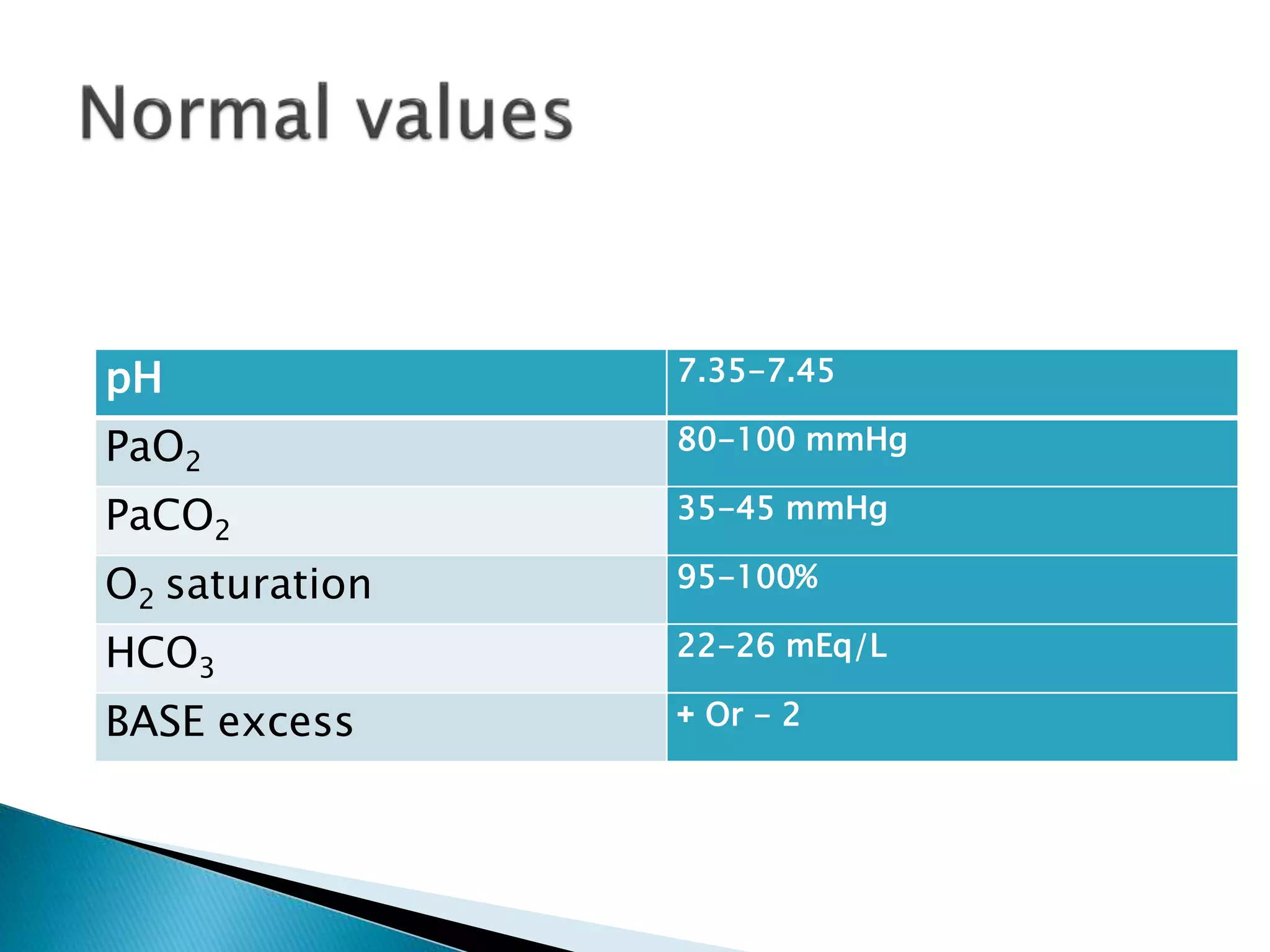
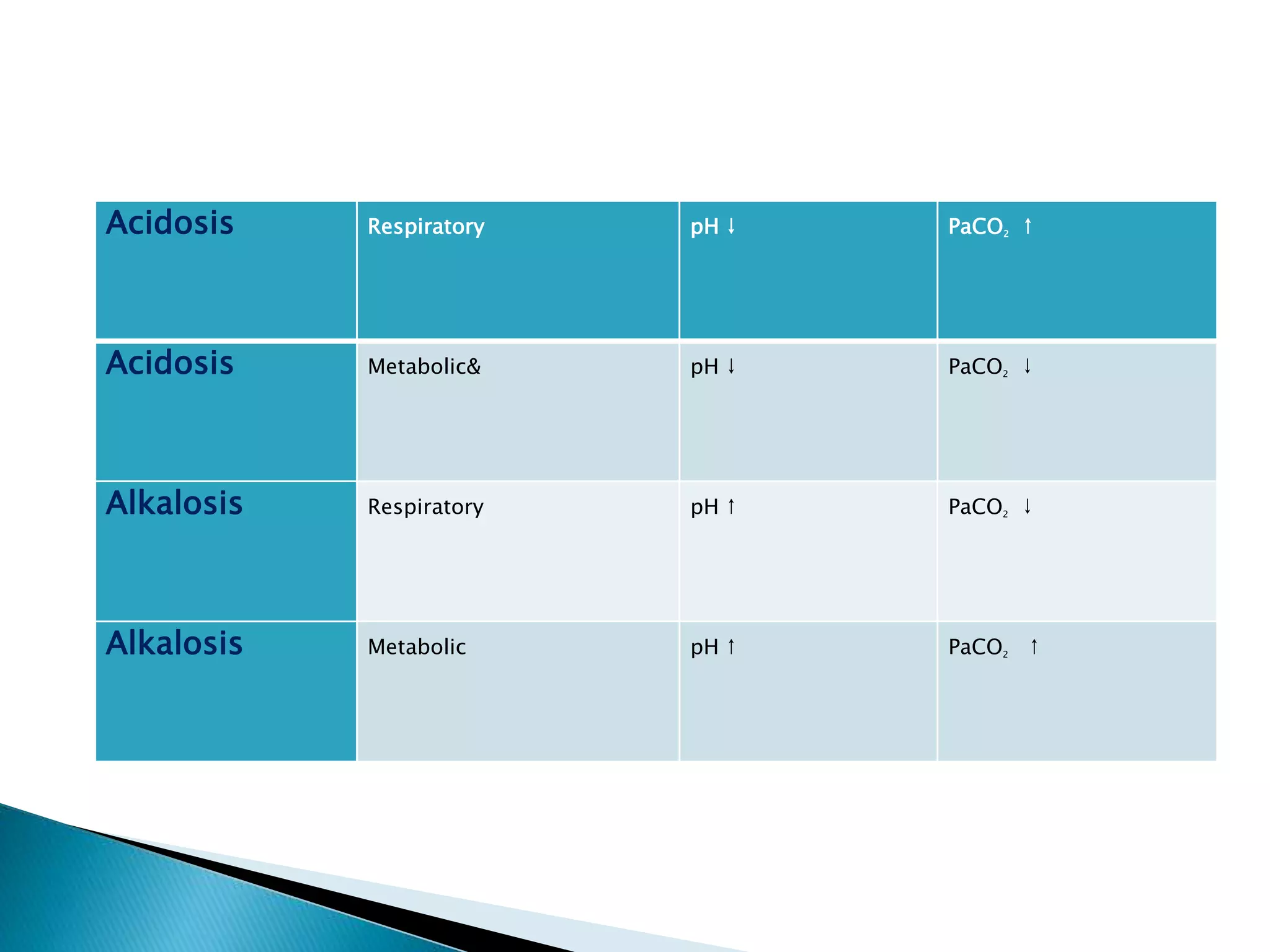
- Tables listing normal ranges for ABG components like pH, PaCO2, HCO3, and bases for common acid-base disorders.
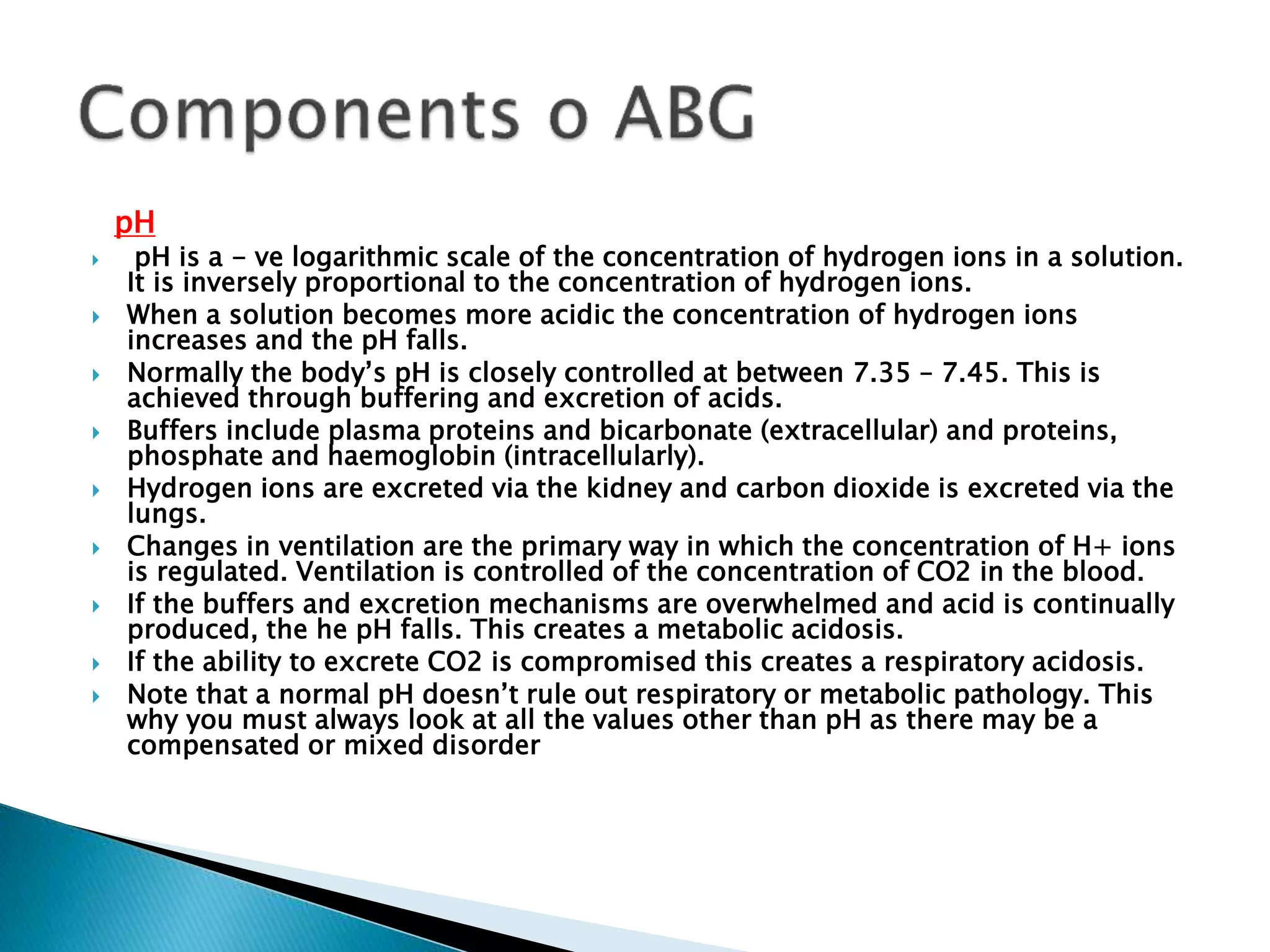
- Explanations of key components like pH, partial pressure, base excess, bicarbonate, and their relationships in respiratory and metabolic acidosis/alkalosis.
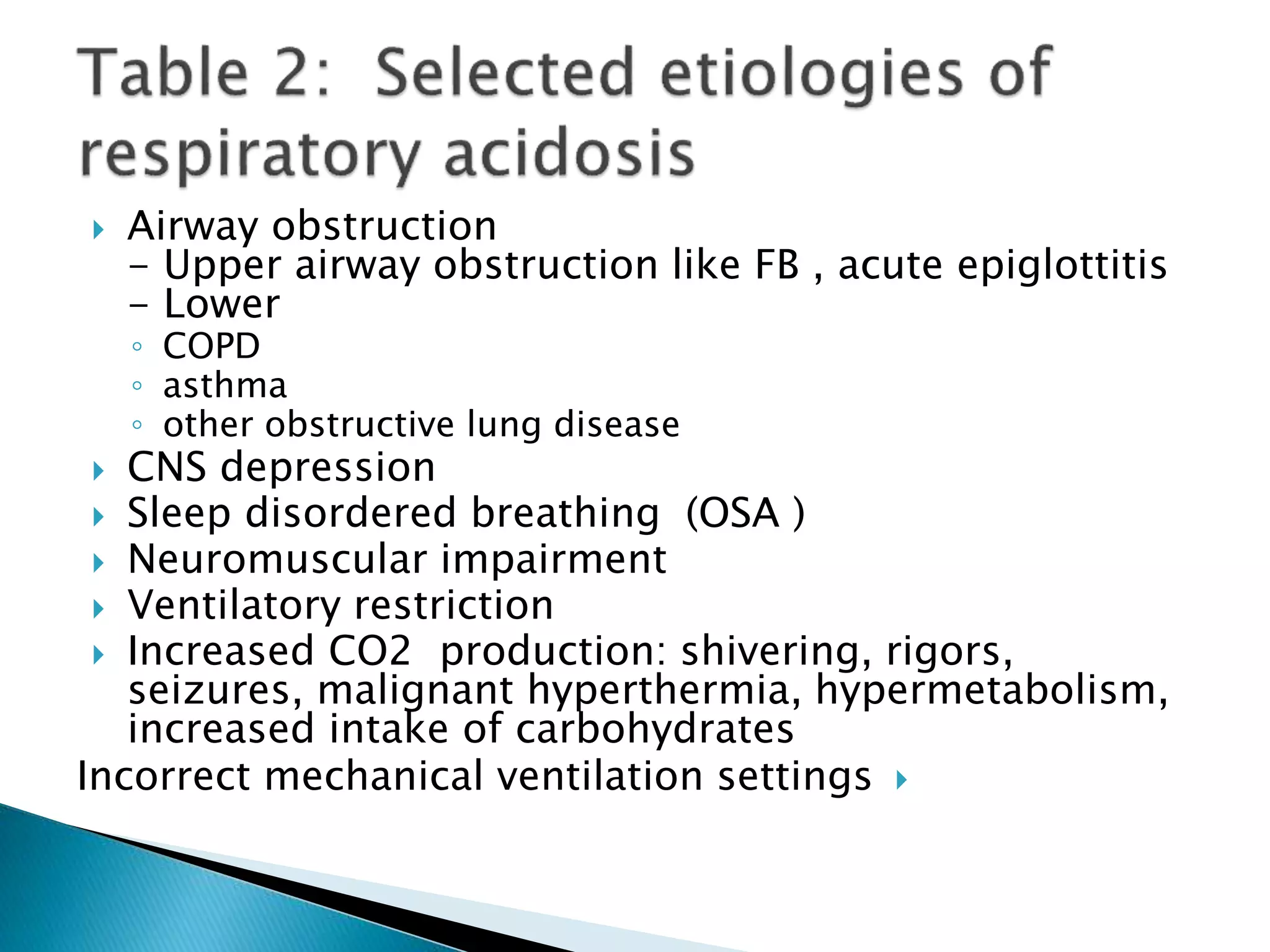
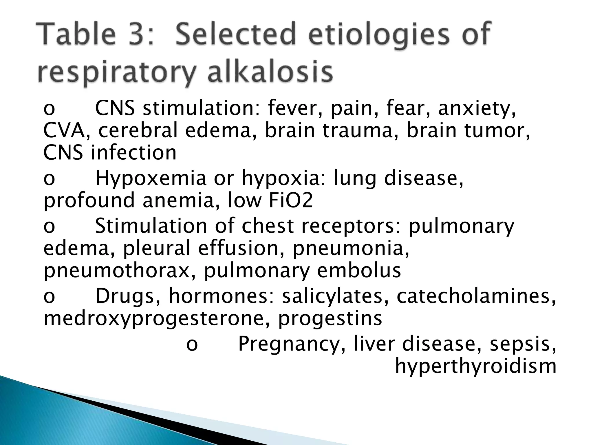
- Causes and mechanisms of respiratory and metabolic acidosis and alkalosis are outlined.
Concepts of acid base balance and its disorders are very important for practice of medicine.It is for the benefit of medical and students of allied fields.
ABGs or VBGs interpretation made simple straight forward easy to remember and easy to apply. The presentation is designed to help the residents and junior ER physicians. The second part will discuss the oxygenation and the third part will review the "Stewart Approach" while fourth and last part is meant for the Experts.
"The body maintains a balance of acids and bases in order to constantly maintain blood pH within a narrow range, despite the continuous generation of metabolic products. In turn, this allows the body to maintain cell enzyme systems in good operation conditions, together with the proper concentration of ionized (active) forms of various electrolytes such as Ca and Mg . This influences the speed of metabolic reactions and trans-membrane transportation systems (pharmacokinetics and pharmacodynamics)." - Luis Núñez Ochoa, Facultad de Medicina Veterinaria y Zootecnia, Unam, Mexico
PH definition and determinants , how to regulate the Acid/base in our body ,ABG's normal values in atrery and vein , how to obtain an arterial blood sample, the interpretation of ABG's , steps to analuse Acid-base, respiratory acidosis and alkalosis and its causes also about metablic acidosis and alkalosis and the causes and some case studies .
Short Review regarding Metabolic Acidosis
The Causes, anion gap,urine osmolal gap, Renal Tubular Acidosis, approach to Metabolic Acidosis in Final Slide
This presentation discuss about acid-base-gas normal ratio and its indication in relation to varying abnormal level and how to manage it. This includes clinical analysis practice.
The normal ranges for arterial blood gas values
Approach to arterial blood gas interpretation
Arterial blood gas abnormalities in special circumstances
TEST BANK for Operations Management, 14th Edition by William J. Stevenson, Ve...kevinkariuki227
TEST BANK for Operations Management, 14th Edition by William J. Stevenson, Verified Chapters 1 - 19, Complete Newest Version.pdf
Anti ulcer drugs and their Advance pharmacology ||
Anti-ulcer drugs are medications used to prevent and treat ulcers in the stomach and upper part of the small intestine (duodenal ulcers). These ulcers are often caused by an imbalance between stomach acid and the mucosal lining, which protects the stomach lining.
||Scope: Overview of various classes of anti-ulcer drugs, their mechanisms of action, indications, side effects, and clinical considerations.
Factory Supply Best Quality Pmk Oil CAS 28578–16–7 PMK Powder in Stockrebeccabio
Factory Supply Best Quality Pmk Oil CAS 28578–16–7 PMK Powder in Stock
Telegram: bmksupplier
signal: +85264872720
threema: TUD4A6YC
You can contact me on Telegram or Threema
Communicate promptly and reply
Free of customs clearance, Double Clearance 100% pass delivery to USA, Canada, Spain, Germany, Netherland, Poland, Italy, Sweden, UK, Czech Republic, Australia, Mexico, Russia, Ukraine, Kazakhstan.Door to door service
Hot Selling Organic intermediates
These simplified slides by Dr. Sidra Arshad present an overview of the non-respiratory functions of the respiratory tract.
Learning objectives:
1. Enlist the non-respiratory functions of the respiratory tract
2. Briefly explain how these functions are carried out
3. Discuss the significance of dead space
4. Differentiate between minute ventilation and alveolar ventilation
5. Describe the cough and sneeze reflexes
Study Resources:
1. Chapter 39, Guyton and Hall Textbook of Medical Physiology, 14th edition
2. Chapter 34, Ganong’s Review of Medical Physiology, 26th edition
3. Chapter 17, Human Physiology by Lauralee Sherwood, 9th edition
4. Non-respiratory functions of the lungs https://academic.oup.com/bjaed/article/13/3/98/278874
Title: Sense of Taste
Presenter: Dr. Faiza, Assistant Professor of Physiology
Qualifications:
MBBS (Best Graduate, AIMC Lahore)
FCPS Physiology
ICMT, CHPE, DHPE (STMU)
MPH (GC University, Faisalabad)
MBA (Virtual University of Pakistan)
Learning Objectives:
Describe the structure and function of taste buds.
Describe the relationship between the taste threshold and taste index of common substances.
Explain the chemical basis and signal transduction of taste perception for each type of primary taste sensation.
Recognize different abnormalities of taste perception and their causes.
Key Topics:
Significance of Taste Sensation:
Differentiation between pleasant and harmful food
Influence on behavior
Selection of food based on metabolic needs
Receptors of Taste:
Taste buds on the tongue
Influence of sense of smell, texture of food, and pain stimulation (e.g., by pepper)
Primary and Secondary Taste Sensations:
Primary taste sensations: Sweet, Sour, Salty, Bitter, Umami
Chemical basis and signal transduction mechanisms for each taste
Taste Threshold and Index:
Taste threshold values for Sweet (sucrose), Salty (NaCl), Sour (HCl), and Bitter (Quinine)
Taste index relationship: Inversely proportional to taste threshold
Taste Blindness:
Inability to taste certain substances, particularly thiourea compounds
Example: Phenylthiocarbamide
Structure and Function of Taste Buds:
Composition: Epithelial cells, Sustentacular/Supporting cells, Taste cells, Basal cells
Features: Taste pores, Taste hairs/microvilli, and Taste nerve fibers
Location of Taste Buds:
Found in papillae of the tongue (Fungiform, Circumvallate, Foliate)
Also present on the palate, tonsillar pillars, epiglottis, and proximal esophagus
Mechanism of Taste Stimulation:
Interaction of taste substances with receptors on microvilli
Signal transduction pathways for Umami, Sweet, Bitter, Sour, and Salty tastes
Taste Sensitivity and Adaptation:
Decrease in sensitivity with age
Rapid adaptation of taste sensation
Role of Saliva in Taste:
Dissolution of tastants to reach receptors
Washing away the stimulus
Taste Preferences and Aversions:
Mechanisms behind taste preference and aversion
Influence of receptors and neural pathways
Impact of Sensory Nerve Damage:
Degeneration of taste buds if the sensory nerve fiber is cut
Abnormalities of Taste Detection:
Conditions: Ageusia, Hypogeusia, Dysgeusia (parageusia)
Causes: Nerve damage, neurological disorders, infections, poor oral hygiene, adverse drug effects, deficiencies, aging, tobacco use, altered neurotransmitter levels
Neurotransmitters and Taste Threshold:
Effects of serotonin (5-HT) and norepinephrine (NE) on taste sensitivity
Supertasters:
25% of the population with heightened sensitivity to taste, especially bitterness
Increased number of fungiform papillae
- Video recording of this lecture in English language: https://youtu.be/lK81BzxMqdo
- Video recording of this lecture in Arabic language: https://youtu.be/Ve4P0COk9OI
- Link to download the book free: https://nephrotube.blogspot.com/p/nephrotube-nephrology-books.html
- Link to NephroTube website: www.NephroTube.com
- Link to NephroTube social media accounts: https://nephrotube.blogspot.com/p/join-nephrotube-on-social-media.html
Prix Galien International 2024 Forum ProgramLevi Shapiro
June 20, 2024, Prix Galien International and Jerusalem Ethics Forum in ROME. Detailed agenda including panels:
- ADVANCES IN CARDIOLOGY: A NEW PARADIGM IS COMING
- WOMEN’S HEALTH: FERTILITY PRESERVATION
- WHAT’S NEW IN THE TREATMENT OF INFECTIOUS,
ONCOLOGICAL AND INFLAMMATORY SKIN DISEASES?
- ARTIFICIAL INTELLIGENCE AND ETHICS
- GENE THERAPY
- BEYOND BORDERS: GLOBAL INITIATIVES FOR DEMOCRATIZING LIFE SCIENCE TECHNOLOGIES AND PROMOTING ACCESS TO HEALTHCARE
- ETHICAL CHALLENGES IN LIFE SCIENCES
- Prix Galien International Awards Ceremony
Explore natural remedies for syphilis treatment in Singapore. Discover alternative therapies, herbal remedies, and lifestyle changes that may complement conventional treatments. Learn about holistic approaches to managing syphilis symptoms and supporting overall health.
Title: Sense of Smell
Presenter: Dr. Faiza, Assistant Professor of Physiology
Qualifications:
MBBS (Best Graduate, AIMC Lahore)
FCPS Physiology
ICMT, CHPE, DHPE (STMU)
MPH (GC University, Faisalabad)
MBA (Virtual University of Pakistan)
Learning Objectives:
Describe the primary categories of smells and the concept of odor blindness.
Explain the structure and location of the olfactory membrane and mucosa, including the types and roles of cells involved in olfaction.
Describe the pathway and mechanisms of olfactory signal transmission from the olfactory receptors to the brain.
Illustrate the biochemical cascade triggered by odorant binding to olfactory receptors, including the role of G-proteins and second messengers in generating an action potential.
Identify different types of olfactory disorders such as anosmia, hyposmia, hyperosmia, and dysosmia, including their potential causes.
Key Topics:
Olfactory Genes:
3% of the human genome accounts for olfactory genes.
400 genes for odorant receptors.
Olfactory Membrane:
Located in the superior part of the nasal cavity.
Medially: Folds downward along the superior septum.
Laterally: Folds over the superior turbinate and upper surface of the middle turbinate.
Total surface area: 5-10 square centimeters.
Olfactory Mucosa:
Olfactory Cells: Bipolar nerve cells derived from the CNS (100 million), with 4-25 olfactory cilia per cell.
Sustentacular Cells: Produce mucus and maintain ionic and molecular environment.
Basal Cells: Replace worn-out olfactory cells with an average lifespan of 1-2 months.
Bowman’s Gland: Secretes mucus.
Stimulation of Olfactory Cells:
Odorant dissolves in mucus and attaches to receptors on olfactory cilia.
Involves a cascade effect through G-proteins and second messengers, leading to depolarization and action potential generation in the olfactory nerve.
Quality of a Good Odorant:
Small (3-20 Carbon atoms), volatile, water-soluble, and lipid-soluble.
Facilitated by odorant-binding proteins in mucus.
Membrane Potential and Action Potential:
Resting membrane potential: -55mV.
Action potential frequency in the olfactory nerve increases with odorant strength.
Adaptation Towards the Sense of Smell:
Rapid adaptation within the first second, with further slow adaptation.
Psychological adaptation greater than receptor adaptation, involving feedback inhibition from the central nervous system.
Primary Sensations of Smell:
Camphoraceous, Musky, Floral, Pepperminty, Ethereal, Pungent, Putrid.
Odor Detection Threshold:
Examples: Hydrogen sulfide (0.0005 ppm), Methyl-mercaptan (0.002 ppm).
Some toxic substances are odorless at lethal concentrations.
Characteristics of Smell:
Odor blindness for single substances due to lack of appropriate receptor protein.
Behavioral and emotional influences of smell.
Transmission of Olfactory Signals:
From olfactory cells to glomeruli in the olfactory bulb, involving lateral inhibition.
Primitive, less old, and new olfactory systems with different path
New Directions in Targeted Therapeutic Approaches for Older Adults With Mantl...i3 Health
i3 Health is pleased to make the speaker slides from this activity available for use as a non-accredited self-study or teaching resource.
This slide deck presented by Dr. Kami Maddocks, Professor-Clinical in the Division of Hematology and
Associate Division Director for Ambulatory Operations
The Ohio State University Comprehensive Cancer Center, will provide insight into new directions in targeted therapeutic approaches for older adults with mantle cell lymphoma.
STATEMENT OF NEED
Mantle cell lymphoma (MCL) is a rare, aggressive B-cell non-Hodgkin lymphoma (NHL) accounting for 5% to 7% of all lymphomas. Its prognosis ranges from indolent disease that does not require treatment for years to very aggressive disease, which is associated with poor survival (Silkenstedt et al, 2021). Typically, MCL is diagnosed at advanced stage and in older patients who cannot tolerate intensive therapy (NCCN, 2022). Although recent advances have slightly increased remission rates, recurrence and relapse remain very common, leading to a median overall survival between 3 and 6 years (LLS, 2021). Though there are several effective options, progress is still needed towards establishing an accepted frontline approach for MCL (Castellino et al, 2022). Treatment selection and management of MCL are complicated by the heterogeneity of prognosis, advanced age and comorbidities of patients, and lack of an established standard approach for treatment, making it vital that clinicians be familiar with the latest research and advances in this area. In this activity chaired by Michael Wang, MD, Professor in the Department of Lymphoma & Myeloma at MD Anderson Cancer Center, expert faculty will discuss prognostic factors informing treatment, the promising results of recent trials in new therapeutic approaches, and the implications of treatment resistance in therapeutic selection for MCL.
Target Audience
Hematology/oncology fellows, attending faculty, and other health care professionals involved in the treatment of patients with mantle cell lymphoma (MCL).
Learning Objectives
1.) Identify clinical and biological prognostic factors that can guide treatment decision making for older adults with MCL
2.) Evaluate emerging data on targeted therapeutic approaches for treatment-naive and relapsed/refractory MCL and their applicability to older adults
3.) Assess mechanisms of resistance to targeted therapies for MCL and their implications for treatment selection
Ozempic: Preoperative Management of Patients on GLP-1 Receptor Agonists Saeid Safari
Preoperative Management of Patients on GLP-1 Receptor Agonists like Ozempic and Semiglutide
ASA GUIDELINE
NYSORA Guideline
2 Case Reports of Gastric Ultrasound
2. The following six-step process helps ensure a
complete interpretation of every ABG. In addition,
you will find tables that list commonly
encountered acid-base disorders.
Many methods exist to guide the interpretation
of the ABG. This discussion does not include
some methods, such as analysis of base excess
or Stewart’s strong ion difference. A summary of
these techniques can be found in some of the
suggested articles. It is unclear whether these
alternate methods offer clinically important
advantages over the presented approach, which
is based on the “anion gap.”
4. pH
pH is a - ve logarithmic scale of the concentration of hydrogen ions in a solution.
It is inversely proportional to the concentration of hydrogen ions.
When a solution becomes more acidic the concentration of hydrogen ions
increases and the pH falls.
Normally the body’s pH is closely controlled at between 7.35 – 7.45. This is
achieved through buffering and excretion of acids.
Buffers include plasma proteins and bicarbonate (extracellular) and proteins,
phosphate and haemoglobin (intracellularly).
Hydrogen ions are excreted via the kidney and carbon dioxide is excreted via the
lungs.
Changes in ventilation are the primary way in which the concentration of H+ ions
is regulated. Ventilation is controlled of the concentration of CO2 in the blood.
If the buffers and excretion mechanisms are overwhelmed and acid is continually
produced, the he pH falls. This creates a metabolic acidosis.
If the ability to excrete CO2 is compromised this creates a respiratory acidosis.
Note that a normal pH doesn’t rule out respiratory or metabolic pathology. This
why you must always look at all the values other than pH as there may be a
compensated or mixed disorder
5. Partial pressure (PP)
Partial pressure is a way of assessing the number of
molecules of a particular gas in a mixture of gases. It is
the amount of pressure a particular gas contributes to the
total pressure. For example, we normally breathe air
which at sea level has a pressure of 100kPa, oxygen
contributes 21% of 100kPa, which corresponds to a partial
pressure of 21kPa.
When used in blood gases, Henry’s law is used to ascertain
the partial pressures of gases in the blood. This law states
that when a gas is dissolved in a liquid the partial pressure
(i.e. concentration of gas) within the liquid is the same as
in the gas in contact with the liquid. Therefore you can
measure the partial pressure of gases in the blood.
PaO2 is the partial pressure of oxygen in arterial blood
PaCO2 is the partial pressure of carbon dioxide in arterial
blood.
NB Kpa = 7.6 x mmHG.
6. Base excess (BE)
• This is the amount of strong base which
would need to be added or subtracted from a
substance in order to return the pH to normal
(7.40).
• A value outside of the normal range (-2 to
+2 mEq/L) suggests a metabolic cause for the
acidosis or alkalosis.
• In terms of basic interpretation
• A base excess more than +2 mEq/L
indicates a metabolic alkalosis.
• A base excess less than -2 mEq/L
indicates a metabolic acidosis.
7. Bicarbonate (HCO3)
• Bicarbonate is produced by the kidneys
and acts as a buffer to maintain a normal pH.
The normal range for bicarbonate is 22 –
26mmol/l.
• If there are additional acids in the blood
the level of bicarbonate will fall as ions are used
to buffer these acids. If there is a chronic
acidosis additional bicarbonate is produced by
the kidneys to keep the pH in range.
• It is for this reason that a raised
bicarbonate may be seen in chronic type 2
respiratory failure where the pH remains normal
despite a raised CO2.
8. pH is closely controlled in the human body and
there are various mechanisms to maintain it at a
constant value. It is important to note that the
body will never overcompensate as the drivers
for compensation cease as the pH returns to
normal. In essence compensation for an
acidosis will not cause an alkalosis or visa
versa.
9. • If a metabolic acidosis develops the change is
sensed by chemoreceptors centrally in the medulla
oblongata and peripherally in the carotid bodies.
• The body responds by increasing depth and rate
of respiration therefore increasing the excretion of
CO2 to try to keep the pH constant.
o The classic example of this is ‘Kussmaul
breathing’ the deep sighing pattern of respiration
seen in severe acidosis including diabetic
ketoacidosis. Here you will see a low pH and a low
pCO2 which would be described as a metabolic
acidosis with partial respiratory compensation (partial
as a normal pH has not been reached).
10. •In response to a respiratory acidosis, for
example in CO2 retention secondary to COPD,
the kidneys will start to retain more HCO3 in
order to correct the pH.
•Here you would see a low normal pH with
a high CO2 and high bicarbonate.
•This process takes place over days.
•It is important to ensure that the
compensation that you see is appropriate, i.e.
as you would expect. If not then you should
start to think about mixed acid base disorders.
11.
12. Step 1: Assess the internal consistency of the
values using the Henderseon-Hasselbach
equation:
[H+] = 24 x (PaCO2/HCO3)
PH = 6.1 X (HCO3/.003XPaCO2)
If the pH and the [H+] are inconsistent, the
ABG is probably not valid.
14. Is there alkalemia or acidemia present?
pH < 7.35 acidemia
pH > 7.45 alkalemia
This is usually the primary disorder
Remember: an acidosis or alkalosis may be
present even if the pH is in the normal range
(7.35 – 7.45)
You will need to check the PaCO2, HCO3-
and anion gap
15. Is the disturbance respiratory or metabolic?
What is the relationship between the direction
of change in the pH and the direction of
change in the PaCO2?
In primary respiratory disorders, the pH and
PaCO2 change in opposite directions;
in metabolic disorders the pH and
PaCO2 change in the same direction
17. Is there appropriate compensation for
the primary disturbance?
Usually, compensation does not
return the pH to normal (7.35 – 7.45).
18. Disorder Expected compensation Correction
factor
Metabolic acidosis PaCO2 = (1.5 x [HCO3-]) +8 ± 2
Acute respiratory
acidosis
Increase in [HCO3-]= ∆
PaCO2/10
± 3
Chronic respiratory
acidosis (3-5 days)
Increase in [HCO3-]= 3.5(∆
PaCO2/10)
Metabolic alkalosis Increase in PaCO2 = 40 +
0.6(∆HCO3-)
Acute respiratory
alkalosis
Decrease in [HCO3-]= 2(∆
PaCO2/10)
Chronic respiratory
alkalosis
Decrease in [HCO3-] = 5(∆
PaCO2/10) to 7(∆ PaCO2/10)
19. Calculate the anion gap (if a metabolic acidosis
exists):
AG= [Na+]-( [Cl-] + [HCO3-] )=12 ± 2
20. A normal anion gap is approximately 12 meq/L.
In patients with hypoalbuminemia, the normal
anion gap is lower than 12 meq/L; the “normal”
anion gap in patients with hypoalbuminemia is
about 2.5 meq/L lower for each 1 gm/dL
decrease in the plasma albumin concentration
(for example, a patient with a plasma albumin of
2.0 gm/dL would be approximately 7 meq/L.)
Corrected anion gap = AG + 2.5(4-albumin)
If the anion gap is elevated, consider calculating
the osmolal gap in compatible clinical situations.
◦ Elevation in AG is not explained by an obvious case
(DKA, lactic acidosis, renal failure
◦ Toxic ingestion is suspected
OSM gap = measured OSM – (2[Na+] -
glucose/18 – BUN/2.8
◦ The OSM gap should be < 10
21. If an increased anion gap is present, assess the
relationship between the increase in the anion gap and
the decrease in [HCO3-].
Assess the ratio of the change in the anion gap
(∆AG ) to the change in [HCO3-] (∆[HCO3-]):
∆AG/∆[HCO3-]
This ratio should be between 1.0 and 2.0 if an
uncomplicated anion gap metabolic acidosis is present.
If this ratio falls outside of this range, then another
metabolic disorder is present:
If ∆AG/∆[HCO3-] < 1.0, then a concurrent non-
anion gap metabolic acidosis is likely to be present.
If ∆AG/∆[HCO3-] > 2.0, then a concurrent metabolic
alkalosis is likely to be present
22. Disorder pH Primary problem Compensation
Metabolic acidosis ↓ ↓ in HCO3- ↓ in PaCO2
Metabolic alkalosis ↑ ↑ in HCO3- ↑ in PaCO2
Respiratory acidosis ↓ ↑ in PaCO2 ↑ in [HCO3-]
Respiratory alkalosis ↑ ↓ in PaCO2 ↓ in [HCO3-]
24. o CNS stimulation: fever, pain, fear, anxiety,
CVA, cerebral edema, brain trauma, brain tumor,
CNS infection
o Hypoxemia or hypoxia: lung disease,
profound anemia, low FiO2
o Stimulation of chest receptors: pulmonary
edema, pleural effusion, pneumonia,
pneumothorax, pulmonary embolus
o Drugs, hormones: salicylates, catecholamines,
medroxyprogesterone, progestins
o Pregnancy, liver disease, sepsis,
hyperthyroidism
25. o Hypovolemia with Cl- depletion
o GI loss of H+
Vomiting, gastric suction, villous adenoma, diarrhea with
chloride-rich fluid
o Renal loss H+
Loop and thiazide diuretics, post-hypercapnia (especially
after institution of mechanical ventilation)
o Hypervolemia, Cl- expansion
o Renal loss of H+: edematous states (heart failure, cirrhosis,
nephrotic syndrome), hyperaldosteronism, hypercortisolism,
excess ACTH, exogenous steroids, hyperreninemia, severe
hypokalemia, renal artery stenosis, bicarbonate administration
27. o Normal anion gap:
will have increase in [Cl-]
o GI loss of HCO3-
Diarrhea, ileostomy, proximal colostomy, ureteral diversion
o Renal loss of HCO3-
proximal RTA
carbonic anhydrase inhibitor (acetazolamide)
o Renal tubular disease
ATN
Chronic renal disease
Distal RTA
Aldosterone inhibitors or absence
NaCl infusion, TPN, NH4+ administration
28.
29. Disorder Characteristics Selected situations
Respiratory
acidosis with
metabolic acidosis
↓in pH
↓ in HCO3
↑ in PaCO2
Cardiac arrest
Intoxications
Multi-organ failure
Respiratory
alkalosis with
metabolic alkalosis
↑in pH
↑ in HCO3-
↓ in PaCO2
Cirrhosis with diuretics
Pregnancy with vomiting
Over ventilation of COPD
Respiratory
acidosis with
metabolic alkalosis
pH in normal
range
↑ in PaCO2,
↑ in HCO3-
COPD with diuretics,
vomiting, NG suction
Severe hypokalemia
Respiratory
alkalosis with
metabolic acidosis
pH in normal
range
↓ in PaCO2
↓ in HCO3
Sepsis
Salicylate toxicity
Renal failure with CHF or
pneumonia
Advanced liver disease
Metabolic acidosis
with metabolic
alkalosis
pH in normal
range
HCO3- normal
Uremia or ketoacidosis with
vomiting, NG suction,
diuretics, etc.
Editor's Notes
If the observed compensation is not the expected compensation, it is likely that more than one acid-base disorder is present.
Most common causes of metabolic acidosis with an elevated anion gapb Frequently associated with an osmolal gap