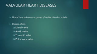
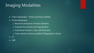
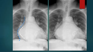
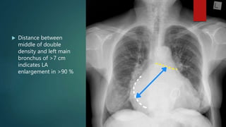
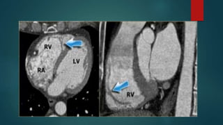
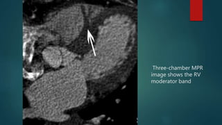
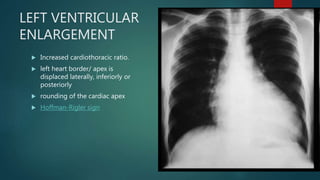
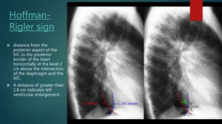
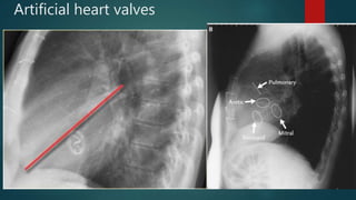
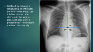
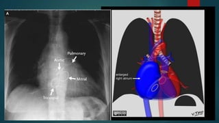
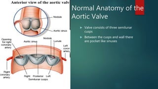
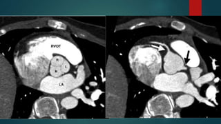
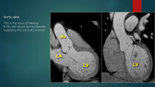
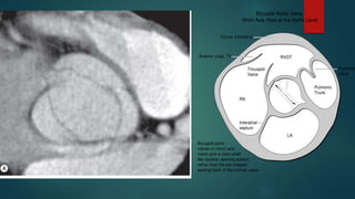
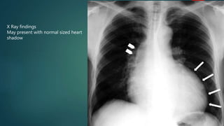
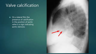
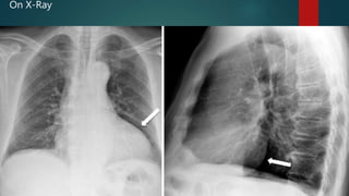
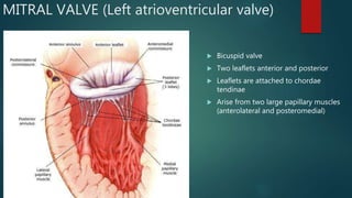
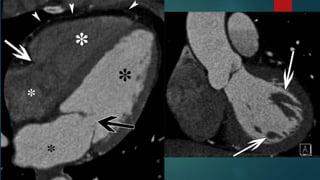
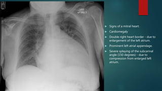
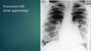
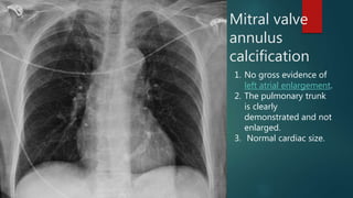
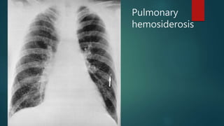
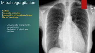
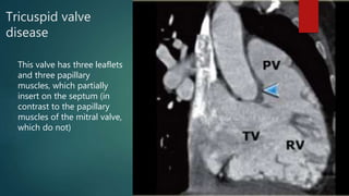
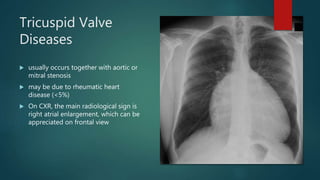
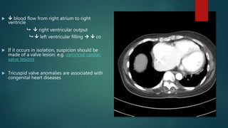
IMAGING OF VALVULAR
HEART DISEASES
DEVKANT LAKHERA
VALVULAR HEART DISEASES
 One of the most common groups of cardiac disorders in India
 Disease affects
 Mitral valve
 Aortic valve
 Tricuspid valve
 Pulmonary valve
Imaging Modalities
 Chest radiograph – Initial screening modality.
 Echocardiography
 Real time evaluation of heart chambers
 Evaluate the stenosis and regurgitation
 Ventricular function ( Size, wall thickness)
 Trans stenotic pressure gradient/ Regurgitant volume
 CT
 MRI
Objectives of Imaging
 Assessment of valves.
 Effects of disease on pulmonary vasculature and ventricular
function.
 Detection of associated pulmonary pathologies.
 Follow up
Assessing the chamber enlargement :
Left atrium
 Has smooth walls
 Receives pulmonary veins
 Forms most of the base of the
heart
LEFT ATRIUM
 At the level of 7th Thoracic
vertebra
Enlargement
 Splaying of carina.
 Double heart border/
Double density sign
 Posterior displacement of
esophagus
 Distance between
middle of double
density and left main
bronchus of >7 cm
indicates LA
enlargement in >90 %
Right atrium
 Forms the right heart
border
 Interior of right atrium is
separated by the crista
terminalis(smooth
muscular ridge)
 Space anterior to crista is
covered by ridges called
pectinate muscle
Crista
terminalis
 right atrium lies the crista
terminalis, a muscular ridge
that runs from the entrance
of the superior- to that of
the inferior vena cava.
 This structure separates the
smooth part of the right
atrium - the sinus venosus -
from the trabecularized
right atrium.
RIGHT ATRIAL
ENLARGEMENT
 Features are more subtle
 Elongation of the right
heart border
 Increased convexity of
right heart border
LEFT VENTRICLE
 Elliptical / Oval shape
 Wall thickness is >1 cm in mid
diastole. Thickest at the base.
 Endocardium is lined by fine
lattice like trabeculations
except in the basal half of iv
septum which is smooth.
 Papillary muscles attach to
free wall only and not septum.
RIGHT VENTRICLE
• Rhomboid in shape
• Papillary muscles attached to both free wall
and septum.
Three-chamber MPR
image shows the RV
moderator band
LEFT VENTRICULAR
ENLARGEMENT
 Increased cardiothoracic ratio.
 left heart border/ apex is
displaced laterally, inferiorly or
posteriorly
 rounding of the cardiac apex
 Hoffman-Rigler sign
Hoffman-
Rigler sign
 distance from the
posterior aspect of the
IVC to the posterior
border of the heart
horizontally at the level 2
cm above the intersection
of the diaphragm and the
IVC.
 A distance of greater than
1.8 cm indicates left
ventricular enlargement.
Right ventricular enlargement
 Frontal view demonstrates:
 rounded left heart border
 uplifted cardiac apex
 Lateral view
demonstrates:
 filling of the retrosternal space
Artificial heart valves
 localized by drawing a
longitudinal line through
the mid sternal body. Use
this line to bisect the
sternum in the sagittal
plane and then draw a
perpendicular line dividing
the heart horizontally.
Normal Anatomy of the
Aortic Valve
 Valve consists of three semilunar
cusps
 Between the cusps and wall there
are pocket like sinuses
Aortic valve
This is the sinus of Valsalva.
It fills with blood during diastole,
supplying the coronary arteries
Aortic Stenosis
Narrowing of the aortic valve
Main causes:
 Congenital bicuspid aortic
valve (younger)
 Degenerative Calcification of
aortic valve (Elder)
 Rheumatic inflammatory
fusion of aortic valve
Pathophysiology
Stiffening/Narrowing of Aortic
Valve
Incomplete emptying of
left atrium
Left ventricular hypertrophy
Pulmonary congestion
Compression of
coronary arteries
Right-sided heart failure
 CO
 Myocardial
O2 needs
Myocardial ischemia
(chest pain)
 O2 supply
X Ray findings
May present with normal sized heart
shadow
Valve calcification
 On a lateral film the
presence of calcification
in the position of aortic
valve is a sign indicating
aortic stenosis.
Aortic Regurgitation
 Congenital:
 Bicuspid valve or disproportionate cusps
 Acquired:
 Rheumatic disease
 Infective endocarditis
 Trauma
 Aortic dilatation (marfan’s syndrome, aneurysm, dissection,
syphilis)
Pathophysiology
Incomplete closure of the
aortic valve
Backflow of blood to Left
ventricle
Left ventricular hypertrophy
& dilation
 Left atrial pressure
Left-sided heart failure
(late stage)
Left atrium hypertrophy
 CO
 Pulmonary pressure
Right-sided heart failure  Right ventricular
pressure
On X-Ray
MITRAL VALVE (Left atrioventricular valve)
 Bicuspid valve
 Two leaflets anterior and posterior
 Leaflets are attached to chordae
tendinae
 Arise from two large papillary muscles
(anterolateral and posteromedial)
MITRAL STENOSIS
 Almost always rheumatic in
origin
 Older people: can be caused
by heavy calcification of
mitral valve congestion
 Congenital (rare)
Pathophysiology
Narrowing of mitral valve
 CO
O2/CO2 exchange
(fatigue, dyspnea,
orthopnea)
Left ventricular
atrophy
pulmonary
congestion
 pulmonary
pressure
 left atrial
pressure
Hypertrophy left
atrium
 blood flow to
left ventricle
Right-sided
failure
Fatigue
 Signs of a mitral heart:
 Cardiomegaly
 Double right heart border - due to
enlargement of the left atrium.
 Prominent left atrial appendage.
 Severe splaying of the subcarinal
angle (150 degrees) - due to
compression from enlarged left
atrium.
Prominent left
atrial appendage
Mitral valve
annulus
calcification
1. No gross evidence of
left atrial enlargement.
2. The pulmonary trunk
is clearly
demonstrated and not
enlarged.
3. Normal cardiac size.
Pulmonary
hemosiderosis
Mitral regurgitation
CAUSES
Congenital anomalies
Degenerative myxomatous changes
Marfan’s syndrome
Left ventricular enlargement is
more common.
Calcification of valve is less
common
Tricuspid valve
disease
This valve has three leaflets
and three papillary
muscles, which partially
insert on the septum (in
contrast to the papillary
muscles of the mitral valve,
which do not)
Tricuspid Valve
Diseases
 usually occurs together with aortic or
mitral stenosis
 may be due to rheumatic heart
disease (<5%)
 On CXR, the main radiological sign is
right atrial enlargement, which can be
appreciated on frontal view
  blood flow from right atrium to right
ventricle
  right ventricular output
  left ventricular filling   co
 If it occurs in isolation, suspicion should be
made of a valve lesion: e.g. carcinoid cardiac
valve lesions
 Tricuspid valve anomalies are associated with
congenital heart diseases
Pulmonary valve disease
 Very rare
 Occasionally seen in carcinoid disease and endocarditis

Valvular heart diseases imaging

  • 1.
    IMAGING OF VALVULAR HEARTDISEASES DEVKANT LAKHERA
  • 2.
    VALVULAR HEART DISEASES One of the most common groups of cardiac disorders in India  Disease affects  Mitral valve  Aortic valve  Tricuspid valve  Pulmonary valve
  • 3.
    Imaging Modalities  Chestradiograph – Initial screening modality.  Echocardiography  Real time evaluation of heart chambers  Evaluate the stenosis and regurgitation  Ventricular function ( Size, wall thickness)  Trans stenotic pressure gradient/ Regurgitant volume  CT  MRI
  • 4.
    Objectives of Imaging Assessment of valves.  Effects of disease on pulmonary vasculature and ventricular function.  Detection of associated pulmonary pathologies.  Follow up
  • 5.
    Assessing the chamberenlargement : Left atrium  Has smooth walls  Receives pulmonary veins  Forms most of the base of the heart
  • 6.
    LEFT ATRIUM  Atthe level of 7th Thoracic vertebra
  • 7.
    Enlargement  Splaying ofcarina.  Double heart border/ Double density sign
  • 9.
  • 10.
     Distance between middleof double density and left main bronchus of >7 cm indicates LA enlargement in >90 %
  • 11.
    Right atrium  Formsthe right heart border  Interior of right atrium is separated by the crista terminalis(smooth muscular ridge)  Space anterior to crista is covered by ridges called pectinate muscle
  • 12.
    Crista terminalis  right atriumlies the crista terminalis, a muscular ridge that runs from the entrance of the superior- to that of the inferior vena cava.  This structure separates the smooth part of the right atrium - the sinus venosus - from the trabecularized right atrium.
  • 13.
    RIGHT ATRIAL ENLARGEMENT  Featuresare more subtle  Elongation of the right heart border  Increased convexity of right heart border
  • 15.
    LEFT VENTRICLE  Elliptical/ Oval shape  Wall thickness is >1 cm in mid diastole. Thickest at the base.  Endocardium is lined by fine lattice like trabeculations except in the basal half of iv septum which is smooth.  Papillary muscles attach to free wall only and not septum.
  • 16.
    RIGHT VENTRICLE • Rhomboidin shape • Papillary muscles attached to both free wall and septum.
  • 18.
    Three-chamber MPR image showsthe RV moderator band
  • 19.
    LEFT VENTRICULAR ENLARGEMENT  Increasedcardiothoracic ratio.  left heart border/ apex is displaced laterally, inferiorly or posteriorly  rounding of the cardiac apex  Hoffman-Rigler sign
  • 20.
    Hoffman- Rigler sign  distancefrom the posterior aspect of the IVC to the posterior border of the heart horizontally at the level 2 cm above the intersection of the diaphragm and the IVC.  A distance of greater than 1.8 cm indicates left ventricular enlargement.
  • 21.
    Right ventricular enlargement Frontal view demonstrates:  rounded left heart border  uplifted cardiac apex
  • 22.
     Lateral view demonstrates: filling of the retrosternal space
  • 23.
  • 24.
     localized bydrawing a longitudinal line through the mid sternal body. Use this line to bisect the sternum in the sagittal plane and then draw a perpendicular line dividing the heart horizontally.
  • 26.
    Normal Anatomy ofthe Aortic Valve  Valve consists of three semilunar cusps  Between the cusps and wall there are pocket like sinuses
  • 28.
    Aortic valve This isthe sinus of Valsalva. It fills with blood during diastole, supplying the coronary arteries
  • 30.
    Aortic Stenosis Narrowing ofthe aortic valve Main causes:  Congenital bicuspid aortic valve (younger)  Degenerative Calcification of aortic valve (Elder)  Rheumatic inflammatory fusion of aortic valve
  • 31.
    Pathophysiology Stiffening/Narrowing of Aortic Valve Incompleteemptying of left atrium Left ventricular hypertrophy Pulmonary congestion Compression of coronary arteries Right-sided heart failure  CO  Myocardial O2 needs Myocardial ischemia (chest pain)  O2 supply
  • 32.
    X Ray findings Maypresent with normal sized heart shadow
  • 33.
    Valve calcification  Ona lateral film the presence of calcification in the position of aortic valve is a sign indicating aortic stenosis.
  • 34.
    Aortic Regurgitation  Congenital: Bicuspid valve or disproportionate cusps  Acquired:  Rheumatic disease  Infective endocarditis  Trauma  Aortic dilatation (marfan’s syndrome, aneurysm, dissection, syphilis)
  • 35.
    Pathophysiology Incomplete closure ofthe aortic valve Backflow of blood to Left ventricle Left ventricular hypertrophy & dilation  Left atrial pressure Left-sided heart failure (late stage) Left atrium hypertrophy  CO  Pulmonary pressure Right-sided heart failure  Right ventricular pressure
  • 36.
  • 37.
    MITRAL VALVE (Leftatrioventricular valve)  Bicuspid valve  Two leaflets anterior and posterior  Leaflets are attached to chordae tendinae  Arise from two large papillary muscles (anterolateral and posteromedial)
  • 39.
    MITRAL STENOSIS  Almostalways rheumatic in origin  Older people: can be caused by heavy calcification of mitral valve congestion  Congenital (rare)
  • 40.
    Pathophysiology Narrowing of mitralvalve  CO O2/CO2 exchange (fatigue, dyspnea, orthopnea) Left ventricular atrophy pulmonary congestion  pulmonary pressure  left atrial pressure Hypertrophy left atrium  blood flow to left ventricle Right-sided failure Fatigue
  • 41.
     Signs ofa mitral heart:  Cardiomegaly  Double right heart border - due to enlargement of the left atrium.  Prominent left atrial appendage.  Severe splaying of the subcarinal angle (150 degrees) - due to compression from enlarged left atrium.
  • 42.
  • 43.
    Mitral valve annulus calcification 1. Nogross evidence of left atrial enlargement. 2. The pulmonary trunk is clearly demonstrated and not enlarged. 3. Normal cardiac size.
  • 44.
  • 45.
    Mitral regurgitation CAUSES Congenital anomalies Degenerativemyxomatous changes Marfan’s syndrome Left ventricular enlargement is more common. Calcification of valve is less common
  • 46.
    Tricuspid valve disease This valvehas three leaflets and three papillary muscles, which partially insert on the septum (in contrast to the papillary muscles of the mitral valve, which do not)
  • 47.
    Tricuspid Valve Diseases  usuallyoccurs together with aortic or mitral stenosis  may be due to rheumatic heart disease (<5%)  On CXR, the main radiological sign is right atrial enlargement, which can be appreciated on frontal view
  • 48.
      bloodflow from right atrium to right ventricle   right ventricular output   left ventricular filling   co  If it occurs in isolation, suspicion should be made of a valve lesion: e.g. carcinoid cardiac valve lesions  Tricuspid valve anomalies are associated with congenital heart diseases
  • 49.
    Pulmonary valve disease Very rare  Occasionally seen in carcinoid disease and endocarditis

Editor's Notes

  • #3 Mainly due to the prevalence of rheumatic heart disease in India. Disease most commonly involves
  • #4 Provides information about cardiac size pulmonary vasculature, arterial and venous hypertension, secondary changes in lungs.. Real time evaluation in multiple planes,…valvular calcification CECT can help assess chamber and valves..Ecg gated CT can help in reconstructing cardiac motion
  • #6 shape of an irregular ellipsoid, with the exception of the right atrial appendage, which arises anteriorly
  • #7 Most superiorly located chamber Lies posteriorly in the midline and enlarges posterior and to the right(not anteriorly) Normally left atrium does not form any cardiac border on PA view.. Lt atrial appendage is a finger like / tubular structure has a pointed end and is trabeculated. Originates supralaterally….. trapezoidal shape of the right atrial appendage
  • #8 Stenotomy sutures and prosthetic valve implant are seen. The cardiac shadow is enlarged. A double right heart border is noted. Prominence of pulmonary vasculature in the upper zones which indicates cephalization. Widening of carinal angle >90 degrees aortic knuckle calcification is seen ...Prominence of mid part of left heart border suggesting enlarged left atrial appendage.
  • #9 White line enlarged left atrium Atria may be massively enlarged crossing the right atrial shadow k/a Atrial escape
  • #10 LA is located just anterior to the mid part of thoracic oesopagus, Enlarges posteriorly. Barium Swallow
  • #12 Recieves the SVC, IVC and the Coronary sinus
  • #14 Distance between right cardiac border convexity and the right lateral vertebral border > 3cms
  • #15  known history of tricuspid regurgitation.
  • #16 Has high pressure function Most muscular chamber conical in shape with an anteroinferiorly projecting apex and is longer with thicker walls than the right ventricle  Internally, there are smooth inflow and outflow tracks and the remainder of the left ventricle (mainly apical) is lined by fine trabeculae carnae.
  • #18 The right ventricle also has a thinner wall which is more trabeculated, especially towards the apex.. moderator band is another distinguishing feature of the right ventricle. It runs from the septum to the lateral wall of the right ventricle, it caries the conducting system of the heart Right bundle branch
  • #19 Multiplaner reconstructon(2d post processing techniques) re-formats images at different planes, defined by the operator, using the pixel data from a stack of planar images (base images). The digital value for each pixel is assigned to a virtual voxel with the same thickness as the slice thickness of the base image. Airways , pulmonary emboli
  • #23 Cardiomegaly with a lifted up cardiac apex.Notice that it is especially the right ventricle that is dilated. This is well seen on the lateral film (yellow arrow). There is a small aortic knob (blue arrow), while the pulmonary trunk and the right lower pulmonary artery are dilated. All these findings are probably the result of a left-to-right shunt with subsequent development of pulmonary hypertension.
  • #24 The location of the cardiac valves is best determined on the lateral radiograph. A line is drawn on the lateral radiograph from the carina to the cardiac apex. The pulmonic and aortic valves generally sit above this line and the tricuspid and mitral valves sit below this line
  • #26 Pulmonary valve is more horizontal others are more oblique..mitral and tricuspid valve are usually seen as rings a line drawn through the valve forms more acute angle with the base of heart..anterior and posterior borders of the aortic valve are superimposed on pa view
  • #27 Edge of these cusps are projecting into the aa, Right left and posterior (also known a non coronary cusp) Blood fills these aortic sinuses and fills the coronaries
  • #28 oblique CT image at the level of the aortic valve demonstrates right (R), left (L), and noncoronary (N) cusps CT image obtained at a slightly more cranial level shows the origins of the right (white arrow) and left (black arrow) main coronary arteries
  • #30 Cardiac CT showing a bicuspid aortic valve ..only two semilunar valves are visible
  • #32 CO fails to meet the demands of heart: syncopy dec blood to brain, angina due to increased demand to lv and dec co, and exertional dysnoea later stages
  • #33 Chest X-ray of aortic stenosis shows rounded profile of left ventricle), with slight enlargement of ascending aorta due to the stenotic jet of blood flow having a pressure effect
  • #37 Frontal and lateral views demonstrate left ventricle enlargement, lateral displacement of apex, as left third cardiac arch widening in the frontal view, and second posterior arch in the lateral view
  • #38 Mitral valve is surrounded by annulus (fibrous ring around mitral leaflets) helps in proper closure of valve.
  • #39 Horizontal long-axis MPR image shows the LV (large black *), RV (large white *), LA (small black *), right atrium (RA) (small white *), MV (black arrow), tricuspid valve (white arrow), and pericardium (arrowheads). The latter structure is normally very thin. Three-chamber MPR image shows the LV papillary muscles (arrow) and chordae tendineae (arrowheads).
  • #40 Very prevalent in developing countries
  • #41 Left atrium is filled with blood…Dysnoea, cough (pulmonary congestion), fatigue ,oedema
  • #43 Rheumatic mitral stenosis. This frontal film shows marked enlargement of the left atrial appendage
  • #44 band of coarse calcification projected over the expected location of the mitral valve, to the left of midline.. mitral annulus is not significantly associated with stenosis of the valve 
  • #45 Pulmonary hemosiderosis due to long-standing mitral stenosis show diffuse small, rounded, “miliary” nodular areas of increased opacity bilaterally 1-4 mm(t.b histo,sarco,silicosis,vp)
  • #46 Myxomatous is pathological weakening of connective tissue
  • #47 crista supraventricularis a thick muscle separates pulmonary valve, from the tricuspid valve by known as the (blue arrow