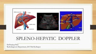
This document provides an overview of Doppler ultrasound of the normal portal system. It discusses adjusting spectral Doppler settings and interpreting normal and abnormal findings. Key points include:
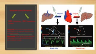
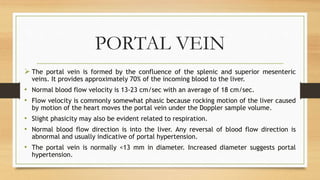
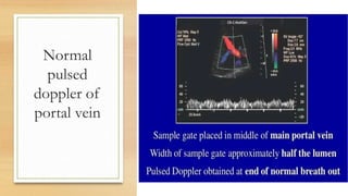
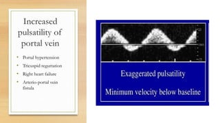
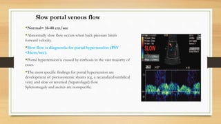
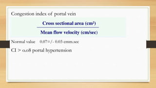
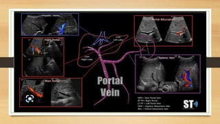
- Normal Doppler findings of the portal vein include a velocity of 13-23 cm/sec and antegrade flow. Increased diameter or retrograde flow indicate portal hypertension.
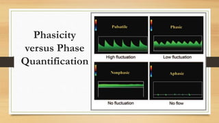
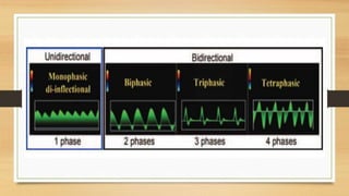
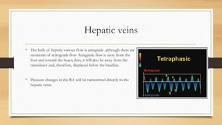
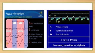
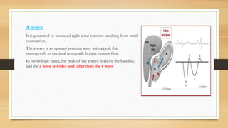
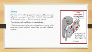
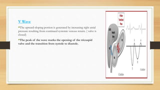
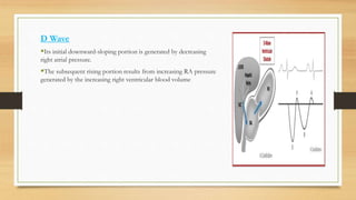
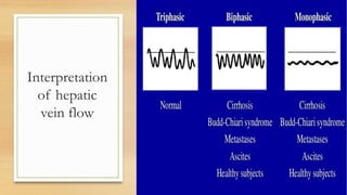
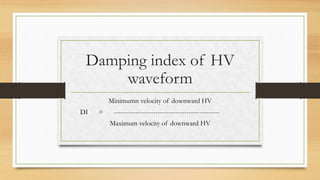
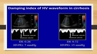
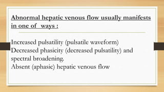
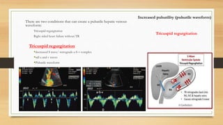
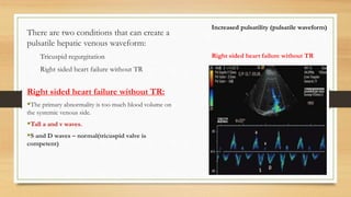
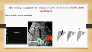
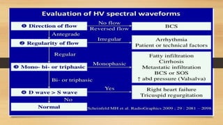
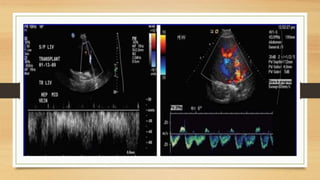
- Hepatic vein Doppler shows a triphasic waveform. Increased pulsatility can be seen with tricuspid regurgitation or right heart failure. Loss of phasicity indicates outflow obstruction.
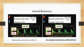
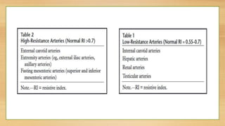
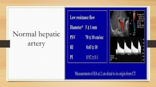
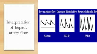
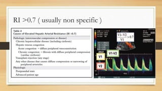
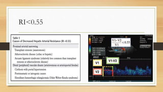
- The hepatic artery has a resistive index over 0.7 normally. Values under 0.55 may indicate stenosis.
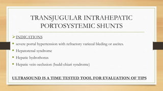
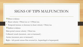
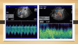
- Transjugular intrahepatic portos
This document discusses renal Doppler ultrasound techniques. It describes three main approaches to imaging the renal arteries - anterior, oblique, and flank. The oblique approach is optimal for Doppler assessment as it allows an angle of 60 degrees. Indirect evaluation of segmental arteries can detect renal artery stenosis seen as absent early systolic peaks or tardus parvus waveforms. The document also reviews renal transplant pathologies like rejection seen as edema, infarction seen as perfusion defects, and arteriovenous fistula seen as arterialized vein flow.
This document discusses the Doppler ultrasound assessment of the portal venous system. It begins with an overview of the sonographic and Doppler evaluation of the portal system's anatomy and normal circulation. It then covers the assessment of specific vessels like the portal vein, hepatic veins, and hepatic artery through grayscale ultrasound and Doppler evaluation. Key aspects like normal vessel diameters, waveforms, and flow direction are defined. The document concludes by outlining the important Doppler assessment techniques and parameters used to evaluate the portal system.
This document discusses renal Doppler ultrasound techniques and findings. It describes three main approaches to imaging the renal arteries - anterior, oblique, and flank. Normal and abnormal Doppler waveforms are presented. Evaluation of renal artery stenosis can be done directly by imaging the renal arteries or indirectly by imaging intrarenal arteries. Findings suggestive of stenosis include increased velocities, renal/aortic ratios over 3.5, absence of the early systolic peak, and tardus parvus waveforms. Pathologies of renal transplants like rejection, infarction, and arterial or venous stenosis are also summarized.
This document discusses Doppler ultrasonography and the analysis of blood flow patterns. It contains the following key points:
1. Doppler spectroscopy provides a time-velocity waveform representing blood flow velocities during the cardiac cycle, showing how velocity and the number of reflecting red blood cells varies over time.
2. Normal vessels have characteristic flow patterns depending on their anatomy and the organ they supply. Widening of the spectral line and filling of the spectral window indicates high flow or small vessels.
3. Pseudoaneurysms are caused by a complete rupture of the artery wall. Doppler can detect the communicating channel showing a "to-and-fro" waveform as blood enters and leaves the pooling cavity.
4.
This document provides information on renal artery anatomy and Doppler ultrasound evaluation of the renal arteries. It describes:
1. The typical origin and course of the right and left renal arteries. Approximately 30% of individuals have variant anatomy with more than one renal artery on each side.
2. How Doppler ultrasound is used to image the renal arteries from different approaches and measure parameters like peak systolic velocity to evaluate for renal artery stenosis.
3. The normal Doppler waveforms expected in the main renal artery and intrarenal arteries, as well as normal values for measured parameters.
4. How a bilateral renal Doppler examination is performed, including evaluating each kidney, the renal arteries and veins, and measuring parameters to identify
This document discusses Doppler ultrasound of the kidneys. It begins with the normal anatomy of the kidneys and renal vasculature. It then describes how to perform grayscale and Doppler ultrasound of the kidneys, including imaging planes and settings. Normal Doppler waveforms of renal arteries are presented. Key measurements like resistive index, acceleration time, and peak systolic velocity of renal arteries are discussed. Variants of renal and renal vein anatomy are also reviewed.
This document discusses non-invasive imaging techniques for evaluating portal hypertension. Ultrasound is the most widely used initial imaging modality to assess for portal vein patency, presence of collaterals, portal blood flow, and changes of cirrhosis. CT and MRI can further evaluate the portal venous system and collateral vessels. Ultrasound Doppler is used to measure portal blood flow direction, velocity, and assess for portosystemic collaterals and changes in hepatic veins. Imaging findings of various etiologies of portal hypertension are described, including cirrhosis, non-cirrhotic portal fibrosis, portal vein thrombosis, Budd-Chiari syndrome, and extrahepatic causes.
Role of Doppler in Liver Cirrhosis & Portal Hypertensionnishit viradia
Doppler ultrasound is useful for assessing portal hypertension and liver cirrhosis. Key findings include increased portal vein diameter (>13mm), decreased increase in splenic or portal vein diameter with respiration, reversed or biphasic portal flow, increased hepatic artery flow and resistive index, altered hepatic vein waveforms, splenomegaly (>13cm), and presence of portosystemic collateral veins. Together these Doppler ultrasound metrics can diagnose and characterize portal hypertension noninvasively.
Doppler ultrasound of carotid arteriesSamir Haffar
This document discusses Doppler ultrasound of carotid arteries. It begins with the anatomy of carotid arteries and then discusses normal Doppler ultrasound findings of the carotid arteries including flow patterns and spectral waveforms. It describes various pathologies that can cause carotid artery disease such as atherosclerosis and other non-atherosclerotic diseases. It also discusses how diseases outside the carotid arteries can affect them. The document provides detailed information on ultrasound techniques for evaluating the carotid arteries and interpreting ultrasound findings for plaque characterization and grading stenosis.
Ultrasound & doppler ultrasound in liver transplantationSamir Haffar
1) Doppler ultrasound is useful for detecting vascular complications following liver transplantation such as hepatic artery thrombosis, stenosis, and pseudoaneurysm as well as portal vein stenosis and thrombosis.
2) Reversible Doppler findings in the immediate postoperative period include elevated hepatic artery velocities and pulsatile portal vein flow that typically resolve within a few days.
3) Biliary cast syndrome is a rare but serious complication characterized by hard casts within the biliary ducts that can cause strictures, dilatation, and abscesses, often requiring surgery or endoscopic removal. Doppler ultrasound may demonstrate associated hepatic artery stenosis.
The liver is the largest solid organ located in the right upper quadrant of the abdomen. It is divided into eight segments based on vascular and biliary anatomy. The document describes the normal anatomy of the liver and common variations. It also discusses ultrasound techniques for imaging the liver and provides details on identifying different liver lesions including cysts, benign and malignant tumors, infections, and vascular anomalies on ultrasound scans.
This document provides information about lower limb venous Doppler ultrasound techniques and findings. It begins with an overview of venous anatomy of the lower limbs. Key points about performing a lower limb venous Doppler exam are provided, including the importance of understanding anatomy, obtaining a thorough patient history, and focusing on Doppler waveforms and symmetry between limbs. Common venous conditions like deep vein thrombosis and varicose veins are also summarized. The document concludes with techniques for performing lower limb venous Doppler ultrasound exams.
This document discusses the use of radiology in evaluating and managing portal hypertension. It begins by defining portal hypertension and describing how ultrasound can be used to diagnose it by measuring portal vein pressure and blood flow. Specific ultrasound findings that indicate portal hypertension are described, including enlarged portal veins, decreased flow, and the presence of collateral blood vessels. The document then discusses how computed tomography and magnetic resonance imaging can further evaluate the portal venous system and collateral vessels. It concludes by covering interventional radiology procedures like TIPS and variceal embolization that can treat portal hypertension by decompressing the portal vein or controlling its complications.
Doppler ultrasound of lower limb arteriesSamir Haffar
This document provides information on Doppler ultrasound of lower limb arteries. It begins with the anatomy of lower limb arteries including the abdominal aorta, iliac arteries, femoral arteries, and crural arteries. It then discusses normal Doppler ultrasound findings of lower limb arteries including normal arterial diameters, waveforms, and velocities. Finally, it covers duplex ultrasound criteria for arterial evaluation and various causes of lower limb arterial diseases such as atherosclerosis, thrombosis, aneurysms, and arterial occlusions.
This document provides information on performing and interpreting renal Doppler ultrasounds. It discusses the optimal approaches for imaging the renal arteries, including the anterior, oblique, and flank approaches. It also outlines criteria for evaluating renal artery stenosis, including peak systolic velocity measurements and the renal-aortic ratio. Common renal pathologies that can be identified with Doppler ultrasound are also summarized, such as fibromuscular dysplasia, atherosclerosis, aneurysms, and hydronephrosis.
The document discusses the anatomy and sonographic appearance of the pancreas. It describes the pancreas' location and relationships to nearby structures like blood vessels. The normal sonographic features include homogeneous echotexture and absence of duct dilation. Common pathologies like pancreatic cancer and pancreatitis are also summarized, noting how they can appear on ultrasound with features like duct obstruction or diffuse swelling. Ultrasound is established as a useful initial imaging method for evaluating the pancreas.
Portal hypertension radiological diagnosis and interventionsSourav Talukder
This document discusses portal hypertension, including its causes, diagnosis, and interventions. It describes how portal hypertension can be diagnosed using ultrasound, CT, MRI, and angiography by evaluating the portal vein diameter and flow, presence of collateral vessels, and hepatic and arterial changes. Interventions to treat portal hypertension aim to reduce portal blood pressure, such as transjugular intrahepatic portosystemic shunt (TIPS) placement, or palliate symptoms like variceal embolization. Recanalization of portal veins and revision of shunts are also discussed.
Imaging of kidny i htn by dr.abd alla shady mdFarragBahbah
Renovascular hypertension is a common cause of secondary hypertension that results from renal artery stenosis. It can be diagnosed through imaging tests like Doppler ultrasound, MRI/CT angiography, and renal scintigraphy. These tests identify anatomical narrowing of the renal arteries and assess renal perfusion and function before and after administration of drugs that stimulate the renin-angiotensin system. Percutaneous angioplasty and stenting are endovascular procedures used to treat renovascular hypertension in select patients with refractory or progressive hypertension related to renal artery stenosis. The choice of imaging test depends on factors like renal function and bilateral versus unilateral involvement to identify and characterize renal artery stenosis safely and effectively.
This document discusses Doppler ultrasound of the kidneys. It begins by describing the normal anatomy of the kidneys and renal vasculature. It then discusses how to perform grayscale and Doppler ultrasound exams of the kidneys, including identifying normal anatomical variants. Technical parameters for optimizing Doppler signals are provided. Normal Doppler waveform indices for the renal arteries are defined, including peak systolic velocity, resistive index, acceleration time and acceleration index.
Doppler ultrasound of the portal system - Normal findingsSamir Haffar
This document provides an overview of Doppler ultrasound of the normal portal system, including:
1. Principles of Doppler ultrasound and how to adjust settings like color box size, velocity scale, gain, and wall filter to optimize the examination.
2. Sites for duplex insonation of the portal system and techniques for obtaining spectral waveforms.
3. Normal Doppler ultrasound findings of the portal vein, hepatic veins, and hepatic artery, including measurements and anatomy.
Ultrasound of the urinary tract - Renal infectionsSamir Haffar
Ultrasound can detect various renal infections including:
1) Acute pyelonephritis seen as renal enlargement, decreased echogenicity, and loss of corticomedullary differentiation on ultrasound.
2) Renal abscesses appear as hypoechoic masses with thick irregular walls that increase in distinctness over time.
3) Pyohydronephrosis is infection of the obstructed collecting system seen as echogenic debris and fluid-fluid levels.
4) Emphysematous pyelonephritis involves gas in the renal parenchyma seen as high amplitude echoes and dirty shadowing.
1) Carotid Doppler ultrasound is used to evaluate the carotid arteries for stenosis or occlusion. It involves using grayscale, color Doppler, and spectral Doppler to examine the anatomy and flow of the carotid arteries.
2) A normal carotid Doppler ultrasound will show triphasic waveforms within the carotid arteries with velocities under 125 cm/sec. The intima-media thickness should be less than 0.8-0.9mm.
3) Carotid artery disease is most commonly caused by atherosclerosis which can be evaluated using Doppler ultrasound parameters like peak systolic velocity, end diastolic velocity, and ICA/CCA velocity ratios to grade the severity of stenosis.
Varicose veins are dilated, elongated superficial veins caused by venous hypertension. They commonly occur in the legs but can affect other areas. The venous system of the lower limb consists of deep and superficial veins that connect via perforating veins. The long saphenous vein originates in the foot and travels up the medial leg and thigh, connecting to the femoral vein. Short saphenous vein originates laterally and connects to the popliteal vein. Valves in the veins normally prevent backflow but can become incompetent, allowing reflux and varicose vein formation. Color duplex ultrasound can evaluate reflux patterns and valve competence.
Doppler ultrasound of normal venous flowSamir Haffar
This document discusses Doppler ultrasound findings of normal venous flow. It notes that normal venous flow is spontaneous without augmentation, changes with respiration, and veins compress fully in the transverse plane. It also describes augmented flow seen with distal compression and abdominal/pelvic vein patency seen during the Valsalva maneuver. The document provides images demonstrating normal venous flow patterns and measurements.
This document provides an overview of performing lower limb doppler examinations to diagnose deep vein thrombosis and other causes of limb pain. It discusses the essential techniques including recognizing the vessels, avoiding pitfalls, applying compression, and following the anatomy. Criteria for diagnosing DVT include vessel expansion, compressibility, presence of thrombus, and absent or reduced blood flow waves. The document also reviews using doppler to diagnose and grade arterial stenosis by analyzing spectral wave patterns and meanings.
This document provides an overview of Doppler ultrasound applications for evaluating various vascular structures and conditions. It discusses Doppler evaluation of the carotid arteries, renal arteries, portal vein, hepatic veins, and arteriovenous fistulas. Key points include diagnostic criteria for stenosis of the carotid, renal and hepatic arteries as well as portal hypertension. Imaging findings and waveforms related to conditions such as subclavian steal, Budd-Chiari syndrome, and arteriovenous fistula maturation/complications are also reviewed. The document emphasizes a thorough ultrasound technique and understanding of normal versus abnormal vascular Doppler evaluations.
This document discusses hepatic Doppler ultrasound waveforms. It defines key terms like antegrade and retrograde flow. The normal hepatic artery waveform is pulsatile with low resistance. The normal hepatic vein waveform is biphasic or tetraphasic. Abnormal portal vein waveforms can be pulsatile, demonstrate slow flow, show retrograde flow, or have absent flow. The document provides detailed descriptions of hepatic artery, hepatic vein, and portal vein waveforms both normal and abnormal.
The document discusses hepatic Doppler ultrasound waveforms. It defines key terms like antegrade and retrograde flow. It describes the normal waveforms seen in the hepatic artery, hepatic veins and portal vein. The hepatic artery normally shows low resistance flow. The hepatic veins show a biphasic or tetraphasic waveform. Increased pulsatility can indicate abnormal portal vein flow. The document provides detailed explanations of waveform features and their clinical significance.
Portal hypertension occurs when pressure in the portal venous system rises, usually due to liver cirrhosis or scarring impeding blood flow into the liver. Ultrasound can detect signs of portal hypertension like ascites, splenomegaly, dilated portal veins and collateral vessels that form around the liver. Specific findings include thrombosed or narrowed portal veins, enlarged arteries supplying the liver, and reversed blood flow away from the liver.
This document discusses normal and pathological findings on Doppler ultrasound of the liver vasculature. It begins with descriptions of normal portal venous circulation and measurements. It then covers pathological findings in portal hypertension, including dilated portal vein diameter and decreased mean velocity. Specific portosystemic collaterals are described along with their Doppler findings. Hepatic vein and artery waveforms and measurements in cirrhosis are also summarized.
This document provides information on portal hypertension, including:
1. It defines portal hypertension and describes types such as cirrhotic and non-cirrhotic portal hypertension.
2. It outlines the portal venous system and portosystemic circulation.
3. It discusses causes, clinical features, investigations, and management of portal hypertension including pharmacotherapy, endoscopic therapy, TIPS procedure, and surgeries.
4. Prevention of recurrent variceal hemorrhage is highlighted through long-term pharmacotherapy, endoscopic therapy, interventional procedures like TIPS, or surgical shunts if other options fail.
This document summarizes ultrasound findings for cirrhosis of the liver. Key findings include coarse echotexture and nodular surface of the liver. Regenerating nodules appear isoechoic or hypoechoic. Portal hypertension is a complication and is suggested by Doppler findings like dilated portal vein and low venous velocity. Findings of portal hypertension include splenomegaly, ascites, and portosystemic venous collaterals visible with ultrasound.
This document summarizes ultrasound findings for cirrhosis of the liver. Key findings include coarse echotexture and nodular surface of the liver. Regenerating nodules appear isoechoic or hypoechoic. Portal hypertension is a complication and is evidenced by dilated portal veins, low portal flow, and portosystemic collaterals visible on ultrasound. Doppler ultrasound can detect changes in portal flow from normal hepatopetal to reversed hepatofugal flow in advanced cases.
This document provides information on various cyanotic heart lesions including Tetralogy of Fallot, Transposition of the Great Arteries, Tricuspid Atresia, Ebstein's Anomaly and Total Anomalous Pulmonary Venous Return. It describes the anatomy, clinical features, diagnosis and management of these conditions. Key cyanotic heart lesions are characterized by mixing of oxygenated and deoxygenated blood resulting in central cyanosis.
Jugular venous pressure (JVP) refers to the height of blood in the jugular veins and provides information about right heart function. An elevated JVP indicates issues such as right heart failure, tricuspid valve problems, superior vena cava obstruction, or fluid overload. The JVP is assessed by looking for visibility, double waves, changes with inspiration, and hepatojugular reflux. Conditions that limit right ventricular filling can also raise JVP.
PORTAL HYPERTENSION by Dr.Abhijeet Majhi.pptxAbhijeet Majhi
The portal vein drains blood from the intestines and organs into the liver. Portal hypertension is defined as increased pressure in the portal vein above normal levels. It can be caused by obstruction anywhere along the portal vein system or within the liver. Complications include gastrointestinal bleeding, ascites, and liver dysfunction. Treatment involves medication to reduce pressure, endoscopic procedures to treat varices, surgical shunts, and liver transplantation in severe cases.
This document discusses fetal circulation and several congenital heart defects that can occur, including their pathophysiology, clinical presentation, and treatment. It begins by describing the normal fetal circulation, where oxygenated blood from the placenta travels through the umbilical vein, ductus venosus, and foramen ovale to supply the fetus. It then covers several defects: atrial septal defect, ventricular septal defect, patent ductus arteriosus, pulmonary stenosis, aortic stenosis, and coarctation of the aorta. For each, it provides an overview of how blood flow is disrupted, typical signs and symptoms, and treatment approaches like surgery, catheterization, or medication management.
Swan-Ganz catheters are balloon-tipped catheters inserted into the heart to measure pressures and collect blood samples from the right atrium, right ventricle, and pulmonary artery. This allows clinicians to assess conditions like shock, respiratory distress, and complications of myocardial infarction. Measurements of pressures, oxygen saturations, and cardiac output can guide therapy for critical illnesses and help evaluate the effects of treatments. While useful for management, the procedure does carry risks of complications if not performed carefully.
The portal vein is formed by the superior mesenteric and splenic veins. It drains blood from the intestines, pancreas, colon and rectum. Blood from the portal and hepatic arteries mixes in the liver sinusoids before draining into the hepatic vein and inferior vena cava. Portal hypertension occurs when pressure increases due to increased resistance in the liver from cirrhosis and increased blood flow. It can cause ascites, variceal bleeding, and other complications.
The document summarizes renal artery stenosis, including its normal anatomy, causes, presentations, diagnostic tests, and treatment. It describes how renal artery stenosis can cause secondary hypertension and deteriorating renal function by narrowing the renal arteries. Diagnostic tests discussed include intravenous urography, captopril scintigraphy, ultrasound Doppler criteria, CTA/MRA, and angiography. Treatment may involve percutaneous angioplasty with or without stent placement to reopen narrowed arteries.
A 22-year-old male presented with complaints of easy fatigability, abdominal discomfort, leg swelling, and shortness of breath with exertion over the past 6-8 months. Examination found edema, elevated jugular venous pressure, hepatomegaly, and cachexia. Testing showed elevated liver enzymes and BNP. Echocardiogram demonstrated thickened pericardium with ventricular interdependence and equal diastolic pressures, consistent with constrictive pericarditis. The patient was diagnosed with constrictive pericarditis likely due to a prior unknown infection or inflammatory process causing thickening and scarring of the pericardium.
Cirrhosis is a diffuse process characterized by liver necrosis and fibrosis that converts the normal liver architecture into abnormal nodules lacking normal structure. It has many causes including viral hepatitis, alcohol, autoimmune conditions, and genetic disorders. Complications of cirrhosis arise from portal hypertension and liver insufficiency, leading to variceal bleeding, ascites, encephalopathy, and jaundice. Cirrhosis is diagnosed based on clinical signs, lab tests, imaging, and may involve liver biopsy. Management focuses on treating complications and screening for hepatocellular carcinoma.
Mammography -A ppt bt J K PATIL, Prof,dept of radiologydypradio
Mammography uses low-dose x-rays to image the breast and detect cancers. Key aspects include using a molybdenum target and filter to produce low-energy x-rays for high soft tissue contrast. Breast compression is important to reduce scatter and motion, separate tissues, and highlight rigid masses. Views include craniocaudal and mediolateral oblique to image the entire breast. Digital mammography systems like computed radiography and direct detectors directly convert x-rays to digital images, improving contrast and allowing post-processing.
Ultrasound Features of Fetal Syndromes Part 1.pptxdypradio
This document lists various fetal syndromes that can be identified on ultrasound and describes their key features. It discusses fetal infections caused by cytomegalovirus, parvovirus B19, rubella virus, syphilis, toxoplasmosis, and varicella zoster virus. For each infection, it provides information on etiology, diagnosis, associated anomalies, and differential diagnosis. Teratogens such as alcohol and medications that can cause fetal syndromes are also mentioned. Numerous structural abnormalities involving the central nervous system, cranium, musculoskeletal system, and other organ systems are detailed. Conditions of fetal overgrowth, growth restriction, and microdeletion syndromes conclude the document.
This document discusses various fetal abnormalities that can be detected on prenatal ultrasound. It describes ectopic and pelvic kidneys, prune belly syndrome, megacystis microcolon intestinal hypoperistalsis syndrome, adrenal glands, neuroblastoma, bladder abnormalities like extrophy and non-visualization, genital abnormalities like hypospadias, congenital adrenal hyperplasia, ovarian cysts, and hydrocolpos/hydrometrocolpos. Differential diagnoses and key sonographic findings for each condition are provided.
This document discusses aortic angiography, a procedure that uses dye and x-rays to examine the aorta and detect any abnormalities. It begins by listing several congenital anomalies of the aorta that can be identified. It then describes how the test is performed, including catheter insertion and dye injection. Potential findings that could indicate problems are outlined, along with risks of the procedure. The document explains that aortic angiography may be recommended if symptoms suggest issues with the aorta.
This document provides an overview of evaluating the fetal central nervous system (CNS) using ultrasound. It describes the normal ultrasound appearance of the cerebral mantle, ventricles, and basal cisterns. It outlines various ultrasound planes and sections used to examine the fetal brain, including measurements of structures like the intracranial translucency and ventricles. The document discusses visualization of sulcation and various sulci as the fetus develops. It also covers evaluation of the spine, identification of neural tube defects, ventriculomegaly, and midline anomalies like holoprosencephaly.
This document provides an overview of carotid artery ultrasound evaluation. It describes the normal anatomy of the carotid arteries and their branches. The protocol for a carotid ultrasound examination is outlined, including patient positioning, transducer selection, scanning sequences, and evaluation of stenosis. Key anatomical structures are defined, such as the intima-media complex. Non-atherosclerotic diseases that can involve the carotid or vertebral arteries, such as fibromuscular dysplasia, dissection, vasospasm, and aneurysms are also reviewed. The limitations of carotid ultrasound are noted.
This document discusses different types of Aspergillus infections including allergic bronchopulmonary aspergillosis (ABPA), semi-invasive aspergillosis, and invasive pulmonary aspergillosis. ABPA is an allergic reaction seen in patients with asthma or cystic fibrosis and results in bronchial wall damage and bronchiectasis. Semi-invasive aspergillosis typically occurs in patients with mild immunosuppression and results in thick-walled cavities in the lungs. Invasive pulmonary aspergillosis is seen in severely immunocompromised patients like those with leukemia and causes multiple or single ill-defined lung opacities or consolidations.
evaluation of fetal anatomy in 1st trimester.pptxdypradio
EVALUATION OF FETAL ANATOMY IN FIRST TRIMESTER .
FETAL DEVELOPMENT IN FIRST YAER.
NORMAL ULTRASOUND FINDINGS IN THE FIRST TRIMESTER.Evaluation of fetal anatomy, including a detailed fetal cardiac examination, is possible in the late first trimester.
Many anatomic abnormalities can be detected in the first trimester, giving families time to make important decisions regarding pregnancy management and the opportunity for early termination of pregnancy to reduce maternal morbidity risks.Week 6: By the 6th week, the limb buds begin to differentiate into upper and lower limbs with large hand plates, which develop primordial digits. The lower extremities lag behind the upper limbs by approximately 4 to 5 days. The primordial ear develops and the eyes become obvious as the retina becomes pigmented. The fetal liver occupies the majority of the abdominal cavity at the 6th week. As the rapid growth of the intestines exceeds the growth of the abdominal cavity the physiologic herniation of the intestines into the umbilical cord occurs. Spontaneous twitching movements and reflex responses to touch begin to take place.
ULTRASOUND EVALUATION OF ANEUPLOIDY IN FIRST AND SECOND TRIMESTERdypradio
This document discusses ultrasound evaluation methods for detecting fetal aneuploidy in the first and second trimesters. It describes various ultrasound markers that can be used, including nuchal translucency measurement, absent nasal bone, ductus venosus Doppler, cystic hygroma, tricuspid regurgitation, and frontomaxillary facial angle in the first trimester. In the second trimester, it discusses markers like thickened nuchal fold, absent/hypoplastic nasal bone, and hyperechoic bowel that can be assessed as part of a genetic sonogram to adjust risk for aneuploidy. The presence of these minor ultrasound findings in addition to biochemical screening can improve detection
This document discusses various chromosomal anomalies including numeric and structural abnormalities of autosomes and sex chromosomes. It describes common trisomies like Down syndrome, Edward syndrome, and Patau syndrome. It also discusses sex chromosome anomalies such as Turner syndrome, Klinefelter syndrome, and Jacob's syndrome. The physical traits, clinical findings, antenatal ultrasound markers, and radiological features of each condition are provided.
This document discusses the arterial supply, venous drainage, and normal and abnormal ultrasound findings of the scrotum and testes. It describes the arterial supply from the testicular artery and other branches. The venous drainage is via the pampiniform plexus draining into the spermatic veins. Normal color Doppler shows low resistance flow in the testes. Common pathologies discussed include epididymitis, varicocele, testicular torsion, cysts and tumors. Features on ultrasound and Doppler help differentiate these conditions.
Diffusion MRI measures the random movement of water molecules in tissues using diffusion gradients. There are two main types of diffusion - isotropic, which is equal in all directions, and anisotropic, which differs with direction. Diffusion weighted imaging uses isotropic diffusion, while diffusion tensor imaging measures anisotropic diffusion. The apparent diffusion coefficient is a quantitative measure of diffusion that is calculated from images acquired with different b-values. Diffusion MRI is useful clinically for early detection of stroke, distinguishing tumor types, and assessing treatment response.
imaging of scrotum [Repaired] [Repaired].pptxdypradio
The scrotum contains the testes and epididymides. On ultrasound, the normal anatomy includes the oval testes with homogeneous echotexture and color flow. Potential pathological findings include infections like epididymitis, tumors such as seminomas which appear hypoechoic and well-defined, and traumatic injuries or torsion which may demonstrate absent flow. Malignancies require evaluation for metastases while infections require treatment with antibiotics. Imaging guides diagnosis and management of scrotal pathologies.
The document discusses the anatomical development and structures of the brain. It covers the main stages of development including neurulation and the formation of the ventricles. It then describes the lobes, sulci, gyri and other structures of the brain, as well as the neurological deficits associated with lesions in different areas. Finally, it discusses the grey matter, white matter, vascular territories and key anatomical regions visible on neuroimaging.
Imaging plays an important role in evaluating female infertility. Hysterosalpingography is commonly used to assess tubal patency and identify uterine anomalies. Transvaginal ultrasound with or without saline contrast is also useful for evaluating the endometrial cavity, ovaries, and other pelvic structures. Common causes of female infertility found on imaging include uterine fibroids, adenomyosis, cervical stenosis, tubal occlusion, polyps, endometriosis, and ovarian dysfunction. Accurate diagnosis of anatomical abnormalities through imaging can guide treatment and improve fertility outcomes.
radiation protection and personnel monitoring in radiology.pptxdypradio
Radiation protection and personnel monitoring are important in radiology. There are different types of radiation including ionizing radiation like alpha particles, beta particles, x-rays, and gamma rays which have enough energy to cause ionization in the medium through which they pass. Proper radiation protection and monitoring are needed when working with ionizing radiation to minimize harmful effects.
This document provides an overview of the anatomy and imaging of the lymphatic system. It describes the main functions and components of the lymphatic system, including lymph, vessels, nodes, tonsils, spleen, thymus and bone marrow. It then explains the pathway of lymph flow and structure of lymphatic vessels. Various invasive and non-invasive imaging techniques for the lymphatic system are outlined, such as pedal lymphangiography, intranodal lymphangiography, and retrograde lymphangiography approaches.
This document discusses various radiological procedures used to examine different parts of the body. It provides information on barium swallow, barium meal, small bowel follow through, barium enema, intravenous urogram, voiding cystourethrogram, retrograde urethrogram, CT enteroclysis, CT urography, hysterosalpingography and their clinical indications and patient preparation requirements.
- Video recording of this lecture in English language: https://youtu.be/kqbnxVAZs-0
- Video recording of this lecture in Arabic language: https://youtu.be/SINlygW1Mpc
- Link to download the book free: https://nephrotube.blogspot.com/p/nephrotube-nephrology-books.html
- Link to NephroTube website: www.NephroTube.com
- Link to NephroTube social media accounts: https://nephrotube.blogspot.com/p/join-nephrotube-on-social-media.html
Osteoporosis - Definition , Evaluation and Management .pdfJim Jacob Roy
Osteoporosis is an increasing cause of morbidity among the elderly.
In this document , a brief outline of osteoporosis is given , including the risk factors of osteoporosis fractures , the indications for testing bone mineral density and the management of osteoporosis
share - Lions, tigers, AI and health misinformation, oh my!.pptxTina Purnat
• Pitfalls and pivots needed to use AI effectively in public health
• Evidence-based strategies to address health misinformation effectively
• Building trust with communities online and offline
• Equipping health professionals to address questions, concerns and health misinformation
• Assessing risk and mitigating harm from adverse health narratives in communities, health workforce and health system
Hiranandani Hospital in Powai, Mumbai, is a premier healthcare institution that has been serving the community with exceptional medical care since its establishment. As a part of the renowned Hiranandani Group, the hospital is committed to delivering world-class healthcare services across a wide range of specialties, including kidney transplantation. With its state-of-the-art facilities, advanced medical technology, and a team of highly skilled healthcare professionals, Hiranandani Hospital has earned a reputation as a trusted name in the healthcare industry. The hospital's patient-centric approach, coupled with its focus on innovation and excellence, ensures that patients receive the highest standard of care in a compassionate and supportive environment.
One health condition that is becoming more common day by day is diabetes.
According to research conducted by the National Family Health Survey of India, diabetic cases show a projection which might increase to 10.4% by 2030.
8 Surprising Reasons To Meditate 40 Minutes A Day That Can Change Your Life.pptxHolistified Wellness
We’re talking about Vedic Meditation, a form of meditation that has been around for at least 5,000 years. Back then, the people who lived in the Indus Valley, now known as India and Pakistan, practised meditation as a fundamental part of daily life. This knowledge that has given us yoga and Ayurveda, was known as Veda, hence the name Vedic. And though there are some written records, the practice has been passed down verbally from generation to generation.
Travel vaccination in Manchester offers comprehensive immunization services for individuals planning international trips. Expert healthcare providers administer vaccines tailored to your destination, ensuring you stay protected against various diseases. Conveniently located clinics and flexible appointment options make it easy to get the necessary shots before your journey. Stay healthy and travel with confidence by getting vaccinated in Manchester. Visit us: www.nxhealthcare.co.uk
Local Advanced Lung Cancer: Artificial Intelligence, Synergetics, Complex Sys...Oleg Kshivets
Overall life span (LS) was 1671.7±1721.6 days and cumulative 5YS reached 62.4%, 10 years – 50.4%, 20 years – 44.6%. 94 LCP lived more than 5 years without cancer (LS=2958.6±1723.6 days), 22 – more than 10 years (LS=5571±1841.8 days). 67 LCP died because of LC (LS=471.9±344 days). AT significantly improved 5YS (68% vs. 53.7%) (P=0.028 by log-rank test). Cox modeling displayed that 5YS of LCP significantly depended on: N0-N12, T3-4, blood cell circuit, cell ratio factors (ratio between cancer cells-CC and blood cells subpopulations), LC cell dynamics, recalcification time, heparin tolerance, prothrombin index, protein, AT, procedure type (P=0.000-0.031). Neural networks, genetic algorithm selection and bootstrap simulation revealed relationships between 5YS and N0-12 (rank=1), thrombocytes/CC (rank=2), segmented neutrophils/CC (3), eosinophils/CC (4), erythrocytes/CC (5), healthy cells/CC (6), lymphocytes/CC (7), stick neutrophils/CC (8), leucocytes/CC (9), monocytes/CC (10). Correct prediction of 5YS was 100% by neural networks computing (error=0.000; area under ROC curve=1.0).
- Video recording of this lecture in English language: https://youtu.be/Pt1nA32sdHQ
- Video recording of this lecture in Arabic language: https://youtu.be/uFdc9F0rlP0
- Link to download the book free: https://nephrotube.blogspot.com/p/nephrotube-nephrology-books.html
- Link to NephroTube website: www.NephroTube.com
- Link to NephroTube social media accounts: https://nephrotube.blogspot.com/p/join-nephrotube-on-social-media.html
2. DOPPLER
OF
NORMAL
PORTAL
SYSTEM
•Normal US of portal system
•Principles of doppler US
•Adjusting spectral doppler US
•Normal portal vein
•Normal hepatic veins
•Normal hepatic artery
3. Antegrade- flow in the forward direction
with respect to its expected direction in the
circulatory system
Retrograde - flow in the reverse direction
with respect to its expected direction in
the circulatory system
Antegrade versus Retrograde
WRT circulatory
system
WRT transducer
9. CAUSES OF SPECTRAL BROADENING
Artifical
• Large sample volume
• High gain
Physiologic
• Normal small vessel (hepatic arteries)
• Normal turbulence (bifurcation)
Pathologic
• Compressed vessels (eg.hepatic veins in cirrhosis)
• Turbulent flow (post stenotic flow)
11. GOALS
OF
DOPPLER
• Detection flow in a vessel
• Detection direction of flow
• Detection type of flow :
-Arterial or venous
-Normal or abnormal
• Measurement of flow velocity
16. Obtain waveform at end of normal breath – out
• Take normal breath
• Take normal breath out
• Stop breathing
• Then obtain a waveform
17. PORTAL VEIN
The portal vein is formed by the confluence of the splenic and superior mesenteric
veins. It provides approximately 70% of the incoming blood to the liver.
• Normal blood flow velocity is 13-23 cm/sec with an average of 18 cm/sec.
• Flow velocity is commonly somewhat phasic because rocking motion of the liver caused
by motion of the heart moves the portal vein under the Doppler sample volume.
• Slight phasicity may also be evident related to respiration.
• Normal blood flow direction is into the liver. Any reversal of blood flow direction is
abnormal and usually indicative of portal hypertension.
• The portal vein is normally <13 mm in diameter. Increased diameter suggests portal
hypertension.
22. Slow portal venous flow
Normal= 16-40 cm/sec
Abnormally slow flow occurs when back pressure limits
forward velocity.
Slow flow is diagnostic for portal hypertension (PSV
<16cm/sec).
Portal hypertension is caused by cirrhosis in the vast majority of
cases.
The most specific findings for portal hypertension are
development of portosystemic shunts (eg, a recanalized umbilical
vein) and slow or reversed (hepatofugal) flow.
Splenomegaly and ascites are nonspecific.
23. Hepatofugal
(retrograde) flow
•Hepatofugal flow occurs when
back pressure exceeds forward
pressure, with flow subsequently
reversing direction.
•This results in a waveform that is
below the baseline.
• As with slow flow, this finding is
diagnostic for portal hypertension
from whatever cause
24. Absent (aphasic) portal
venous flow
• Absent flow in the portal vein may be due to
stagnant flow (portal hypertension) or occlusive
disease.
• Not all cases of absent flow represent occlusive
disease-like in portal HTN.
25. • Another feature of occlusive portal vein thrombosis (especially the non acute variety) is the development of
collateral vessels in or around the occluded portal vein; this condition is referred to as cavernous transformation.
• Cavernous transformation tends to be a marker for bland thrombus, since these collateral vessels usually take a long
time to develop.
26. Congestion index of portal vein
Normal value 0.07=/- 0.03 cmm.sec
CI > o.o8 portal hypertension
28. Hepatic veins
• The bulk of hepatic venous flow is antegrade ,although there are
moments of retrograde flow Antegrade flow is away from the
liver and toward the heart; thus, it will also be away from the
transducer and, therefore, displayed below the baseline.
• Pressure changes in the RA will be transmitted directly to the
hepatic veins.
29.
30. A wave
It is generated by increased right atrial pressure resulting from atrial
contraction.
The a wave is an upward-pointing wave with a peak that
corresponds to maximal retrograde hepatic venous flow.
In physiologic states, the peak of the a wave is above the baseline,
and the a wave is wider and taller than the v wave
31. S wave
Its initial downward-sloping portion is generated by decreasing
right atrial pressure, as a result of the “sucking” effect created by
the downward motion of the atrioventricular septum.
Note that the tricuspid valve remains closed.
The lowest point occurs in midsystole and is the point at which
negative pressure is minimally opposed and antegrade velocity is
maximal.
32. V Wave
The upward-sloping portion is generated by increasing right atrial
pressure resulting from continued systemic venous return. ( valve is
closed)
The peak of the wave marks the opening of the tricuspid
valve and the transition from systole to diastole.
33. D Wave
Its initial downward-sloping portion is generated by decreasing
right atrial pressure.
The subsequent rising portion results from increasing RA pressure
generated by the increasing right ventricular blood volume
35. Damping index of HV
waveform
Minimumn velocity of downward HV
DI = ------------------------------------------------
Maximum velocity of downward HV
36.
37. Abnormal hepatic venous flow usually manifests
in one of ways :
Increased pulsatility (pulsatile waveform)
Decreased phasicity (decreased pulsatility) and
spectral broadening.
Absent (aphasic) hepatic venous flow
38. There are two conditions that can create a pulsatile hepatic venous
waveform:
Tricuspid regurgitation
Right sided heart failure without TR
Tricuspid regurgitation
decreased S wave/ retrograde a-S-v complex
tall a and v waves
Pulsatile waveform
Increased pulsatility (pulsatile waveform)
Tricuspid regurgitation
39. There are two conditions that can create a
pulsatile hepatic venous waveform:
Tricuspid regurgitation
Right sided heart failure without TR
Right sided heart failure without TR:
The primary abnormality is too much blood volume on
the systemic venous side.
Tall a and v waves.
S and D waves – normal(tricuspid valve is
competent)
Increased pulsatility (pulsatile waveform)
Right sided heart failure without TR
40. • During late systole, when there should normally be continued systemic venous return against a closed tricuspid
valve (rising portion of the v wave), the incompetent valve allows large amounts of retrograde flow. This results in
the other finding in tricuspid regurgitation, namely, an abnormally tall v wave.
• Toward end diastole, when the right atrium contracts, there is a much higher blood volume (and thus, pressure)
than normal, resulting in a tall a wave
41. Decreased phasicity (decreased pulsatility) and spectral
broadening.
• Pathologic causes of nonphasicity - cirrhosis, hepatic vein thrombosis (Budd-Chiari syndrome), hepatic
veno-occlusive disease, and hepatic venous outflow obstruction.
• As disease severity progresses and the veins become more compressed by fibrotic constriction or
parenchymal edema, they lose their ability to accommodate retrograde flow.
• Decreased venous compliance is seen as a waveform with a proportional loss of phasicity.
• Spectral broadening is due to the narrowed caliber of compressed hepatic veins
42. This finding is diagnostic for venous outflow obstruction (Budd-Chiari
syndrome).
Absent (aphasic) hepatic venous flow
48. TRANSJUGULAR INTRAHEPATIC
PORTOSYSTEMIC SHUNTS
INDICATIONS
severe portal hypertension with refractory variceal bleding or ascites.
Hepatorenal syndrome
Hepatic hydrothorax
Hepatic vein occlusion (budd-chiari syndrome)
ULTRASOUND IS A TIME TESTED TOOL FOR EVALUATION OF TIPS
49. SIGNS OF TIPS MALFUNCTION
Direct evidence
• Shunt velocity <90cm/sec or >190cm/sec.
• Temporal increase or decrease in shunt velocity >50cm/sec.
Indirect evidence
Main portal venous velocity <30cm/sec.
Collateral vessels (recurrent , new or increased )
Ascites (recurrent ,new or increased )
Right – left portal venous flow reversal (ie , hepatofugal to hepatopetal)
50.
51. EVALUATION OF HEPATIC VEIN IN
LIVER TRANSPLANT
• Standard modality for evaluating the liver after transplantation to quickly and cost –
effectively diagnose complications and prevent graft loss.
• The presence of a triphasic waveform had a 98% negative predictive value for
hepatic vein stenosis.
• A persistent triphasic hepatic vein waveform virtually excludes hepatic vein stenosis.
52. EVALUATION OF HEPATIC VEIN IN
LIVER TRANSPLANT
Loss of a triphasic waveform was found to be nonspecific for rejection .
• Cholangitis
• Hepatitis
• Fibrosis
• Lymphoproliferative disorder
• Juxta hepatic fluid collections
Transient spectral blunting may be seen in immediate post op period because of edema .
53.
54. MAIN INDICATIONS OF SPLENIC
DOPPLER
Differential diagnosis of splenomegaly (acute and chronic infections, haematological and immunological
diseases, portal hypertension, storage diseases)
Differential diagnosis of reduced splenic size (hyposplenia/asplenia)
Diffuse alterations of the spleen (diffuse benign or malign infiltration, systemic inflammatory or infectious
diseases)
Vascular alterations (thrombosis, infarction, aneurysm)
Trauma
Focal lesions of the spleen
55. SPLENIC VEIN
• The splenic vein drains the spleen and receives inflow from the inferior
mesenteric vein. The splenic vein joins the superior mesenteric vein
posterior to the neck of the pancreas to form the portal vein.
• The splenic vein shows low velocity forward flow toward the liver.
Reversal of blood flow direction is seen with advanced portal
hypertension.
• Slight respiratory variation is common.
• Normal diameter of the splenic vein is <10 mm. Increase in diameter is a
sign of portal hypertension.
56.
57.
58. TAKE HOME MESSAGE
• An understanding of the basic principles of vascular doppler US is required to
suuessfully perform liver doppler US
• Pathologic conditions such portal hypertension , right sided heart failure , and
tricuspid regurgitation have characteristic effects on doppler waveforms.
• Doppler US remains the “ WORKHORSE” modality for the evaluation of TIPS
patency.
• Standard modality for evaluating the liver after transplantation to quickly and cost –
effectively diagnose complications and prevent graft loss.
Editor's Notes
For example, antegrade flow moves away from the heart in the systemic arteries and toward the heart in the systemic veins.
antegrade flow may be either toward or away from the transducer, depending on the spatial relationship of the transducer to the vessel; therefore, antegrade flow may be displayed above or below the baseline, depending on the vessel being interrogated.
Phasic is another word for cyclic; its absence or presence (and degree) may be qualified…..NOT QUANTIFIED
Phase is a stage, or portion, of a phasic process; the number of phases may be quantified
As long as there is flow, there is some form of phasicity.
If there is mild undulation (shallow slopes and a small vertical range between inflections), as in normal veins, the waveform is described as phasic. If there is marked undulation (steep slopes and a wide vertical range between inflections), as in normal arteries, the waveform is described as pulsatile
the flow pattern is described as “biphasic” if two sounds are heard during each cycle and as “triphasic” if three sounds are heard. More recently, sonologists have held that phase is defined in terms of discrete flow components in either direction
Spectral broadening is seen when the waveform is no longer traceable with a marker or pencil
In other words the spectral window starts to fill in.
1)Artificially 2)physiologically(in small vessel)
3)pathologically
From the perspective of the stenosis, transducer A is located upstream. At the position of transducer A, a downstream stenosis is detected. From the perspective of the stenosis, transducer B is located downstream. At the position of transducer B, an upstream stenosis is perceived
Sos – sinosidual obstruction syndrome
Tips – trans juglular intrahepatic porto-systemic shunt
In severe portal hypertension, there is a period of time during the disease course when flow is neither hepatopetal nor hepatofugal, but stagnant. This results in absent portal venous flow and puts the patient at increased risk for portal vein thrombosis.
The most reliable distinguishing gray-scale US feature of malignant thrombus is the combination of an echogenic filling defect with an adjacent liver mass
Arterialization (of the portal venous waveform)
Even in pathologic states, the a wave remains wider than the v wave,bwhich represents the best way to initially orient oneself on the waveform. The only time this rule breaks down is in cases of severe tricuspid regurgitation.
The position of the peak of the v wave varies from above to below the baseline in normal states
Normal value : <0.6
Severe portal hypertension : >0.6
In early systole, when the atrioventricular septum is descending and would normally create a large burst of negative right atrial pressure, creating the deepest antegrade wave (S wave), the incompetent valve instead relieves all or part of the vacuum effect. The result is an S wave that is no longer as deep as the D wave.
When severe TR, flow can switch to retrograde, resulting in an S wave that is above the baseline, merging with the a and v waves
The tall a wave is due to increased right atrial pressure toward end diastole, generated by the larger-than-normal volume contained by the RA as it contracts.
The tall v wave is due to increased right atrial pressure toward end systole, due to the larger-than-normal volume the right atrium contains while still trying to accommodate continued systemic venous return
Types OTBO obstruction: ( Obstruction d/t thrombus – hypercoagulable state)
type (I) with obstruction of the IVC (±HV), radicular type (II) with obstruction of HV, venoocclusive type (III) with obstruction of small centrilobular veins.
large intrahepatic collateral vein bypassing the occluded hepatic veins.
End stage liver disease
hepatic arterial RI is not useful for diagnosing cirrhosis or predicting its severity
inflammatory edema, arterial compression by regenerative nodules, and arterial compression by stiff noncompliant (fibrotic) parenchyma, have been thought to increase resistance
hepatic arterial buffer response” (compensatory small artery proliferation and increased numbers of arteriolar beds) and arteriovenous shunting, are thought to decrease resistance
Normal functioning TIPS . On a spectral doppler US image , the colour doppler image shows the cephalic end of a TIPS in blue
The waveform is below the baseline , a finding that corresponds to antegrade flow.
Normally functioning TIPS spectral doppler image shows the caudal end of the TIPS in red . The waveform is above the baseline (antegrade flow)
Transient dampening of the hepatic vein waveform in 51 yr old man after orthotopic liver transplantation .
Duplex doppler image obtained 1 day after surgery shows the waveform of the middle hepatic vein . The spectrum is in the correct direction , posteriorly toward the IVC : however , the waveform is blunted the pt continued to do well clinically
Duplex doppler image obtained 1 day later shows a normal waveform of the middle hepatic vein.
(a) The splenic vein can be imaged behind the body and tail of the pancreas in B-mode. Transverse section through the upper abdomen. (b) Colour Doppler of the flow in the splenic vein in a transverse section through the upper abdomen. The flow in the splenic vein (SV) along the pancreatic tail is directed towards the transducer and therefore displayed red. Close to the pancreatic head, the flow in the splenoportal confluence (SPC) is directed away from the transducer and therefore displayed blue. The aorta (DAO), the inferior vena cava (IVC), right renal artery (RA) and the superior mesenteric artery (SMA) are shown.
(c) Spectral Doppler of the flow in the splenic vein shows an antegrade flow with a time average maximal velocity (TAMAX) of 30.7 cm/s and a mean time average velocity (TAMEAN) of 16.6 cm/s. Transverse section through the upper abdomen. (d) Longitudinal section through the middle upper abdomen shows the pancreatic body (P) and the splenic vein (SV). The stomach is marked with “S”