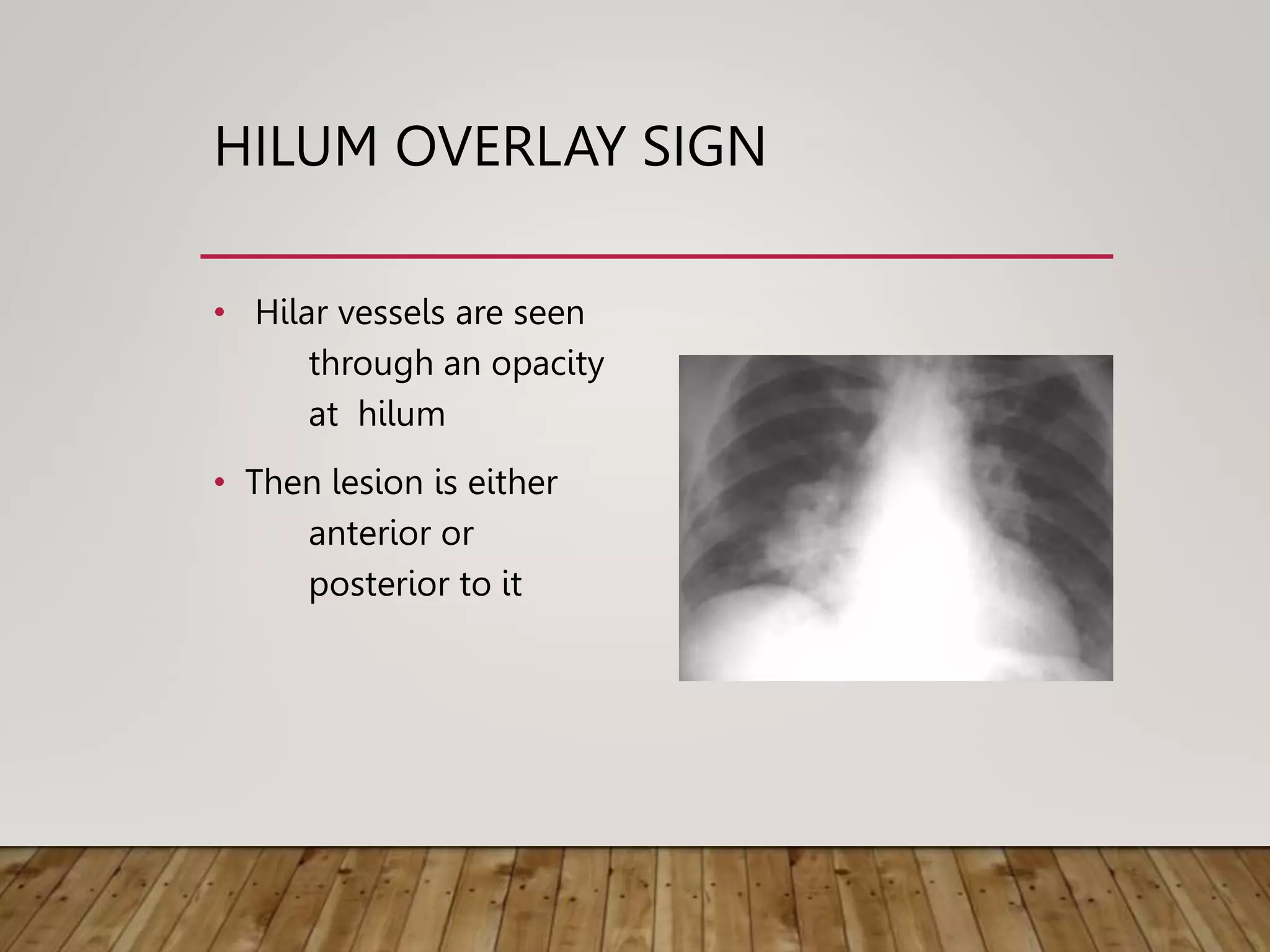
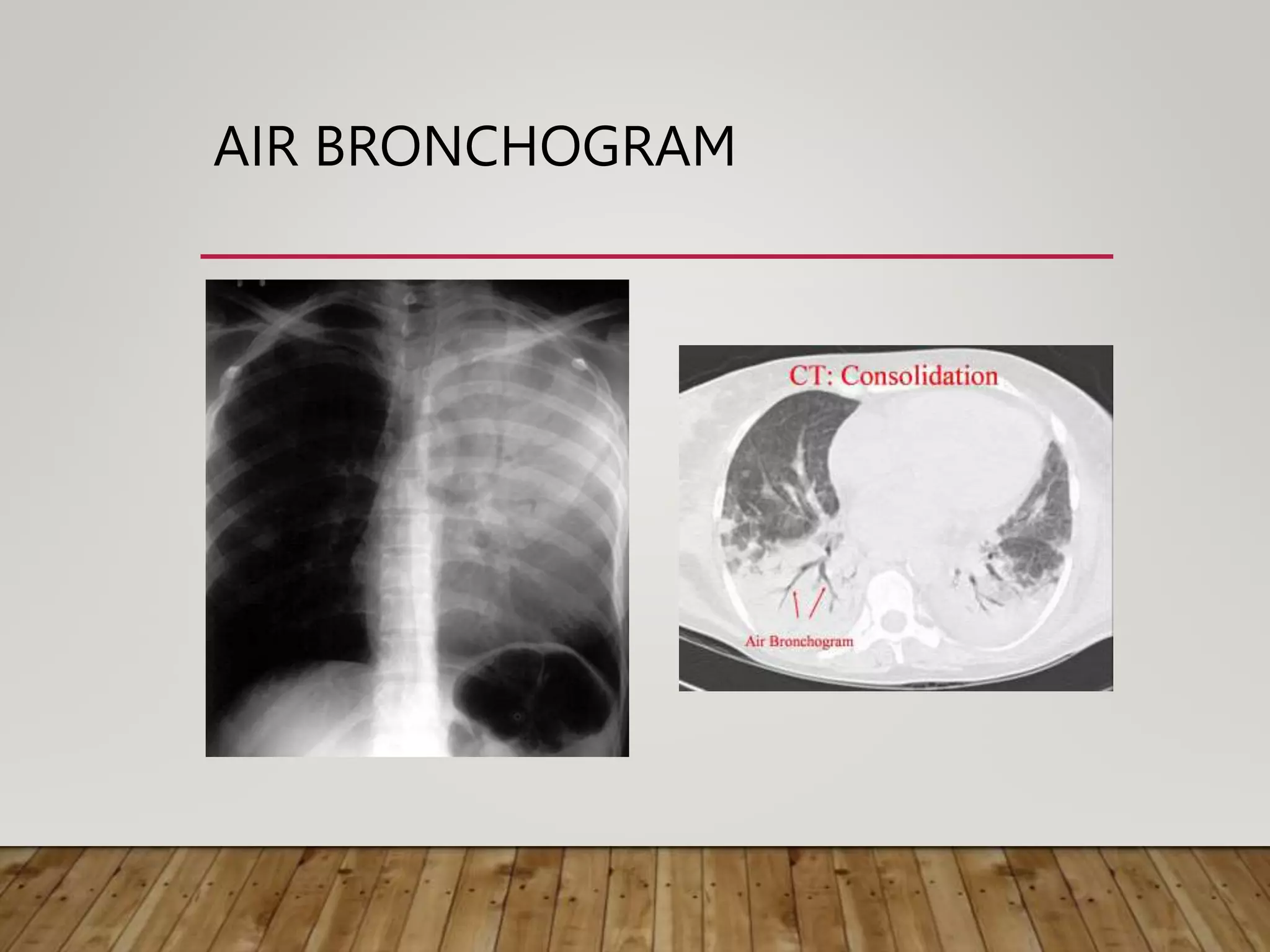
3. HOW DOES THE PICTURE FORMED
• AMOUNT OF X RAY ABSORBED
• Capo4 -22
• Ca co3- 15
• Water—1
• Carbon- 0.7
• Fat -0.5
• Paraffin -0.4
• Air- 0.0001
4. PICTURE ...
Different tissues absorb X-rays at different extents:
• Bone- high absorption (white)
• Tissue- somewhere in the middle absorption (grey)
• Air- low absorption (black)
5. VIEWS
• PA
• AP
• LATERAL
• DECUBITUS
• APICAL tube angled up 50-60 degree -Apex
LORDOTIC tube angled 50-60 degree downwards---middle lobe
collapse
• OBLIQUE
• Supine
6. QUALITY OF FILM
• Name ,age, sex, date , id no
• Side
• Inspiratory (sixth rib / tenth rib)
• No rotation
• Penetration visible down to T8 -T9
• Scapula
• Full film (All area seen)
18. RIB
• Companion shadow
• Indistinct inferior border is normal for middle and lower
rib(when we look for rib erosion)
Dense First costal cartilage calcification may obscure
UZnotch
• Normally on inferior surface just lateral to tubercle
• confused with pathological notch
21. BORDERS OF MEDIASTINUM
AND HEART
1Edge of
Superior Vena Cava
2. Right Atrium
3. Aortic knuckle
4. Edge of Main
Pulmonary Artery
5. Left Atrial Appendage
6. Left Ventricle
22.
23.
24.
25. DIAPHRAGM
• Right is higher than left( ½ space)
• –Lt is pushdown by heart
• May lie at same level
• Left is higher – in 3%
• Difference > 3 cm is significant
26. Lymphatics
• Normally not seen
• Thickening of lymphatics and surrounding connective
tissue produces Kerley lines
• Enlarged broncho pulmonary node - lobulated hilar
mass
27. KERLEY A
• 1-2 mm thick
• non branching
• radiating from hilum
• 2-6 cm long
• thickened deep interlobular
septa
KERLEY B
• 1-2 mm thick
• non branching
• Transverse line at lung base
perpendicular to pleura
• 1-3 cm long
• thickened interlobular septa
28. CP ANGLE
• Normally –acute angle
• Obliterated when diaphragm becomes flat
• Low density ill defined opacity-normal due to Fat pad
29. HILUM
• Left hilum at higher level than right-97%Left PA alies aboveLMB
• Same level -3%
• Both hila –equal density similar size
• Concave lateral border-superior pulmonary v meet basal
pulmonary artery
• Normally LN are not seen
32. SILHOUETTE SIGN
• Described by Felson in 1950
“Loss of an interface by adjacent disease helps in localization
of a lesion by studying the diaphragm, cardiac and aortic
outlines”
44. ANTERIOR MASS
• contact with soft
tissues rather than the
aerated lung
• cephalic border of a
shadow disappears as it
approaches the clavicle
45. HILUM CONVERGENCE SIGN
• Pulmonary artery
branches converge
towards the hilar mass
• Then the hilar mass
may be an enlarged
pulmonary artery
46. HILUM OVERLAY SIGN
• Hilar vessels are seen
through an opacity
at hilum
• Then lesion is either
anterior or
posterior to it
47.
48. AIR BRONCHOGRAM
• Described by Fleischner-1941
• Named by Felson-1973
• A sign that indicate lesion is intra pulmonary (reverse
is not true)
• When lung parenchyma are opacified , surrounding
bronchi will be visualised due to contrast between air
and opaque lung
• supplying bronchus should not be occluded
55. BATS WING SHADOW
• Perihilar distribution with peripheral lung remaining
translucent
• Pulmonary edema
may be unilateral
clear rapidly with treatment
• PCP,Goodpasture’s syndrome ,Alveolar cell
cacinoma,Alveolar proteinosis
56.
57.
58.
59. MILIARY SHADOWING
• Any discrete opacity 2- 4 mm in diameter
• Uniform size
• Wide spread
• Common in TB
• Dense calcification seen in metallic dust disease
61. GROUND GLASS APPEARANCE
• Finely granular pattern of pulmonary opacity in which
normal anatomical details are partially obscured
• Seen with interstitial or air space pattern
79. RETICULAR PATTERN
• Numerous interlacing line shadows
• fine reticulation--simulates a very fine mesh
• coarse reticulation--characterized by large cystic spaces 1 cm or more
in diameter that are ringed by soft tissue
• medium reticulation--characterized by 3-10 mm spaces between the
reticular mesh
85. HONEY COMBING
• Thin walled cysts representing air spaces 5-10 mm diameter with
walls 2-3 mm thick
• Occurs in parenchymal destruction leading to end stage fibrosis
• Dilatation of respiratory bronchioles and alveolar ducts
• IPF,CTD,Sarcoidosis
• Increased risk of pneumothorax
86.
87.
88. LINE & BAND SHADOWS
• Linear opacities < 5 mm in diameter are line shadows
• Linear densities with diameter >5 mm in diameter are band
shadows
92. PULMONARY INFARCT
• Irregular thick wedge shaped line with base adjacent
to pleura or
• Non specific area of peripheral consolidation at lung
base
• Splinting of diaphragm , pleural reaction
• Resolution slower than infection
93.
94. SOLITARY PULMONARY
NODULE
• Single discrete pulmonary opacity
• Surrounded by normal lung tissue
• Not associated with atelecatasis or adenopathy
• Approximately circular
• 2-30 mm in diameter (<3 cm )
95. Calcification
• indicate benign lesion
• hamartoma—Popcorn
“Carcinoma may arise from old calcified
focus”
“Metastasis from bone or cartilagenous
tumor may have calcification”