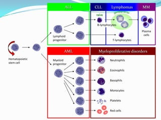
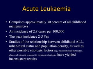
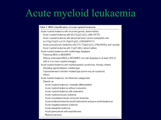
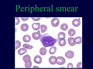
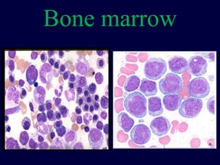
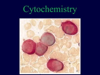
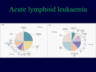
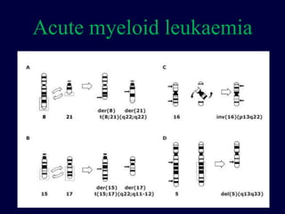
Acute leukemia
- Dr Dinesh Bhurani, DM, FRCPA
Senior bone marrow transplant physician
Rajiv Gandhi Cancer Inst & RC, Delhi
ALL MMCLL Lymphomas
Hematopoietic
stem cell
Neutrophils
Eosinophils
Basophils
Monocytes
Platelets
Red cells
Myeloid
progenitor
Myeloproliferative disordersAML
Lymphoid
progenitor T-lymphocytes
Plasma
cells
B-lymphocytes
naïve
Incidence
Acute myeloid leukaemia
ALL
Cytopenia
Production failure
• Stem cell defect
– Apastic anaemia
• Bone marrow
suppression
– Drugs
– Infections eg
malaria, dengue, HIV
• Ineffective
haemopoiesis
– Megaloblastic anaemia
– Myelodysplastic syndrome
Production failure
• Bone marrow infiltration
– Haematological malignancy
– Non haematological malignancy
• Marrow fibrosis
– Primary
– Secondary
• Haemophagocytic syndrome
Increased consumption
• Hypersplenism
• Immune cytopenia
Peripheral smear
• Oval macrocytes, hypersegmented neutrophils –
megaloblastic anaemia
• Leuko-erythroblastic blood picture – marrow
infiltration
• Leuko-erythroblastic picture with tear drop
cells – marrow fibrosis
• Blast or atypical lymphoid cells – haematological
malignancy
• Dysplastic feature – MDS, drugs or HIV infection
Leukaemia
• Pancytopenia or bicytopenia – Can it be
haematological malignancy? CBC &
peripheral smear
• Avoid steroids prior to confirmation of
diagnosis
Bone marrow aspirate and
biopsy both are essential.
Acute Leukaemia
• Comprises approximately 30 percent of all childhood
malignancies
• An incidence of 2.8 cases per 100,000
• The peak incidence 2-5 Yrs
• Studies of the relationship between childhood ALL,
urban/rural status and population density, as well as
other possible etiologic factors (eg, environmental exposures,
abnormal immune response to common infections) have yielded
inconsistent results
Acute leukaemia
• Persistent or progressive lymphadenopathy
that does not respond to antibiotic therapy
suggests the need for more extensive
evaluation.
Differential diagnosis
• Juvenile idiopathic arthritis
• Osteomyelitis
• Epstein-Barr virus
• Idiopathic thrombocytopenic purpura
• Pertussis, parapertussis
• Aplastic anemia
• Acute infectious lymphocytosis
• Other malignancies with bone marrow involvement
(eg, neuroblastoma, retinoblastoma, rhabdomyosarcoma, and Ewing sarcoma
Acute lymphoblastic leukaemia
Etiology
Leukaemogenesis
Leukaemogenesis
Classification
Acute myeloid leukaemia
Diagnosis
Peripheral smear
Bone marrow
Cytochemistry
Flowcytometry
Cytogenetics
FISH
PCR
Prognosis
Prognostic factors
• Age
• Initial WBC count
• Cytogenetics
• Immunologic subtype
• Response to initial therapy
Risk groups
• Low risk — 92 percent
• Standard risk — 82 percent
• High risk — 73 percent
• Very high risk — 46 percent
Acute lymphoid leukaemia
Acute myeloid leukaemia
Treatment
Acute lymphoblastic leukaemia
• INDUCTION THERAPY
– To eradicate more than 99 percent of the initial burden.
– to restore normal hematopoiesis
– Usually last for 4-5 weeks
• Intensification (consolidation) therapy
– High dose methotrexate
– Reinduction therapy
• CNS targeted therapy
– Intrathecal chemo, cranial RT, High Mtx
• Maintenance therapy
– Oral 6MP and Mtx
• Allogenic bone marrow transplantation
Acute myeloid leukaemia
• Induction therapy
– Cytarabine (7 days) & Daunorubicine (3days)
• Consolidation therapy
– 3-4 cycles of high dose cytarabine
• Autologous and allogenic bone marrow
transplantation
Acute myeloid leukaemia
Future
Minimal residual disease
Technique and criterion Blasts per 100,000 nucleated cells
Standard microscopy ("complete
remission")
5,000
Karyotype analysis (20 mitotic
figures)
5,000
Microscopy, expert 1,000
Immunophenotyping: multi-
parameter flow cytometry
10
Colony growth 1
Polymerase chain reaction 0.1

Slidecast