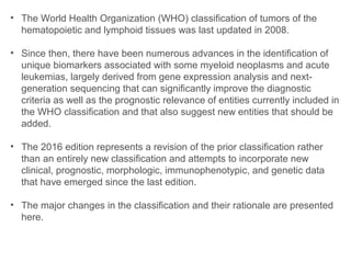
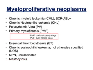
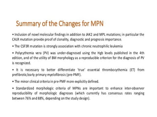
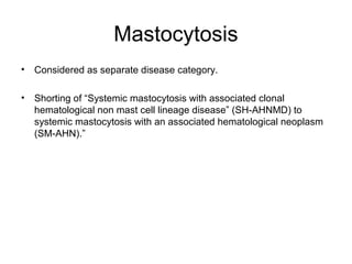
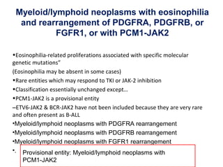
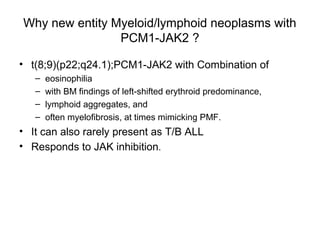
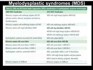
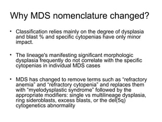
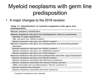
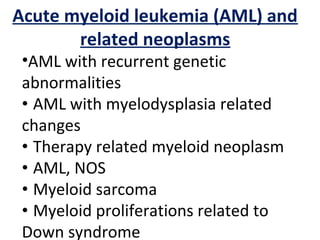
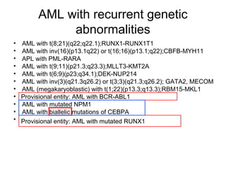
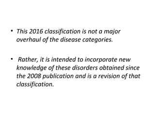
Recent advances to the World Health
Organization classification of
myeloid neoplasms and acute leukemia.
DR. NOOPUR PATIL
JR1 PATHOLOGY
MIMSR,LATUR
• The World Health Organization (WHO) classification of tumors of the
hematopoietic and lymphoid tissues was last updated in 2008.
• Since then, there have been numerous advances in the identification of
unique biomarkers associated with some myeloid neoplasms and acute
leukemias, largely derived from gene expression analysis and next-
generation sequencing that can significantly improve the diagnostic
criteria as well as the prognostic relevance of entities currently included in
the WHO classification and that also suggest new entities that should be
added.
• The 2016 edition represents a revision of the prior classification rather
than an entirely new classification and attempts to incorporate new
clinical, prognostic, morphologic, immunophenotypic, and genetic data
that have emerged since the last edition.
• The major changes in the classification and their rationale are presented
here.
• This revision has been influenced by several
factors :
1. The discovery of recently identified molecular features has yielded
new perspectives regarding diagnostic and prognostic markers
2. Improved characterization and standardization of morphological
features aiding in the reliability and reproducibility of diagnosis.
3. A number of clinical-pathological studies have now validated the
WHO postulate of an integrated approach that includes
hematologic, morphologic, cytogenetic, and molecular genetic
findings
Myeloproliferative neoplasms
• Chronic myeloid leukemia (CML), BCR-ABL+
• Chronic Neutrophilic leukemia (CNL)
• Polycythemia Vera (PV)
• Primary myelofibrosis (PMF)
• Essential thrombocythemia (ET)
• Chronic eosinophilic leukemia, not otherwise specified
(NOS)
• MPN, unclassifiable
• Mastocytosis
•PMF, prefibrotic /early stage
•PMF, overt fibrotic stage
CML:-
•Largely unchanged since 2008 WHO
•BMAT (Bone marrow aspiration and trephine biopsy)at
diagnosis still recommended
Complete work-up of karyotype and morphology
•Criteria for AccPhase provisionally includes response to
Tyrosine kinase inhibitor therapy
•Lymphoblasts in peripheral blood should trigger thorough
investigation for impending lymphoid blast crisis
CML
CNL:-
•G-CSF receptor mutations which allow G-CSF independent granulocyte proliferation and
differentiation are found in over 80% of cases conforming to the WHO definition of CNL.
•CSF3R T6181 is the most common mutation
Removed hepatosplenomegaly as criteria
Polycythemia vera:-
•The Hb level at which we suspect PV has been
substantially lowered in men irrespective of age. (It is well
known that the Hb in males drops during the 7th and 8th
decades.)
•BMAT with panmyelosis (pleomorphic megakaryocytes)
promoted to a major criteria.
•Exclusion of secondary causes is not mentioned.
•In the absence of the JAK 2’s, subnormal EPO is acceptable
as a 3rd criteria.
PV2016
2008
ET
Pre PMF
OVERT PMF:-
Mastocytosis
• Considered as separate disease category.
• Shorting of “Systemic mastocytosis with associated clonal
hematological non mast cell lineage disease” (SH-AHNMD) to
systemic mastocytosis with an associated hematological neoplasm
(SM-AHN).”
Myeloid/lymphoid neoplasms with eosinophilia
and rearrangement of PDGFRA, PDGFRB, or
FGFR1, or with PCM1-JAK2
•Eosinophilia-related proliferations associated with specific molecular
genetic mutations”
(Eosinophilia may be absent in some cases)
•Rare entities which may respond to TKI or JAK-2 inhibition
•Classification essentially unchanged except…
•PCM1-JAK2 is a provisional entity
–ETV6-JAK2 & BCR-JAK2 have not been included because they are very rare
and often present as B-ALL
•Myeloid/lymphoid neoplasms with PDGFRA rearrangement
•Myeloid/lymphoid neoplasms with PDGFRB rearrangement
•Myeloid/lymphoid neoplasms with FGFR1 rearrangement
•. Provisional entity: Myeloid/lymphoid neoplasms with
PCM1-JAK2
Why new entity Myeloid/lymphoid neoplasms with
PCM1-JAK2 ?
• t(8;9)(p22;q24.1);PCM1-JAK2 with Combination of
– eosinophilia
– with BM findings of left-shifted erythroid predominance,
– lymphoid aggregates, and
– often myelofibrosis, at times mimicking PMF.
• It can also rarely present as T/B ALL
• Responds to JAK inhibition.
Myelodysplastic/myeloproliferati
ve neoplasms (MDS/MPN)
• Chronic myelomonocytic leukemia (CMML)
• Atypical chronic myeloid leukemia (aCML), BCR-
ABL-
• Juvenile myelomonocytic leukemia (JMML)
• .
• MDS/MPN, unclassifiable
MDS/MPN with ring sideroblasts and
thrombocytosis (MDS/MPN-RS-T)
CMML
• Due to the discovery of molecular and clinical differences
between the so-called
– “Proliferative type” of CMML (WBC count>= 13X10’9/L) and the
– “Dysplastic type” (WBC <13X10’9/L), particularly those
differences related to aberrancies in the RAS/MAPK signaling
pathways
• Recent evidence has shown that a more precise
prognostication can be obtained with 3 blast-based
groupings:
– CMML-0, a category for cases with < 2% blasts in PB
and <5% blasts in BM;
– CMML-1 for cases with 2% to 4% blasts in PB and/or
5% to 9% blasts in BM; and
– CMML-2 for cases with 5% to 19% blasts in PB, 10%
to 19% in BM, and/or when any Auer rods are present
Atypical CML:-
• No changes in criteria.
• Not CML
–BCR-ABL negative
–No Basophilia
–Dysplastic
•Not CNL
–Significant left shift
–Dysplastic
–Unlikely to have an isolated CSF3R mutation (<10%)
•Not MPN
–JAK2,CALR,MPL,PDGFR etc typically absent
•~33% have either SETBP1 and/or ETNK1 mutations
Myelodysplastic/myeloproliferative neoplasm
with ring sideroblasts and thrombocytosis
• Considered full entity under MDS/MPN
• The name RARS-T is changed to MDS/MPN-RS-T.
• Must have >/= 15% ringed sideroblasts
• Megakaryocyte proliferation
• Often associated with the spliceosome gene SF3B1+/- co-mutated with JAK2,
CALR or MPL
MDS like MPN like
Clinical
• Macrocytic anemia
• Transfusion requirement
• Thrombocytosis
• Need for cytoreduction
Morphological
• Erythroid dysplasia
• Ring sideroblasts
• Large megakaryocytes
with bulbous nuclei
Genetic
SF3B1 mutation (80-90%) JAK2 mutation (50-60%)
Rarely CALR/MPL
Juvenile myelomonocytic
leukemia
>90% will have one of:-
Myelodysplastic syndromes (MDS)
Why MDS nomenclature changed?
• Classification relies mainly on the degree of dysplasia
and blast % and specific cytopenias have only minor
impact.
• The lineage's manifesting significant morphologic
dysplasia frequently do not correlate with the specific
cytopenias in individual MDS cases
• MDS has changed to remove terms such as “refractory
anemia” and “refractory cytopenia” and replaces them
with “myelodysplastic syndrome” followed by the
appropriate modifiers: single vs multilineage dysplasia,
ring sideroblasts, excess blasts, or the del(5q)
cytogenetics abnormality
New handling of MDS with ring
sideroblasts
• MDS with multilineage dysplasia and ring
sideroblast will be reinstated (MLD-RS)
• MDS with SF3B1 mutation can be
classified as SLD- RS/ MLD-RS if >5%
ring sideroblasts are present
– Will not require >15% RS
• SF3B1 mutation will not affect MDS –EB
or isolated del(5q)
Changes in MDS del(5q)
• Allow one additional cytogenetic
abnormality
• Excluding high risk abnormalities(del (7q)
or monosomy 7 )
• TP53 mutation study or p53 immunostain
• Exclusions:-
– >5% blasts in PB/BM
– Significant granulocytic dysplasia
Myeloid neoplasms with germ line
predisposition
• A major changes to the 2016 revision
Germ line mutations:
•Trisomy 21
•Neurofibromatosis and Noonan’s syndrome
•Bone marrow failure syndromes / telomeres
•Familial thrombocytopenia (3 types)
•CEBPA/DDX41
•Other hereditary cancer syndromes eg TP53 mutations
Which patients should we test?
•Family history? Haematological +/- solid cancers
•Children and young adults (11% to24% in small series –
MDS/AL/AA)
•Transplant candidates?
•Biallelic CEBPA mutations? (~10% germ-line)
•Any of the mutations which could be germ line?
•History / FH of thrombocytopenia or “ITP”?
•? Syndromic ? History of unusual infections
Acute myeloid leukemia (AML) and
related neoplasms
•AML with recurrent genetic
abnormalities
• AML with myelodysplasia related
changes
• Therapy related myeloid neoplasm
• AML, NOS
• Myeloid sarcoma
• Myeloid proliferations related to
Down syndrome
AML with recurrent genetic
abnormalities
• AML with t(8;21)(q22;q22.1);RUNX1-RUNX1T1
• AML with inv(16)(p13.1q22) or t(16;16)(p13.1;q22);CBFB-MYH11
• APL with PML-RARA
• AML with t(9;11)(p21.3;q23.3);MLLT3-KMT2A
• AML with t(6;9)(p23;q34.1);DEK-NUP214
• AML with inv(3)(q21.3q26.2) or t(3;3)(q21.3;q26.2); GATA2, MECOM
• AML (megakaryoblastic) with t(1;22)(p13.3;q13.3);RBM15-MKL1
• .
• AML with mutated NPM1
• AML with biallelic mutations of CEBPA
• .
Provisional entity: AML with BCR-ABL1
Provisional entity: AML with mutated RUNX1
NOTABLE CHANGES:-
Mostly unchanged
–MLL(mixed lineage leukemia) renamed KMT2A
–APL with PML-RARA includes cryptic / complex rearrangements
which cause the fusion
–AML with BCR-ABL1 provisional entity
•Difficult to distinguish from CML presenting in blast phase but
possibly more likely if IKZF1 / CDKN2a or deletion of IgH/TCR
•CEBPA must be biallelic to confer good prognosis.
• Presence of NPM1 and biallelic CEBPA trump morphological
evidence of multilineage dysplasia .
• AML with RUNX1 mutation (? Poor prognosis) but trumped by MDS
related cytogenetic abnormalities – (AML with RUNX1 mutation is a
provisional entity)
AML with myelodysplasia-related changes
Therapy-related myeloid
neoplasms
• No Changes
AML, NOS
• AML with minimal differentiation
• AML without maturation
• AML with maturation
• Acute myelomonocytic leukemia
• Acute monoblastic/monocytic leukemia
• Pure erythroid leukemia
• Acute megakaryoblastic leukemia
• Acute basophilic leukemia
• Acute panmyelosis with myelofibrosis
Myeloid sarcoma
• No changes
Myeloid proliferations related to
Down syndrome
• No changes.
Acute leukemias of ambiguous lineage
• Acute undifferentiated leukemia
• Mixed phenotype acute leukemia (MPAL) with t(9;22)(q34.1;q11.2);
BCR-ABL1
• MPAL with t(v;11q23.3); KMT2A rearranged
• MPAL, B/myeloid, NOS
• MPAL, T/myeloid, NOS
• Essentially unchanged
• If it is clearly B,T or myeloid, the MPAL(mixed phenotype acute
leukemia) specific markers do not apply .
• Flow cytometry is the preferred method of antigen detection
• When two blast populations are easily identified. If they can be
characterised as B,T or Myeloid they should be.
• The significance of weak MPO expression on an otherwise typical
B cell population is uncertain.
B-lymphoblasticleukemia/lymphoma
• B-lymphoblastic leukemia/lymphoma, NOS
• B-lymphoblastic leukemia/lymphoma with recurrent genetic
abnormalities
• B-lymphoblastic leukemia/lymphoma with t(9;22)(q34.1;q11.2);BCR-
ABL1
• B-lymphoblastic leukemia/lymphoma with t(v;11q23.3);KMT2A
rearranged
• B-lymphoblastic leukemia/lymphoma with t(12;21)(p13.2;q22.1);
ETV6-RUNX1
• B-lymphoblastic leukemia/lymphoma with hyperdiploidy
• B-lymphoblastic leukemia/lymphoma with hypodiploidy
• B-lymphoblastic leukemia/lymphoma with t(5;14)(q31.1;q32.3) IL3-IGH
• B-lymphoblastic leukemia/lymphoma with t(1;19)(q23;p13.3);TCF3-
PBX1
• .
• .
Provisional entity: B-lymphoblastic leukemia/lymphoma, BCR-ABL1–like
Provisional entity: B-lymphoblastic leukemia/lymphoma with iAMP21
B-lymphoblastic leukaemia/lymphoma:-
•Low hypodiploidy (32-39 chromosomes) is associated with TP53 mutations in
~90% of cases.
•Nearly half found to be germ line in paediatric cases.
•B-ALL with intrachromosomal amplification of chromosome 21
–Portion of 21 containing RUNX1
–5+ copies (or 3+ on one allele)
–2% childhood ALL (tend to be older with low WCC)
–Poor prognosis but respond to aggressive therapy.
BCR-ABL-like ALL :-
B-ALL with translocations involving TKI or cytokine receptors
•Poor prognosis but may respond to TKI
–Include CRLF2 (Cytokine receptor-like factor 2)
•CRLF2 often associated with JAK mutations
•Common in trisomy 21
–EPOR (truncated and activated EPO receptor)
–TK genes may involve ABL1, ABL2, PDGFRB, NTRK3, TYK2, CSF1R and JAK2
–Also frequent loss of IKZF1 and CDKN2A/B
T-Lymphoblastic leukemia/lymphoma
• .
• .
Provisional entity: Early T-cell precursor lymphoblastic
leukemia
Provisional entity: Natural killer (NK) cell lymphoblastic
leukemia/lymphoma
Indolent T-lymphoblastic proliferation:-
–Often cervical lymphadenopathy
–Proliferation of normal non-clonal T-lymphoblasts
–Immature thymic phenotype
Early T-Precursor ALL :-
–Early T phenotype (CD1a negative) CD7+
–1 or more myeloid/stem cell marker
•CD34,CD117,HLADR,CD13,CD33,CD11b or CD56
•Often have myeloid associated mutations
-FLT3,NRAS/KRAS,DNMT3A,IDH1and IDH2
–Prognostic significance uncertain
• This 2016 classification is not a major
overhaul of the disease categories.
• Rather, it is intended to incorporate new
knowledge of these disorders obtained since
the 2008 publication and is a revision of that
classification.
REFERENCES:-
•Robins and Cotran, Pathologic basis of disease 7th
edititon.
•www.google.com
•WHO Classification of Tumours of Haematopoietic and
Lymphoid Tissue.
•Atlas and text of hematology- Dr. Tejinder Singh(tables)
•Hematology today by M.B. Agarwal.(2008 classification)
Recent advances in leukemia (2019)

Recent advances in leukemia (2019)

  • 1.
    Recent advances tothe World Health Organization classification of myeloid neoplasms and acute leukemia. DR. NOOPUR PATIL JR1 PATHOLOGY MIMSR,LATUR
  • 2.
    • The WorldHealth Organization (WHO) classification of tumors of the hematopoietic and lymphoid tissues was last updated in 2008. • Since then, there have been numerous advances in the identification of unique biomarkers associated with some myeloid neoplasms and acute leukemias, largely derived from gene expression analysis and next- generation sequencing that can significantly improve the diagnostic criteria as well as the prognostic relevance of entities currently included in the WHO classification and that also suggest new entities that should be added. • The 2016 edition represents a revision of the prior classification rather than an entirely new classification and attempts to incorporate new clinical, prognostic, morphologic, immunophenotypic, and genetic data that have emerged since the last edition. • The major changes in the classification and their rationale are presented here.
  • 3.
    • This revisionhas been influenced by several factors : 1. The discovery of recently identified molecular features has yielded new perspectives regarding diagnostic and prognostic markers 2. Improved characterization and standardization of morphological features aiding in the reliability and reproducibility of diagnosis. 3. A number of clinical-pathological studies have now validated the WHO postulate of an integrated approach that includes hematologic, morphologic, cytogenetic, and molecular genetic findings
  • 4.
    Myeloproliferative neoplasms • Chronicmyeloid leukemia (CML), BCR-ABL+ • Chronic Neutrophilic leukemia (CNL) • Polycythemia Vera (PV) • Primary myelofibrosis (PMF) • Essential thrombocythemia (ET) • Chronic eosinophilic leukemia, not otherwise specified (NOS) • MPN, unclassifiable • Mastocytosis •PMF, prefibrotic /early stage •PMF, overt fibrotic stage
  • 5.
    CML:- •Largely unchanged since2008 WHO •BMAT (Bone marrow aspiration and trephine biopsy)at diagnosis still recommended Complete work-up of karyotype and morphology •Criteria for AccPhase provisionally includes response to Tyrosine kinase inhibitor therapy •Lymphoblasts in peripheral blood should trigger thorough investigation for impending lymphoid blast crisis
  • 6.
  • 7.
    CNL:- •G-CSF receptor mutationswhich allow G-CSF independent granulocyte proliferation and differentiation are found in over 80% of cases conforming to the WHO definition of CNL. •CSF3R T6181 is the most common mutation Removed hepatosplenomegaly as criteria
  • 8.
    Polycythemia vera:- •The Hblevel at which we suspect PV has been substantially lowered in men irrespective of age. (It is well known that the Hb in males drops during the 7th and 8th decades.) •BMAT with panmyelosis (pleomorphic megakaryocytes) promoted to a major criteria. •Exclusion of secondary causes is not mentioned. •In the absence of the JAK 2’s, subnormal EPO is acceptable as a 3rd criteria.
  • 9.
  • 10.
  • 11.
  • 12.
  • 14.
    Mastocytosis • Considered asseparate disease category. • Shorting of “Systemic mastocytosis with associated clonal hematological non mast cell lineage disease” (SH-AHNMD) to systemic mastocytosis with an associated hematological neoplasm (SM-AHN).”
  • 15.
    Myeloid/lymphoid neoplasms witheosinophilia and rearrangement of PDGFRA, PDGFRB, or FGFR1, or with PCM1-JAK2 •Eosinophilia-related proliferations associated with specific molecular genetic mutations” (Eosinophilia may be absent in some cases) •Rare entities which may respond to TKI or JAK-2 inhibition •Classification essentially unchanged except… •PCM1-JAK2 is a provisional entity –ETV6-JAK2 & BCR-JAK2 have not been included because they are very rare and often present as B-ALL •Myeloid/lymphoid neoplasms with PDGFRA rearrangement •Myeloid/lymphoid neoplasms with PDGFRB rearrangement •Myeloid/lymphoid neoplasms with FGFR1 rearrangement •. Provisional entity: Myeloid/lymphoid neoplasms with PCM1-JAK2
  • 16.
    Why new entityMyeloid/lymphoid neoplasms with PCM1-JAK2 ? • t(8;9)(p22;q24.1);PCM1-JAK2 with Combination of – eosinophilia – with BM findings of left-shifted erythroid predominance, – lymphoid aggregates, and – often myelofibrosis, at times mimicking PMF. • It can also rarely present as T/B ALL • Responds to JAK inhibition.
  • 18.
    Myelodysplastic/myeloproliferati ve neoplasms (MDS/MPN) •Chronic myelomonocytic leukemia (CMML) • Atypical chronic myeloid leukemia (aCML), BCR- ABL- • Juvenile myelomonocytic leukemia (JMML) • . • MDS/MPN, unclassifiable MDS/MPN with ring sideroblasts and thrombocytosis (MDS/MPN-RS-T)
  • 19.
    CMML • Due tothe discovery of molecular and clinical differences between the so-called – “Proliferative type” of CMML (WBC count>= 13X10’9/L) and the – “Dysplastic type” (WBC <13X10’9/L), particularly those differences related to aberrancies in the RAS/MAPK signaling pathways • Recent evidence has shown that a more precise prognostication can be obtained with 3 blast-based groupings: – CMML-0, a category for cases with < 2% blasts in PB and <5% blasts in BM; – CMML-1 for cases with 2% to 4% blasts in PB and/or 5% to 9% blasts in BM; and – CMML-2 for cases with 5% to 19% blasts in PB, 10% to 19% in BM, and/or when any Auer rods are present
  • 20.
    Atypical CML:- • Nochanges in criteria. • Not CML –BCR-ABL negative –No Basophilia –Dysplastic •Not CNL –Significant left shift –Dysplastic –Unlikely to have an isolated CSF3R mutation (<10%) •Not MPN –JAK2,CALR,MPL,PDGFR etc typically absent •~33% have either SETBP1 and/or ETNK1 mutations
  • 21.
    Myelodysplastic/myeloproliferative neoplasm with ringsideroblasts and thrombocytosis • Considered full entity under MDS/MPN • The name RARS-T is changed to MDS/MPN-RS-T. • Must have >/= 15% ringed sideroblasts • Megakaryocyte proliferation • Often associated with the spliceosome gene SF3B1+/- co-mutated with JAK2, CALR or MPL MDS like MPN like Clinical • Macrocytic anemia • Transfusion requirement • Thrombocytosis • Need for cytoreduction Morphological • Erythroid dysplasia • Ring sideroblasts • Large megakaryocytes with bulbous nuclei Genetic SF3B1 mutation (80-90%) JAK2 mutation (50-60%) Rarely CALR/MPL
  • 22.
  • 23.
  • 24.
    Why MDS nomenclaturechanged? • Classification relies mainly on the degree of dysplasia and blast % and specific cytopenias have only minor impact. • The lineage's manifesting significant morphologic dysplasia frequently do not correlate with the specific cytopenias in individual MDS cases • MDS has changed to remove terms such as “refractory anemia” and “refractory cytopenia” and replaces them with “myelodysplastic syndrome” followed by the appropriate modifiers: single vs multilineage dysplasia, ring sideroblasts, excess blasts, or the del(5q) cytogenetics abnormality
  • 26.
    New handling ofMDS with ring sideroblasts • MDS with multilineage dysplasia and ring sideroblast will be reinstated (MLD-RS) • MDS with SF3B1 mutation can be classified as SLD- RS/ MLD-RS if >5% ring sideroblasts are present – Will not require >15% RS • SF3B1 mutation will not affect MDS –EB or isolated del(5q)
  • 28.
    Changes in MDSdel(5q) • Allow one additional cytogenetic abnormality • Excluding high risk abnormalities(del (7q) or monosomy 7 ) • TP53 mutation study or p53 immunostain • Exclusions:- – >5% blasts in PB/BM – Significant granulocytic dysplasia
  • 29.
    Myeloid neoplasms withgerm line predisposition • A major changes to the 2016 revision
  • 30.
    Germ line mutations: •Trisomy21 •Neurofibromatosis and Noonan’s syndrome •Bone marrow failure syndromes / telomeres •Familial thrombocytopenia (3 types) •CEBPA/DDX41 •Other hereditary cancer syndromes eg TP53 mutations
  • 31.
    Which patients shouldwe test? •Family history? Haematological +/- solid cancers •Children and young adults (11% to24% in small series – MDS/AL/AA) •Transplant candidates? •Biallelic CEBPA mutations? (~10% germ-line) •Any of the mutations which could be germ line? •History / FH of thrombocytopenia or “ITP”? •? Syndromic ? History of unusual infections
  • 32.
    Acute myeloid leukemia(AML) and related neoplasms •AML with recurrent genetic abnormalities • AML with myelodysplasia related changes • Therapy related myeloid neoplasm • AML, NOS • Myeloid sarcoma • Myeloid proliferations related to Down syndrome
  • 33.
    AML with recurrentgenetic abnormalities • AML with t(8;21)(q22;q22.1);RUNX1-RUNX1T1 • AML with inv(16)(p13.1q22) or t(16;16)(p13.1;q22);CBFB-MYH11 • APL with PML-RARA • AML with t(9;11)(p21.3;q23.3);MLLT3-KMT2A • AML with t(6;9)(p23;q34.1);DEK-NUP214 • AML with inv(3)(q21.3q26.2) or t(3;3)(q21.3;q26.2); GATA2, MECOM • AML (megakaryoblastic) with t(1;22)(p13.3;q13.3);RBM15-MKL1 • . • AML with mutated NPM1 • AML with biallelic mutations of CEBPA • . Provisional entity: AML with BCR-ABL1 Provisional entity: AML with mutated RUNX1
  • 34.
    NOTABLE CHANGES:- Mostly unchanged –MLL(mixedlineage leukemia) renamed KMT2A –APL with PML-RARA includes cryptic / complex rearrangements which cause the fusion –AML with BCR-ABL1 provisional entity •Difficult to distinguish from CML presenting in blast phase but possibly more likely if IKZF1 / CDKN2a or deletion of IgH/TCR •CEBPA must be biallelic to confer good prognosis. • Presence of NPM1 and biallelic CEBPA trump morphological evidence of multilineage dysplasia . • AML with RUNX1 mutation (? Poor prognosis) but trumped by MDS related cytogenetic abnormalities – (AML with RUNX1 mutation is a provisional entity)
  • 36.
  • 37.
  • 38.
    AML, NOS • AMLwith minimal differentiation • AML without maturation • AML with maturation • Acute myelomonocytic leukemia • Acute monoblastic/monocytic leukemia • Pure erythroid leukemia • Acute megakaryoblastic leukemia • Acute basophilic leukemia • Acute panmyelosis with myelofibrosis
  • 39.
  • 40.
    Myeloid proliferations relatedto Down syndrome • No changes.
  • 41.
    Acute leukemias ofambiguous lineage • Acute undifferentiated leukemia • Mixed phenotype acute leukemia (MPAL) with t(9;22)(q34.1;q11.2); BCR-ABL1 • MPAL with t(v;11q23.3); KMT2A rearranged • MPAL, B/myeloid, NOS • MPAL, T/myeloid, NOS • Essentially unchanged • If it is clearly B,T or myeloid, the MPAL(mixed phenotype acute leukemia) specific markers do not apply . • Flow cytometry is the preferred method of antigen detection • When two blast populations are easily identified. If they can be characterised as B,T or Myeloid they should be. • The significance of weak MPO expression on an otherwise typical B cell population is uncertain.
  • 42.
    B-lymphoblasticleukemia/lymphoma • B-lymphoblastic leukemia/lymphoma,NOS • B-lymphoblastic leukemia/lymphoma with recurrent genetic abnormalities • B-lymphoblastic leukemia/lymphoma with t(9;22)(q34.1;q11.2);BCR- ABL1 • B-lymphoblastic leukemia/lymphoma with t(v;11q23.3);KMT2A rearranged • B-lymphoblastic leukemia/lymphoma with t(12;21)(p13.2;q22.1); ETV6-RUNX1 • B-lymphoblastic leukemia/lymphoma with hyperdiploidy • B-lymphoblastic leukemia/lymphoma with hypodiploidy • B-lymphoblastic leukemia/lymphoma with t(5;14)(q31.1;q32.3) IL3-IGH • B-lymphoblastic leukemia/lymphoma with t(1;19)(q23;p13.3);TCF3- PBX1 • . • . Provisional entity: B-lymphoblastic leukemia/lymphoma, BCR-ABL1–like Provisional entity: B-lymphoblastic leukemia/lymphoma with iAMP21
  • 43.
    B-lymphoblastic leukaemia/lymphoma:- •Low hypodiploidy(32-39 chromosomes) is associated with TP53 mutations in ~90% of cases. •Nearly half found to be germ line in paediatric cases. •B-ALL with intrachromosomal amplification of chromosome 21 –Portion of 21 containing RUNX1 –5+ copies (or 3+ on one allele) –2% childhood ALL (tend to be older with low WCC) –Poor prognosis but respond to aggressive therapy. BCR-ABL-like ALL :- B-ALL with translocations involving TKI or cytokine receptors •Poor prognosis but may respond to TKI –Include CRLF2 (Cytokine receptor-like factor 2) •CRLF2 often associated with JAK mutations •Common in trisomy 21 –EPOR (truncated and activated EPO receptor) –TK genes may involve ABL1, ABL2, PDGFRB, NTRK3, TYK2, CSF1R and JAK2 –Also frequent loss of IKZF1 and CDKN2A/B
  • 44.
    T-Lymphoblastic leukemia/lymphoma • . •. Provisional entity: Early T-cell precursor lymphoblastic leukemia Provisional entity: Natural killer (NK) cell lymphoblastic leukemia/lymphoma
  • 45.
    Indolent T-lymphoblastic proliferation:- –Oftencervical lymphadenopathy –Proliferation of normal non-clonal T-lymphoblasts –Immature thymic phenotype Early T-Precursor ALL :- –Early T phenotype (CD1a negative) CD7+ –1 or more myeloid/stem cell marker •CD34,CD117,HLADR,CD13,CD33,CD11b or CD56 •Often have myeloid associated mutations -FLT3,NRAS/KRAS,DNMT3A,IDH1and IDH2 –Prognostic significance uncertain
  • 46.
    • This 2016classification is not a major overhaul of the disease categories. • Rather, it is intended to incorporate new knowledge of these disorders obtained since the 2008 publication and is a revision of that classification.
  • 47.
    REFERENCES:- •Robins and Cotran,Pathologic basis of disease 7th edititon. •www.google.com •WHO Classification of Tumours of Haematopoietic and Lymphoid Tissue. •Atlas and text of hematology- Dr. Tejinder Singh(tables) •Hematology today by M.B. Agarwal.(2008 classification)

Editor's Notes

  • #16 This rare entity is characterized by a combination of eosinophilia with BM findings of left-shifted erythroid predominance, lymphoid aggregates, and often myelofibrosis, at times mimicking PMF and responds to JAK inhibition.