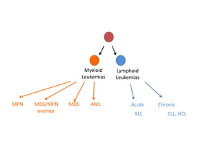
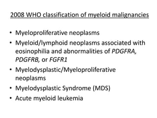
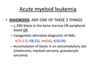
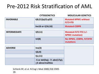
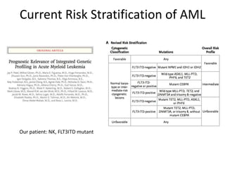
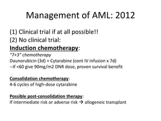
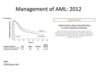
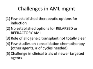
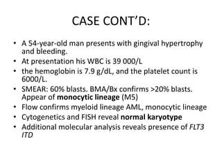
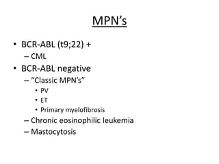
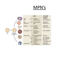
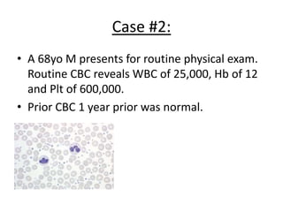
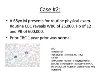
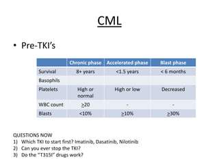
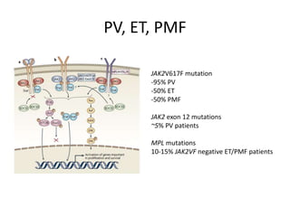
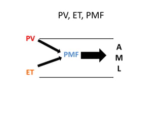
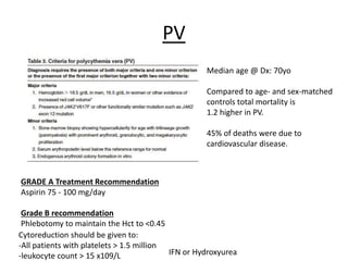
Overview of the Myeloid
Malignancies
Omar Abdel-Wahab, MD
MSK Housestaff Lecture
Myeloid
Leukemias

MPN

MDS/MPN
overlap

MDS

AML

Lymphoid
Leukemias

Acute

ALL

Chronic
CLL, HCL
Overview of the myeloid malignancies
• The most up-to-date classfication is the 2008
WHO CLASSIFICATION
• The FAB Classification of AML (“M0” to “M7”),
although still used is out-dated and does not
take into new molecular understanding of
AML.
Overview of the myeloid malignancies

MYELOPROLIFERATIVE
NEOPLASMS

MYELODYSPLASTIC
SYNDROME

ACUTE MYELOID
LEUKEMIA

ALL ARE CLONAL DISORDERS OF HEMATOPOIESIS
•Increased mature-appearing
Cells
•Less fulminant clinical course
than AML in many instances
(chronic)
•Variable risk of transformation
To AML

•Decreased circulating
Peripheral blood cells
•Abnormal differentiation of
Blood cells in marrow
•Less fulminant clinical course
than AML in many instances
(chronic)
•Variable risk of transformation
To AML

•Decreased circulating mature
Peripheral blood cells
•Presence of immature cells
In BM and/or periphery
•Fulminant clinical course,
Almost invariably lethal without
Therapy.
2008 WHO classification of myeloid malignancies

• Myeloproliferative neoplasms
• Myeloid/lymphoid neoplasms associated with
eosinophilia and abnormalities of PDGFRA,
PDGFRB, or FGFR1
• Myelodysplastic/Myeloproliferative
neoplasms
• Myelodysplastic Syndrome (MDS)
• Acute myeloid leukemia
Acute myeloid leukemia
• DIAGNOSIS: ANY ONE OF THESE 3 THINGS
– > 20% blasts in the bone marrow OR peripheral
blood OR
– Cytogenetic alteration diagnostic of AML:
t(15;17), t(8;21), inv(16), t(16;16)
– Accumulation of blasts in an extramedullary site
(choloroma, myeloid sarcoma, granulocytic
sarcoma)
Acute myeloid leukemia

The best current guideline on how to diagnose, classify (based on 2008 WHO),
And treat AML in adults.
Types of AML
• Former, out of date, classification: FAB CLASS

M3 AML = ACUTE PROMYELOCYTIC LEUKEMIA (APL)
A specific subtype of AML, genetically and clinically
Distinct from any other myeloid malignancy.
A medical emergency; VERY CURABLE!!

Highest mortality is in initial Dx due to DIC.
Except for M3AML the
FAB Subtype of AML
Does not inform prognosis
or management!!

Risk of death from DIC reduced massively with ATRA.
CASE:
• A 54-year-old man presents with gingival
hypertrophy and bleeding.
• At presentation his WBC is 39 000/L
• the hemoglobin is 7.9 g/dL, and the platelet
count is 6000/L.
M3 AML

Majority of patients:
t(15;17)
PML-RARa translocation

Responsive to ATRA.
-Check Smear and DIC labs (PT, PTT,
Fibrinogen, D-dimer) whenever AML
Is suspected
-Start ATRA asap. Oral medication,
Minimal side-effects in short-term
Administration of a few days.

Current standard of care:
ATRA + Cytotoxic chemotherapy
Arsenic Trioxide consolidation
 ATRA maintenance
Very high rate of cure
M6 AML
M6 AML
-acute leukemia of eryrthroid
Precursors
-”Di Guglielmo” disease for
Person who first described it
M7 AML
M7 AML
-acute leukemia of
Megakaryocyte lineage
~1-3% of adult AML cases
-often accompanied by
Fibrosis in BM
-poor px histologic subtype
-more common in children
-Some important associations:
1/3 patients with M7 AML
Have Down Syndrome
Another 1/3 have a
t(1;22)(p13;q13) translocation
The last 1/3 have a novel
Fusion not even published
Yet.
CASE CONT’D:
• A 54-year-old man presents with gingival
hypertrophy and bleeding.
• At presentation his WBC is 39 000/L
• the hemoglobin is 7.9 g/dL, and the platelet
count is 6000/L.
• SMEAR: 60% blasts. BMA/Bx confirms >20%
blasts. Appear of monocytic lineage (M5)
M4/M5 AML

Classically associated with soft-tissue infiltration, especially infiltration of gums.
CASE CONT’D:
• A 54-year-old man presents with gingival hypertrophy
and bleeding.
• At presentation his WBC is 39 000/L
• the hemoglobin is 7.9 g/dL, and the platelet count is
6000/L.
• SMEAR: 60% blasts. BMA/Bx confirms >20% blasts.
Appear of monocytic lineage (M5)
• Flow confirms myeloid lineage AML, monocytic lineage
• Cytogenetics and FISH reveal normal karyotype
• Additional molecular analysis reveals presence of FLT3
ITD
2008 WHO CLASSIFICATION OF AML
• AML with recurrent genetic abnormalities
• AML with myelodysplasia-related changes
• Therapy-related myeloid neoplasms (t-MDS or
t-AML)
• AML not otherwise specified
• Myeloid sarcoma (granulocytic sarcoma,
chloroma)
2008 WHO CLASSIFICATION OF AML
• AML with recurrent genetic abnormalities
–
–
–
–
–
–
–
–
–

T(8;21)
Inv(16)
T(16;16)
T(15;17)
T(9;11)
T(6;9)
Inv(3)
t(1;22)
AML with mutated NPM1 or mutated CEBPA
Genetic abnormalities in AML
• Why do we care?
– Risk stratification of patients (good, intermediate,
adverse risk)
– Clinical management of AML
Pre-2012 Risk Stratification of AML
CYTOGENETICS
FAVORABLE

MOLECULAR GENETICS

Mutated CEBPA

t(9;11)

Mutated FLT3 ITD (+/NPM1 mutation)

tri(8)

ADVERSE

Mutated NPM1 without
FLT3-ITD

Inv16 or t(16;16)
INTERMEDIATE

t(8;21)(q22;q22)

No NPM1, CEBPA, FLT3ITD
mutations

Inv(3)
t(6;9)
t(v;11)
-5 or del(5q); -7; abn(17p);
>3 abnormalities

Schlenk RF, et al. N Engl J Med 2008;358:190918.
Current Risk Stratification of AML

Our patient: NK, FLT3ITD mutant
Management of AML: 2012
(1) Clinical trial if at all possible!!
(2) No clinical trial:
Induction chemotherapy:
“7+3” chemotherapy
Daunorubicin (3d) + Cytarabine (cont IV infusion x 7d)
--If <60 give 90mg/m2 DNR dose, proven survival benefit
Consolidation chemotherapy:
4-6 cycles of high-dose cytarabine
Possible post-consolidation therapy:
If intermediate risk or adverse risk  allogeneic transplant
Management of AML: 2012

AML
16-60 years old
Challenges in AML mgmt
(1) Few established therapeutic options for
induction
(2) No established options for RELAPSED or
REFRACTORY AML
(3) Role of allogeneic transplant not totally clear
(4) Few studies on consolidation chemotherapy
(other agents, # of cycles needed)
(5) Challenge in clinical trials of newer targeted
agents
CASE CONT’D:
• A 54-year-old man presents with gingival hypertrophy
and bleeding.
• At presentation his WBC is 39 000/L
• the hemoglobin is 7.9 g/dL, and the platelet count is
6000/L.
• SMEAR: 60% blasts. BMA/Bx confirms >20% blasts.
Appear of monocytic lineage (M5)
• Flow confirms myeloid lineage AML, monocytic lineage
• Cytogenetics and FISH reveal normal karyotype
• Additional molecular analysis reveals presence of FLT3
ITD
MSKCC AML CLINICAL TRIALS
Overview of the myeloid malignancies

MYELOPROLIFERATIVE
NEOPLASMS

MYELODYSPLASTIC
SYNDROME

ACUTE MYELOID
LEUKEMIA

ALL ARE CLONAL DISORDERS OF HEMATOPOIESIS
•Increased mature-appearing
Cells
•Less fulminant clinical course
than AML in many instances
(chronic)
•Variable risk of transformation
To AML

•Decreased circulating
Peripheral blood cells
•Abnormal differentiation of
Blood cells in marrow
•Less fulminant clinical course
than AML in many instances
(chronic)
•Variable risk of transformation
To AML

•Decreased circulating mature
Peripheral blood cells
•Presence of immature cells
In BM and/or periphery
•Fulminant clinical course,
Almost invariably lethal without
Therapy.
MPN’s
• BCR-ABL (t9;22) +
– CML

• BCR-ABL negative
– “Classic MPN’s”
• PV
• ET
• Primary myelofibrosis

– Chronic eosinophilic leukemia
– Mastocytosis
MPN’s
Case #2:
• A 68yo M presents for routine physical exam.
Routine CBC reveals WBC of 25,000, Hb of 12
and Plt of 600,000.
• Prior CBC 1 year prior was normal.
Case #2:
• A 68yo M presents for routine physical exam.
Routine CBC reveals WBC of 25,000, Hb of 12
and Plt of 600,000.
• Prior CBC 1 year prior was normal.
W/U:
-Differential
-Iron studies (ferriting, Fe, TIBC)
-Smear
-BMA/Bx for review, FISH/cytogenetics,
BCR-ABL translocation testing by qRTPCR,
and JAK2V617F mutation (possibly also MPL
Mutation)
CML
• Pre-TKI’s
Chronic phase Accelerated phase
Survival

Blast phase

8+ years

<1.5 years

< 6 months

High or
normal

High or low

Decreased

>20

-

-

<10%

>10%

>30%

Basophils
Platelets
WBC count
Blasts

QUESTIONS NOW
1) Which TKI to start first? Imatinib, Dasatinib, Nilotinib
2) Can you ever stop the TKI?
3) Do the “T315I” drugs work?
PV, ET, PMF
JAK2V617F mutation
-95% PV
-50% ET
-50% PMF
JAK2 exon 12 mutations
~5% PV patients
MPL mutations
10-15% JAK2VF negative ET/PMF patients
PV, ET, PMF
PV

PMF
ET

A
M
L
PV
Median age @ Dx: 70yo
Compared to age- and sex-matched
controls total mortality is
1.2 higher in PV.

45% of deaths were due to
cardiovascular disease.

GRADE A Treatment Recommendation
Aspirin 75 - 100 mg/day
Grade B recommendation
Phlebotomy to maintain the Hct to <0.45
Cytoreduction should be given to:
-All patients with platelets > 1.5 million
IFN or Hydroxyurea
-leukocyte count > 15 x109/L
ET

It is unclear if ET shortens
Life expectancy on
average.
Studies which found a shorter life
expectancy:
Fenaux et al: 73.5% survival 7
years after diagnosis
Jensen et al: 76% survival
at 5 years

REACTIVE THROMBOCYTOSIS CAUSES:
iron deficiency, splenectomy, surgery,
infection, inflammation, connective tissue
disease, metastatic cancer, and
lymphoproliferative disorders
Management of ET
Grade A recommendation
• Platelet lowering therapy should be given to;
- all patients over 60 years of age
- all patients with earlier thromboembolic complications
-all patients with platelets >1.5million
• Aspirin 75-100 mg daily to all ET patients except when platelets are >1500 x 109/L, in
patients with bleeding symptoms or in patients with other contraindications to aspirin.

• The goal of platelet lowering therapy should be platelets < 400 x 109/L
(Grade B recommendation)

Choice of cytoreductive therapy in ET:
• Hydroxyurea is the best documented therapy in ET
• However, due to the concern of possible increased risk of leukemia transformation with
long-term use it is not recommended as 1st line therapy in younger patients
• <60 years: 1st line interferon-α or anagrelide, 3rd line Hydroxyurea
• >75 years: 1st line Hydroxyurea, 2nd line consider combination therapy (HU-Ana, HU-IFN),
PMF
PMF
SPLENOMEGALY
EARLY SATIETY
FATIGUE
CYTOPENIAS
HIGH RISK OF AML
TRANSFORMATION
2012 FDA-APPROVAL FOR JAK1/2
INHIBITOR Ruxolitinib for MF
2012 FDA-APPROVAL FOR JAK1/2
INHIBITOR Ruxolitinib for MF
Questions now in MPN’s
1) Do JAK inhibitors change the natural history of PMF?
2) Should we use combination strategies of JAKi + something else?
3) Many newer JAK inhibitors coming are they better than ruxolitinib? (JAK2 specific,
JAK1 specific, JAK2V617F specific)
4) What is the genetic abnormality present in the 50% of ET and PMF without
JAK2 or MPL mutations?
5) Are JAK2 mutations really driving the disease?
Myelodysplastic Syndrome
MDS with low blast % can be very difficult to diagnose and the true incidence of
MDS is likely not known.
Definition: >10% of the cells >1 myeloid BM lineage (erythroid, granulocytic, megakaryocytic
must show morphologic dysplasia.
Also, in setting of persistent cytopenia, if the patient has one of the following cytogenetic
Abnormalities, MDS may be diagnosed:
Case #3
78y F presents with CC of slight increase in DOE over last few months. She has a
h/o HTN and type II DM. Physical exam is unrevealing. CBC reveals Hb 8.2g/dL,
WBC 1.8, and Plt 170,000.
What is dysplasia?
Some common morphologic features characteristic of MDS:
-hypolobated neutrophils in peripheral blood
-increased reticulocytes in peripheral blood
-abnormal contours of erythroid precursors in bone marrow
-micromegakaryocytes in bone marrow
-increased RBC precursors with rings of iron around the nuclei
(ringed sideroblasts)
-Also MDS usually characterized by a hypercellular bone marrow
(but not always)
2008 WHO CLASSIFICATION OF MDS
Disease

Blood findings

BM findings

Refractory cytopenia with
unilineage dysplasia

1-2 cytopenias

>10% of the cells in one myeloid
lineage dysplasia;
<5% blasts
<15% RBC prec with ringed
sideroblasts

Refractory anemia with
ringed sideroblasts (RARS)

Anemia

>15% ringed sideroblasts

Erythroid dysplasia only
<5% blasts

Refractory cytopenia with
multilineage dysplasia
(RCMD)

Cytopenias

Dysplasia, <5% blasts

Refractory anemia with
excess blasts 1 (RAEB-1)

Cytopenias

5-9% blasts

Refractory anemia with
excess blasts 2 (RAEB-2)

Cytopenias

10-19% blasts

MDS with isolated del 5q

Anemia with normal to
High plt count

Isolated deletion 5q
Assessing risk in MDS

MDS CLASSIFICATION SCORING SYSTEM
IPSS
R-IPSS
WHO PSS (takes into account WHO category of MDS)
MDACC MDS Scoring System (takes into account peformance status)
Newer systems incorporating molecular mutations coming….
Assessing risk in MDS
Treatment options for MDS
1) Supportive care: transfusions, ESA, GCSF, ESA + GCSF
2) Lenalidomide  especially for MDS with Isolated deletion 5q
3) Hypomethylating agents  Decitabine or 5-Azacitidine
4) If RAEB-1 or -2 in young patient, sometimes treated as if AML with induction
Chemotherapy +/- allogeneic transplant
MPN, MPN/MDS overlap, MDS
TET2
ASXL1
SRSF2

JAK2
MPL
BCR-ABL

MPN

PV
ET
CML

PMF

RARS-t

EZH2

MDS/MPN

MDS with
fibrosis

del5q

MDS

RA
RARS--SF3B1
MDS/MPN overlap syndromes
Chronic myelomonocytic leukemia
Atypical CML, BCR-ABL1 negative
Juvenile myelomonocytic leukemia
MDS/MPN-U
RARS-t
CMML
CMML
Monocytosis of >1 X 10⁹/l + dyplasia
(less than 20% of cells in bone marrow).
CMML-1: <10% blasts in BM
CMML-2: 10-19% blasts in BM

RARS-t
RA + Ringed Sideroblasts + Thrombocytosis
Mixed MPN (JAK2 or MPL mutation) and MDS
>15% ringed sideroblasts + Plt count >450K

myeloid malignancy overview

  • 1.
    Overview of theMyeloid Malignancies Omar Abdel-Wahab, MD MSK Housestaff Lecture
  • 2.
  • 3.
    Overview of themyeloid malignancies • The most up-to-date classfication is the 2008 WHO CLASSIFICATION • The FAB Classification of AML (“M0” to “M7”), although still used is out-dated and does not take into new molecular understanding of AML.
  • 4.
    Overview of themyeloid malignancies MYELOPROLIFERATIVE NEOPLASMS MYELODYSPLASTIC SYNDROME ACUTE MYELOID LEUKEMIA ALL ARE CLONAL DISORDERS OF HEMATOPOIESIS •Increased mature-appearing Cells •Less fulminant clinical course than AML in many instances (chronic) •Variable risk of transformation To AML •Decreased circulating Peripheral blood cells •Abnormal differentiation of Blood cells in marrow •Less fulminant clinical course than AML in many instances (chronic) •Variable risk of transformation To AML •Decreased circulating mature Peripheral blood cells •Presence of immature cells In BM and/or periphery •Fulminant clinical course, Almost invariably lethal without Therapy.
  • 5.
    2008 WHO classificationof myeloid malignancies • Myeloproliferative neoplasms • Myeloid/lymphoid neoplasms associated with eosinophilia and abnormalities of PDGFRA, PDGFRB, or FGFR1 • Myelodysplastic/Myeloproliferative neoplasms • Myelodysplastic Syndrome (MDS) • Acute myeloid leukemia
  • 6.
    Acute myeloid leukemia •DIAGNOSIS: ANY ONE OF THESE 3 THINGS – > 20% blasts in the bone marrow OR peripheral blood OR – Cytogenetic alteration diagnostic of AML: t(15;17), t(8;21), inv(16), t(16;16) – Accumulation of blasts in an extramedullary site (choloroma, myeloid sarcoma, granulocytic sarcoma)
  • 7.
    Acute myeloid leukemia Thebest current guideline on how to diagnose, classify (based on 2008 WHO), And treat AML in adults.
  • 8.
    Types of AML •Former, out of date, classification: FAB CLASS M3 AML = ACUTE PROMYELOCYTIC LEUKEMIA (APL) A specific subtype of AML, genetically and clinically Distinct from any other myeloid malignancy. A medical emergency; VERY CURABLE!! Highest mortality is in initial Dx due to DIC. Except for M3AML the FAB Subtype of AML Does not inform prognosis or management!! Risk of death from DIC reduced massively with ATRA.
  • 9.
    CASE: • A 54-year-oldman presents with gingival hypertrophy and bleeding. • At presentation his WBC is 39 000/L • the hemoglobin is 7.9 g/dL, and the platelet count is 6000/L.
  • 10.
    M3 AML Majority ofpatients: t(15;17) PML-RARa translocation Responsive to ATRA. -Check Smear and DIC labs (PT, PTT, Fibrinogen, D-dimer) whenever AML Is suspected -Start ATRA asap. Oral medication, Minimal side-effects in short-term Administration of a few days. Current standard of care: ATRA + Cytotoxic chemotherapy Arsenic Trioxide consolidation  ATRA maintenance Very high rate of cure
  • 11.
    M6 AML M6 AML -acuteleukemia of eryrthroid Precursors -”Di Guglielmo” disease for Person who first described it
  • 12.
    M7 AML M7 AML -acuteleukemia of Megakaryocyte lineage ~1-3% of adult AML cases -often accompanied by Fibrosis in BM -poor px histologic subtype -more common in children -Some important associations: 1/3 patients with M7 AML Have Down Syndrome Another 1/3 have a t(1;22)(p13;q13) translocation The last 1/3 have a novel Fusion not even published Yet.
  • 13.
    CASE CONT’D: • A54-year-old man presents with gingival hypertrophy and bleeding. • At presentation his WBC is 39 000/L • the hemoglobin is 7.9 g/dL, and the platelet count is 6000/L. • SMEAR: 60% blasts. BMA/Bx confirms >20% blasts. Appear of monocytic lineage (M5)
  • 14.
    M4/M5 AML Classically associatedwith soft-tissue infiltration, especially infiltration of gums.
  • 15.
    CASE CONT’D: • A54-year-old man presents with gingival hypertrophy and bleeding. • At presentation his WBC is 39 000/L • the hemoglobin is 7.9 g/dL, and the platelet count is 6000/L. • SMEAR: 60% blasts. BMA/Bx confirms >20% blasts. Appear of monocytic lineage (M5) • Flow confirms myeloid lineage AML, monocytic lineage • Cytogenetics and FISH reveal normal karyotype • Additional molecular analysis reveals presence of FLT3 ITD
  • 16.
    2008 WHO CLASSIFICATIONOF AML • AML with recurrent genetic abnormalities • AML with myelodysplasia-related changes • Therapy-related myeloid neoplasms (t-MDS or t-AML) • AML not otherwise specified • Myeloid sarcoma (granulocytic sarcoma, chloroma)
  • 17.
    2008 WHO CLASSIFICATIONOF AML • AML with recurrent genetic abnormalities – – – – – – – – – T(8;21) Inv(16) T(16;16) T(15;17) T(9;11) T(6;9) Inv(3) t(1;22) AML with mutated NPM1 or mutated CEBPA
  • 18.
    Genetic abnormalities inAML • Why do we care? – Risk stratification of patients (good, intermediate, adverse risk) – Clinical management of AML
  • 19.
    Pre-2012 Risk Stratificationof AML CYTOGENETICS FAVORABLE MOLECULAR GENETICS Mutated CEBPA t(9;11) Mutated FLT3 ITD (+/NPM1 mutation) tri(8) ADVERSE Mutated NPM1 without FLT3-ITD Inv16 or t(16;16) INTERMEDIATE t(8;21)(q22;q22) No NPM1, CEBPA, FLT3ITD mutations Inv(3) t(6;9) t(v;11) -5 or del(5q); -7; abn(17p); >3 abnormalities Schlenk RF, et al. N Engl J Med 2008;358:190918.
  • 20.
    Current Risk Stratificationof AML Our patient: NK, FLT3ITD mutant
  • 21.
    Management of AML:2012 (1) Clinical trial if at all possible!! (2) No clinical trial: Induction chemotherapy: “7+3” chemotherapy Daunorubicin (3d) + Cytarabine (cont IV infusion x 7d) --If <60 give 90mg/m2 DNR dose, proven survival benefit Consolidation chemotherapy: 4-6 cycles of high-dose cytarabine Possible post-consolidation therapy: If intermediate risk or adverse risk  allogeneic transplant
  • 22.
    Management of AML:2012 AML 16-60 years old
  • 23.
    Challenges in AMLmgmt (1) Few established therapeutic options for induction (2) No established options for RELAPSED or REFRACTORY AML (3) Role of allogeneic transplant not totally clear (4) Few studies on consolidation chemotherapy (other agents, # of cycles needed) (5) Challenge in clinical trials of newer targeted agents
  • 24.
    CASE CONT’D: • A54-year-old man presents with gingival hypertrophy and bleeding. • At presentation his WBC is 39 000/L • the hemoglobin is 7.9 g/dL, and the platelet count is 6000/L. • SMEAR: 60% blasts. BMA/Bx confirms >20% blasts. Appear of monocytic lineage (M5) • Flow confirms myeloid lineage AML, monocytic lineage • Cytogenetics and FISH reveal normal karyotype • Additional molecular analysis reveals presence of FLT3 ITD
  • 25.
  • 26.
  • 27.
    Overview of themyeloid malignancies MYELOPROLIFERATIVE NEOPLASMS MYELODYSPLASTIC SYNDROME ACUTE MYELOID LEUKEMIA ALL ARE CLONAL DISORDERS OF HEMATOPOIESIS •Increased mature-appearing Cells •Less fulminant clinical course than AML in many instances (chronic) •Variable risk of transformation To AML •Decreased circulating Peripheral blood cells •Abnormal differentiation of Blood cells in marrow •Less fulminant clinical course than AML in many instances (chronic) •Variable risk of transformation To AML •Decreased circulating mature Peripheral blood cells •Presence of immature cells In BM and/or periphery •Fulminant clinical course, Almost invariably lethal without Therapy.
  • 28.
    MPN’s • BCR-ABL (t9;22)+ – CML • BCR-ABL negative – “Classic MPN’s” • PV • ET • Primary myelofibrosis – Chronic eosinophilic leukemia – Mastocytosis
  • 29.
  • 30.
    Case #2: • A68yo M presents for routine physical exam. Routine CBC reveals WBC of 25,000, Hb of 12 and Plt of 600,000. • Prior CBC 1 year prior was normal.
  • 31.
    Case #2: • A68yo M presents for routine physical exam. Routine CBC reveals WBC of 25,000, Hb of 12 and Plt of 600,000. • Prior CBC 1 year prior was normal. W/U: -Differential -Iron studies (ferriting, Fe, TIBC) -Smear -BMA/Bx for review, FISH/cytogenetics, BCR-ABL translocation testing by qRTPCR, and JAK2V617F mutation (possibly also MPL Mutation)
  • 32.
    CML • Pre-TKI’s Chronic phaseAccelerated phase Survival Blast phase 8+ years <1.5 years < 6 months High or normal High or low Decreased >20 - - <10% >10% >30% Basophils Platelets WBC count Blasts QUESTIONS NOW 1) Which TKI to start first? Imatinib, Dasatinib, Nilotinib 2) Can you ever stop the TKI? 3) Do the “T315I” drugs work?
  • 33.
    PV, ET, PMF JAK2V617Fmutation -95% PV -50% ET -50% PMF JAK2 exon 12 mutations ~5% PV patients MPL mutations 10-15% JAK2VF negative ET/PMF patients
  • 34.
  • 35.
    PV Median age @Dx: 70yo Compared to age- and sex-matched controls total mortality is 1.2 higher in PV. 45% of deaths were due to cardiovascular disease. GRADE A Treatment Recommendation Aspirin 75 - 100 mg/day Grade B recommendation Phlebotomy to maintain the Hct to <0.45 Cytoreduction should be given to: -All patients with platelets > 1.5 million IFN or Hydroxyurea -leukocyte count > 15 x109/L
  • 36.
    ET It is unclearif ET shortens Life expectancy on average. Studies which found a shorter life expectancy: Fenaux et al: 73.5% survival 7 years after diagnosis Jensen et al: 76% survival at 5 years REACTIVE THROMBOCYTOSIS CAUSES: iron deficiency, splenectomy, surgery, infection, inflammation, connective tissue disease, metastatic cancer, and lymphoproliferative disorders
  • 37.
    Management of ET GradeA recommendation • Platelet lowering therapy should be given to; - all patients over 60 years of age - all patients with earlier thromboembolic complications -all patients with platelets >1.5million • Aspirin 75-100 mg daily to all ET patients except when platelets are >1500 x 109/L, in patients with bleeding symptoms or in patients with other contraindications to aspirin. • The goal of platelet lowering therapy should be platelets < 400 x 109/L (Grade B recommendation) Choice of cytoreductive therapy in ET: • Hydroxyurea is the best documented therapy in ET • However, due to the concern of possible increased risk of leukemia transformation with long-term use it is not recommended as 1st line therapy in younger patients • <60 years: 1st line interferon-α or anagrelide, 3rd line Hydroxyurea • >75 years: 1st line Hydroxyurea, 2nd line consider combination therapy (HU-Ana, HU-IFN),
  • 38.
  • 39.
  • 40.
    2012 FDA-APPROVAL FORJAK1/2 INHIBITOR Ruxolitinib for MF
  • 41.
    2012 FDA-APPROVAL FORJAK1/2 INHIBITOR Ruxolitinib for MF
  • 42.
    Questions now inMPN’s 1) Do JAK inhibitors change the natural history of PMF? 2) Should we use combination strategies of JAKi + something else? 3) Many newer JAK inhibitors coming are they better than ruxolitinib? (JAK2 specific, JAK1 specific, JAK2V617F specific) 4) What is the genetic abnormality present in the 50% of ET and PMF without JAK2 or MPL mutations? 5) Are JAK2 mutations really driving the disease?
  • 43.
    Myelodysplastic Syndrome MDS withlow blast % can be very difficult to diagnose and the true incidence of MDS is likely not known. Definition: >10% of the cells >1 myeloid BM lineage (erythroid, granulocytic, megakaryocytic must show morphologic dysplasia. Also, in setting of persistent cytopenia, if the patient has one of the following cytogenetic Abnormalities, MDS may be diagnosed:
  • 44.
    Case #3 78y Fpresents with CC of slight increase in DOE over last few months. She has a h/o HTN and type II DM. Physical exam is unrevealing. CBC reveals Hb 8.2g/dL, WBC 1.8, and Plt 170,000.
  • 45.
    What is dysplasia? Somecommon morphologic features characteristic of MDS: -hypolobated neutrophils in peripheral blood -increased reticulocytes in peripheral blood -abnormal contours of erythroid precursors in bone marrow -micromegakaryocytes in bone marrow -increased RBC precursors with rings of iron around the nuclei (ringed sideroblasts) -Also MDS usually characterized by a hypercellular bone marrow (but not always)
  • 46.
    2008 WHO CLASSIFICATIONOF MDS Disease Blood findings BM findings Refractory cytopenia with unilineage dysplasia 1-2 cytopenias >10% of the cells in one myeloid lineage dysplasia; <5% blasts <15% RBC prec with ringed sideroblasts Refractory anemia with ringed sideroblasts (RARS) Anemia >15% ringed sideroblasts Erythroid dysplasia only <5% blasts Refractory cytopenia with multilineage dysplasia (RCMD) Cytopenias Dysplasia, <5% blasts Refractory anemia with excess blasts 1 (RAEB-1) Cytopenias 5-9% blasts Refractory anemia with excess blasts 2 (RAEB-2) Cytopenias 10-19% blasts MDS with isolated del 5q Anemia with normal to High plt count Isolated deletion 5q
  • 47.
    Assessing risk inMDS MDS CLASSIFICATION SCORING SYSTEM IPSS R-IPSS WHO PSS (takes into account WHO category of MDS) MDACC MDS Scoring System (takes into account peformance status) Newer systems incorporating molecular mutations coming….
  • 48.
  • 49.
    Treatment options forMDS 1) Supportive care: transfusions, ESA, GCSF, ESA + GCSF 2) Lenalidomide  especially for MDS with Isolated deletion 5q 3) Hypomethylating agents  Decitabine or 5-Azacitidine 4) If RAEB-1 or -2 in young patient, sometimes treated as if AML with induction Chemotherapy +/- allogeneic transplant
  • 50.
    MPN, MPN/MDS overlap,MDS TET2 ASXL1 SRSF2 JAK2 MPL BCR-ABL MPN PV ET CML PMF RARS-t EZH2 MDS/MPN MDS with fibrosis del5q MDS RA RARS--SF3B1
  • 51.
    MDS/MPN overlap syndromes Chronicmyelomonocytic leukemia Atypical CML, BCR-ABL1 negative Juvenile myelomonocytic leukemia MDS/MPN-U RARS-t
  • 52.
  • 53.
    CMML Monocytosis of >1X 10⁹/l + dyplasia (less than 20% of cells in bone marrow). CMML-1: <10% blasts in BM CMML-2: 10-19% blasts in BM RARS-t RA + Ringed Sideroblasts + Thrombocytosis Mixed MPN (JAK2 or MPL mutation) and MDS >15% ringed sideroblasts + Plt count >450K