2. Leukemia is the commonest
pediatric malignancy,
accounting for 1/3 of all cancers
Def: Maliganant clonal
proliferation of lymphoid or
myeloid precursor cells in the
bone marrow as well as
infilteration of other organs
with these cells.
3.
4. SUB-CLASSIFICATION OF LEUKEMIA
Other less common variants, such as mature B-cell and T-cell leukemias,
and NK cell-related leukemias, to name a few, arise from mature white
blood cells
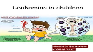
7. General Systemic Effects
1. Fever (60%).
2. Lassitude (50%)
3. Pallor (40%)
Hematologic Effects Arising from Bone Marrow Invasion
1. Anaemia
– pallor, fatigability, tachycardia, dyspnoea & CHF
2. Neutropenia
– fever, ulceration of buccal mucosa and infection.
3. Thrombocytopenia
– petechial, purpura, easy bruisability, bleeding from
mucous membrane and internal bleeding.
CLINICAL FEATURES
8. Clinical Manifestations Arising from Lymphoid
System Infiltration
1. Lymphadenopathy
2. Splenomegaly.
3. Hepatomegaly
CLINICAL FEATURES
9. Clinical Manifestations of Extramedullary Invasion
• Mediastianal mass: SVC compression, dysphagia and dyspnea
• CNS‐ ICT symptoms, seizures
• Genitourinary ‐ painless testicular swelling
• Bone joints‐ bone pain
• Skin ‐ bleeds
• Git ‐ bleeds
CLINICAL FEATURES
10. CONTINUOUS FEVER, WEIGHT LOSS
HEADACHES, EARLY MORNING VOMITION
INCREASED SWELLING OR PERSISTENT PAIN IN BONES, JOINTS, BACK OR LEGS
LUMP OR MASS – ABDO, NECK, CHEST, PELVIS, ARMPITS
DEVELOPMENT OF RASH, BLEEDING, BRUISION
CONSTANT / RECURENT INFECTIONS
AWHITISH COLOR BEHIND PUPIL
NAUSEA – PERSISTANT OR VOMITING WITHO OR W/O SEIZURE
CONSTANT TIREDNESS
EYE OR VISON CHANGES
RECURRENT OR PERSISTENT FEVER
CLINICAL FEATURES – “Childhood Cancer”
11. • Blood count
• Haemoglobin: Moderate to marked reduction
• Blood smear: Blasts are present on blood smear. Very few to none (in
patients with leukopenia).
• White blood cell count: Low, normal, or increased
• Thrombocytopenia: 92% of patients have platelet counts below normal.
Very few to none (in patients with leukopenia).
INVESTIGATION
12. INVESTIGATION – BONE MARROW
• Leukemia must be suspected when the bone marrow contains more than
20 % blasts.
• The hallmark of the diagnosis of acute leukemia is the blast cell, are
relatively undifferentiated cell with diffusely distributed nuclear chromatin,
one or more nucleoli and basophilic cytoplasm.
14. • Chest radiograph: Mediastinal mass in T‐cell leukemia.
• Blood chemistry: Electrolytes, blood urea, uric acid,
• Liver function tests, Immuno globulin levels.
• Coagulation profile: Decreased coagulation factors that frequently occur
with AML are: hypofibrinogenemia, factors V, IX and X.
INVESTIGATION
15. • Three phases:
1. remission induction,
2. consolidation (or intensification), and
3. continuation (or maintenance).
• Protocol adopted depends on the institution
• Modified BFM or COG protocol is often the choice
TREATMENT
16. • Prednisolone 60 mg/m2/day
• Inj.VCR 1.5mg/m2 (weekly, max 2mg)
• IM Asparginase 10,000 U/m2 (bi-weekly)
• PO Prednisolone 40mg/m2 (daily)
INDUCTION
18. • Inj. VCR 1.5 mg/m2 one in a month
• PO PREDNISOLONE 60 mg/m2 for one week
• PO 6MP (mercaptopurine) 50 mg/m2 daily
• PO MTX 20 mg/m2 p.o weekly
The optimal duration of therapy remains unknown. Most investigators
continue to treat patients for 2 to 3 years, based on results of older studies
MAINTENANCE
19. If the patient completes chemotherapy for 2 years without
relapse-stop chemo and follow up.
No relapse within 5 years-can be declared as cured.
FOLLOW UP
20. • Maintain adequate nutrtion and hydration
• Correct fluid and electrolyte imbalance
• Correct severe anemia : use of packed red cells
• When high fever and possible septicemia occur in the presence of neutropenia,
antibiotic therapy should be started after taking appropriate blood cultures
and a chest radiograph.(NEUTROPENIA REGIME)
• Platelet transfusions should be administered to patients with overt bleeding or
when the platelet count is below 10,000/mm3.
• Treatment of metabolic complication eg hyperuricemia (allopurinol)
• Monitering and treatment of chemotherapy related adverse effect
• Psychological support to patient and family
SUPPORTIVE CARE
22. FACTOR FAVOURABLE UNFAVOURABLE
Age (yrs) 1 – 9 < 1 OR > 10
WBC count < 20,000 > 50,000
Immunophenotype Precursor B Cell T Cell
Genetics Hyperploidy Hypoploidy , translocation
CNS Status CNS 1 CNS 3
MRD (end of induction) < 0.01% 0.5 or 1%
Testicular / CNS involvement Absent Present
Remission Early Delayed or early relapse
Ethnicity White Black
RISK STRATIFICATION
23. • ITP‐
• isolated thrombocytopenia,
• well child with no lymph node enlargement or spleenomegaly
• Aplastic Anaemia
• Pancytopenia with
• no organ enlargement
• Juvenile Rheumatoid Arthritis
• Infectious mononucleosis
• Atypical lymphocytes
• Metastatic solid tumours
DD
24. • Despite current intensive front‐line treatments, 20% of children with ALL
experience bone marrow relapse.
• Relapse may be an isolated event in the bone marrow or may be
combined with relapse in other sites
RELAPSE
25. SUMMARY
ALL :
• 3-4 Times more common in males
• Etiology: genetic/ environmental factor
• Pathology : increased number of lymphoblasts in bone marrow with or without their presence in peripheral
blood
• Lymphoblast characterised:
Typical morphology: high nucleus-cytoplasma ratio,dense nuclear chromatin, basophilic non-granular
cytoplasma
Cytochemical characteristics: coarse granules/clumps on PAS staining,absence of peroxidase or sudan- black
positive granules
Biological marker: +terminal desoxynucleotidyl transferase (TdT)
• Classification : WHO CLASSIFICATION (based on lineage of blast cells and presence of immunological marker)
B-Lymphoblastic leukemia, T-Lymphoblastic leukemia, Burkitt leukemia (arising from mature B Cell)
• Clinical features: signs of primary bone marrow failure, signs of secondary organ infilteration
• Diagnosis: PS, BM, CBC, CHEST XRAY, Skeletal XRAY, CSF, Pre- Treatment workup
• Treatment :
Induction (4-6 weeks): IV Vincristin +IM Asperginase+ PO Prednisolone
INTRATHECAL CNS prophylaxis (weekely during induction, than 8 weekly for 2 years): IT Methotrexate+ IT
Hydrcortisone+ IT Ara-C (Cytarabin)
Maintenance therapy (for 2 years): PO 6-Mercaptopurine (daily), PO Methoteraxate (weekly )
Reinforcement therapy (Every 4 weekly during maintenance): IV Vincristine + PO Prednisolone
26. AML:
• 15-20% CHILDHOOD LEUKEMIA
• ASSOCIATED – Genetic disorders: downs, fanconi anemia,bloom syndrome, neurofibromatosis or
developed secondary malignancy after chemotherapy or radiation for other malignancy.
• Clinical presentation : S/S ALL+
severe anemia, bleeding and CHLOROMA (a localized mass of leukemic cells on choroid and epidural
region.)
ACUTE PROMYELOCYTIC TYPE of AML – gum hypertrophy, gum bleeding or severe DIC ( release of
pro-coagulant substance from leukemic blast cell. )
Subcutaneous nodules (BLUEBERRY MUFFINS ) are common in infants.
• Diagnosis: who classification (next slide)
• Treatment :
Anthracyclione and cytosine arabinoside with or without other agents + retinoic acid in induction
phase
Initial remission may be achieved in > 80%, though relapse are common
Supportive treatment is most important in AML
• AML with Downs has favorable prognosis while bad prognosis: severe sepsis/ bleeding on presentation,
Secondary AML, early relapse
SUMMARY
27. Acute myeloid leukemia with recurrent genetic abnormalities
Acute myeloid leukemia with t(8;21)(q22;q22), (AML1/ETO)
Acute myeloid leukemia with abnormal bone marrow eosinophils and inv(16)(p13q22) or t(16;16)(p13;q22), (CBFβ/MYH11)
Acute promyelocytic leukemia with t(15;17)(q22;q12), (PML/RARα) and variants
Acute myeloid leukemia with 11q23 (MLL) abnormalities
Acute myeloid leukemia with multilineage dysplasia
Following MDS or MDS/MPD
Without antecedent MDS or MDS/MPD, but with dysplasia in at least 50% of cells in 2 or more myeloid lineages
Acute myeloid leukemia and myelodysplastic syndromes, therapy related
Alkylating agent/radiation–related type
Topoisomerase II inhibitor–related type (some may be lymphoid)
Others
Acute myeloid leukemia, not otherwise categorized
Classify as:
Acute myeloid leukemia, minimally differentiated
Acute myeloid leukemia without maturation
Acute myeloid leukemia with maturation
Acute myelomonocytic leukemia
Acute monoblastic/acute monocytic leukemia
Acute erythroid leukemia (erythroid/myeloid and pure erythroleukemia)
Acute megakaryoblastic leukemia
Acute basophilic leukemia
Acute panmyelosis with myelofibrosis
Myeloid sarcoma
28. JUVENILE CML
• Aka JUVENILE MYELOMONOCYTIC LEUKEMIA
• <5 YEARS with neurofibromatosis
• Clinically : splenohepatomegaly, lymphadenopathy, skin lesions( eczema, xanthoma,
cafeau lait spots)and signs of bone marrow failure (pallor, bleeding )
• Pathologically:
Increased monocyte precursors in bone marrow with
Typical monocytosis in peripheral blood
Increased fetal Hb
• Treatment: bone marrow as chemotherapy is not effective