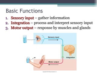
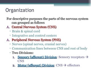
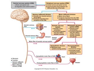
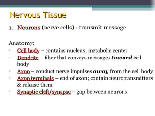
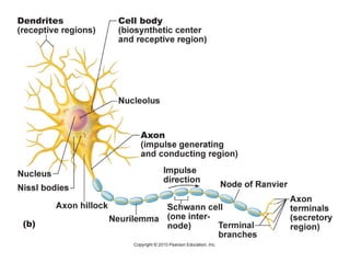
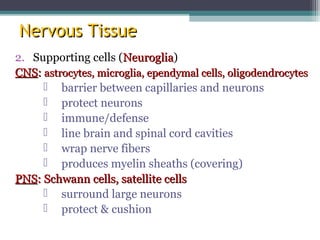
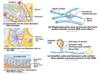
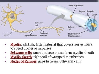
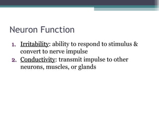
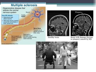
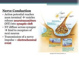
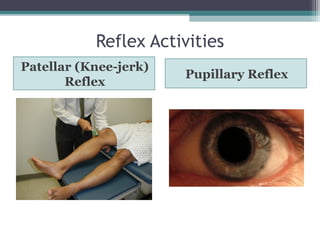
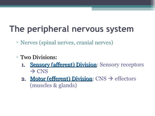
The Nervous System
DR MWAIHOJO JUSTIN
MD, BMH
• Detects and responds to changes inside and
outside the body.
• Provide immediate response to stimuli.
• It consist of the brain, spinal cord and peripheral
nerves.
Basic Functions
1.1. Sensory inputSensory input – gather information
2.2. IntegrationIntegration – process and interpret sensory input
3.3. Motor outputMotor output – response by muscles and glands
Organization
For descriptive purposes the parts of the nervous systemFor descriptive purposes the parts of the nervous system
can grouped as followscan grouped as follows
A.A. Central Nervous System (CNS)Central Nervous System (CNS)
▫ Brain & spinal cord
▫ Integrative and control centers
A.A. Peripheral Nervous System (PNS)Peripheral Nervous System (PNS)
▫ Nerves (spinal nerves, cranial nerves)
▫ Communication lines between CNS and rest of body
▫ Two Divisions:Two Divisions:
1.1. Sensory (afferent) DivisionSensory (afferent) Division: Sensory receptors 
CNS
2.2. Motor (efferent) DivisionMotor (efferent) Division: CNS  effectors
Motor Division
• Somatic nervous systemSomatic nervous system (voluntary) – control
skeletal muscles
• Autonomic nervous systemAutonomic nervous system (ANS) (involuntary)
– regulate smooth muscles, cardiac, glands
▫ Subdivisions: sympatheticsympathetic & parasympatheticparasympathetic
Nervous System
• Master controlling and communicating system
Nervous TissueNervous Tissue
1.1. NeuronsNeurons (nerve cells) - transmit message
Anatomy:
▫ Cell bodyCell body – contains nucleus; metabolic center
▫ DendriteDendrite – fiber that conveys messages toward cell
body
▫ AxonAxon – conduct nerve impulses away from the cell body
▫ Axon terminalsAxon terminals – end of axon; contain neurotransmitters
& release them
▫ Synaptic cleft/synapseSynaptic cleft/synapse – gap between neurons
Nervous TissueNervous Tissue
2. Supporting cells (NeurogliaNeuroglia)
CNSCNS:: astrocytes, microglia, ependymal cells, oligodendrocytesastrocytes, microglia, ependymal cells, oligodendrocytes
 barrier between capillaries and neurons
 protect neurons
 immune/defense
 line brain and spinal cord cavities
 wrap nerve fibers
 produces myelin sheaths (covering)
PNSPNS: Schwann cells, satellite cells: Schwann cells, satellite cells
 surround large neurons
 protect & cushion
• MyelinMyelin:: whitish, fatty material that covers nerve fibers
to speed up nerve impulses
• Schwann cellsSchwann cells:: surround axons and form myelin sheath
• Myelin sheathMyelin sheath:: tight coil of wrapped membranes
• Nodes of RanvierNodes of Ranvier: gaps between Schwann cells
• GangliaGanglia: collections of cell bodies
• Bundles of nerve fibers = tractstracts (CNS) or nervesnerves (PNS)
• White matterWhite matter: dense collections of myelinated fibers
• Gray matterGray matter: unmyelinated fibers & cell bodies
Classification of Neurons
1. Functional Classification: direction nerve
impulse is traveling
Sensory
neurons
Motor
neurons
Interneurons
carry impulses
from sensory
receptors to CNS
carry impulses
from CNS to
muscles & glands
connect sensory &
motor neurons
Vision, hearing,
equilibrium, taste,
smell, pain,
pressure, heat
2. Structural Classification: # processes
extending from cell body
Multipolar Bipolar Unipolar
1 axon, several
dendrites
1 axon, 1 dendrite 1 process
Most common
(99%)
Rare
Short with 2
branches (sensory,
CNS)
Eg. Motor
neurons,
interneurons
Eg. retina, nose,
ear
Eg. PNS ganglia
Nerve Impulses
Neuron Function
1.1. IrritabilityIrritability: ability to respond to stimulus &
convert to nerve impulse
2.2. ConductivityConductivity: transmit impulse to other
neurons, muscles, or glands
Exciting a Neuron:
• Cell membrane at rest = polarizedpolarized
▫ Na+
outside cell, K+
inside cell
▫ Inside is (-) compared to outside
• Stimulus  excited neuron (Na+
rushes in) 
becomes depolarizeddepolarized
• Depolarization activates neuron to transmit an
action potentialaction potential (nerve impulse)
▫ All-or-none response
▫ Impulse conducts down entire axon
• K+ diffuses out  repolarizationrepolarization of membrane
• Na+/K+ ion concentrations restored by sodium-sodium-
potassium pumppotassium pump (uses ATP)
Resting membrane potential (-70mV)
Gated Ion Channels (Na+
and K+
)
Depolarization
• Saltatory conductionSaltatory conduction: electrical signal jumps from
node to node along myelinated axon (30x faster!)
Multiple Sclerosis (MS)
• Autoimmune disease
• Myelin sheaths destroyed  reduced to
hardened lesions (scleroses)
• Blindness, muscle weakness, speech
disturbance, urinary incontinence
• Treatment: interferons, glatiramer (hold off
attacks)
Nerve Conduction
• Action potential reaches
axon terminal  vesicles
release neurotransmittersneurotransmitters
(NT)(NT) into synaptic cleftsynaptic cleft
• NT diffuse across synapse
 bind to receptors of
next neuron
• Transmission of a nerve
impulse = electrochemicalelectrochemical
eventevent
Neurotransmitters
• 50+ identified
• ExcitatoryExcitatory: cause depolarization
• InhibitoryInhibitory: reduce ability to cause action potential
• Eg. acetylcholine, serotonin, endorphins
Neurotransmitters
NeurotransmitterNeurotransmitter ActionAction Affected by:Affected by:
Acetylcholine muscle contraction botulism, curare
(paralytic), nicotine
Dopamine “feeling good” cocaine,
amphetamines
Serotonin sleep, appetite, nausea,
mood, migraines
Prozac, LSD,
ecstasy
Endorphins inhibit pain morphine, heroin,
methadone
GABA main inhibitory NT alcohol, Valium,
barbiturates
Reflexes
• Rapid, predictable, involuntary responses to stimuli
1.Somatic Reflexes: stimulate skeletal muscles
▫ Eg. jerking away hand from hot object
1.Autonomic Reflexes: regulate smooth muscles,
heart, glands
▫ Eg. salivation, digestion, blood pressure, sweating
Reflex Arc (neural pathway)
Five elements:
1. Receptor – reacts to stimulus
2. Sensory neuron
3. CNS integration center
4. Motor neuron
5. Effector organ – muscle or gland
Reflex Activities
Patellar (Knee-jerk)
Reflex
Pupillary Reflex
Patellar (Knee-jerk)
Reflex
Pupillary Reflex
• Stretch reflex
• Tapping patellar ligament
causes quadriceps to
contract  knee extends
• Help maintain muscle
tone, posture, & balance
• Optic nerve  brain stem
 muscles constrict pupil
• Useful for checking brain
stem function and drug
use
Flexor (withdrawal) reflex:
painful stimulus  withdrawal of
threatened body part
▫ Pin prick
Plantar reflex:
draw object down sole of foot 
curling of toes
▫ Babinski’s sign: check to see if
motor cortex or corticospinal tract
is damaged
Voluntary Reactions
• More neurons and synapses are involved 
longer response times
Reflex = Involuntary Reaction Voluntary Reaction
CNS
• THE HUMAN BRAIN
4 Major Regions
1. Cerebral
Hemispheres
2. Diencephalon
3. Brain stem
4. Cerebellum
1. Cerebral Hemispheres (Cerebrum)
L & R hemispheres
Corpus callosum: large fiber tract; connects 2
hemispheres
Lobes: major regions (named for cranial bones)
 ParietalParietal, frontalfrontal, occipitaloccipital, temporaltemporal
Gyri (gyrus) = elevated ridges of tissue
Sulci (sulcus) = shallow grooves
Fissures = deeper grooves, separate large
regions of brain
• Motor & sensory function: opposite
hemispheres
• Responsible for:
▫ reasoning, thought, memory, speaking, sensesation,
sight, hearing, voluntary body movement
▫ reasoning, thought, memory, speaking, sensstion,
sight, hearing, voluntary body movement
Cerebral Cortex
• Grey matter
• “Executive suite”  conscious mind
2. Diencephalon (interbrain)
3 main structures:
1.Thalamus: relay station for incoming info
2.Hypothalamus:
A. Autonomic control center (heart rate, BP, digestion)
B. Emotional response (limbic system)
C. Body temperature regulation
D. Regulate food intake
E. Sleep-wake cycles
F. Control endocrine system  pituitary gland at
base
3.Epithalamus: pineal gland (sleep-wake cycle)
Diencephalon
3. Brain Stem
• Programmed, automatic behaviors for survival
• 3 regions:
1. Midbrain: vision, hearing, reflex
2. Pons: breathing
3. Medulla oblongata: heart rate, BP, breathing,
swallowing, vomiting, coughing, sneezing
Brain Stem
4. Cerebellum
• Balance, equilibrium, timing of skeletal muscle
activity. Responsible for:
▫ coordination of muscles, balance, posture, &
muscle tone
Protection of CNS
• Meninges: connective tissue covering CNS
structures
▫ Dura mater (leathery outer), arachnoid mater
(web-like middle), pia mater (surface of brain)
▫ Meningitis: inflammation of meninges; bacterial
or viral infection
▫ Lumbar (spinal) tap – test for infection, tumors,
multiple sclerosis
CSF
Cerebrospinal fluid (CSF): watery cushion to
protect NS from trauma .Circulates continuously
contains: glucose, proteins,lactic acid, urea, cations,
anions, WBC .app 150mls in adult
Produced in choroid plexus
FUNCTION
• Serves as shock absorber to protect brain and spinal
• Carries nurients to parts of brain and spinal cord
• helps remove metabolic products & wastes
after circulation, absorbed into the blood vessels of
the dura mater.
Hydrocephalus occurs when this balance is disrupted. Although there are
many factors that can disrupt this balance, the most common is a blockage,
or obstruc­tion, somewhere along the circulatory pathway of CSF. The
obstruction may develop from a variety of causes, such as brain tumors,
cysts, scarring and infection.
Treatment for Meningitis
• Bacterial  antibiotics
• Herpes meningitis  antiviral meds
• IV fluids
• Prevention: vaccines for bacterial infections
(HiB)
Traumatic Brain Injuries
• Concussion
• Contusion
• Subdural or subarachnoid
hemorrhage
• Contrecoup injury
Cerebrovascular Accidents (CVAs)
• Ischemia
• Thrombus
• Embolism
• Arteriosclerosis
• Stroke
Degenerative brain diseases
• Alzheimer’s
• Down’s
• Parkinson’s
• Huntington’s Chorea
• MS
• Epilepsy
• Schizophrenia
The peripheral nervous system
▫ Nerves (spinal nerves, cranial nerves)
▫ Two Divisions:Two Divisions:
1.1. Sensory (afferent) DivisionSensory (afferent) Division: Sensory receptors
 CNS
2.2. Motor (efferent) DivisionMotor (efferent) Division: CNS  effectors
(muscles & glands)
Motor Division
• Somatic nervous systemSomatic nervous system (voluntary) – control
skeletal muscles
• Autonomic nervous systemAutonomic nervous system (ANS) (involuntary)
– regulate smooth muscles, cardiac, glands
▫ Subdivisions: sympatheticsympathetic & parasympatheticparasympathetic
On Old Olympus Towering Tops A Fat Voracious German
Viewed A Hop
1. Olfactory- smell
2. Optic- vision
3. Oculomotor- 4 of the 6 extrinsic eye muscles
4. Trochlear- extrinsic eye muscles
5. Trigeminal- sensory fibers to the face and motor fibers to the
chewing muscles
6. Abducens- controls eye muscles that turn the eye laterally
7. Facial- facial expression
8. Vestibulocochlear- hearing and balance
9. Glosopharyngeal- tongue and pharynx
10.Vagus- from medulla- acetylcholine slows heart & breathing
11.Accessory- accessory part of vagus nerve
12.Hypoglossal- moves muscles under tongue
Olfactory
Optic
Oculomotor
Trochlear
Trigeminal
Abducens
Vestibulocochlear
Glossopharyngeal
Vagus
Accessory Hypoglossa
Facial
13-71
Points to Remember
• Cranial nerves are part of the peripheral nervous
system.
• Carry sensory or motor information or a
combination and function in parasympathetic
nervous system.
• Cranial nerves I, II and VIII are purely sensory.
• Cranial nerves III, IV, VI, XI and XII are motor
(although also function for proprioception).
Spinal Nerves-
There are 31 pairs of nerves exiting the spinal column: 8
cervical, 12 thoracic, 5 lumbar, 5 sacral, and 1
coccygeal.
• They innervate the body in sections
• Each nerve has a dorsal (sensory) and ventral root
(motor) that attach to the spinal cord at the rootlets.
• Each spinal nerve also has dorsal and ventral ramus that
carries motor and sensory nerves.
• The ventral ramus connects to rami commicantes that
connect to symphathetic chain ganglia. The dorsal rami
supplies the posterior parts of the body and ventral rami
supplies the lateral and anterior sides of the body.
• A. Innervation of the back- the nerves follow a
neat and simple pattern.
B. Innervation of the anterior thoracic an
abdominal wall- supply intercostals muscles, skin
or anterior and lateral thorax and abdomen.
C. Introduction to nerve
plexuses- these are
networks of nerve
clusters formed by
ventral rami from
different spinal nerves.
Plexuses serve the limbs
and are designed to
prevent paralysis of a
limb muscle by the
distruction of just one
spinal nerve.
1. The cervical plexus
and innervation of
the neck- formed by
C1-C4 nerves, most
branches are cutaneous
nerves and anterior neck
muscles and diaphragm.
2. The brachial plexus and
innervation of the upper
limb- formed by C5-C8
nerves, it supplies the upper
limbs. The plexus’ extremely
complex lies between the
cervical and axillary regions.
Roots run deep of the
sternocleidomastoid, they
unite to form trunks which
divide into anterior and
posterior divisions that break
into lateral, medial, and
posterior cords that divide
into the terminal branches
(around axilla) that
innervate the arm The
nerves of the arms are:
axillary nerve,
musculocutaneous nerve,
median nerve, ulnar nerve,
and radial nerves.
3. The lumbar plexus and innervation of the lower limb- formed by
L1-L4 nerves the main branches innervate the anterior thigh via the
femoral nerve. Medial thigh and adductor muscles are innervated by the
obturator nerve.
4. The sacral
plexus and
innervation
on the lower
limb- formed by
L4-S4 nerves, its
many branches
innervate the
buttock, lower
limb, pelvis,
and perineum.
The largest
branch is the
sciatic nerve
that supplies
lateral and
posterior limb
regions. It
branches into
the tibial and
fibial nerve.
5. Innervation of joints of the body- As a
health professional you need to know the nerves
that innervate the joints. Use Hilton’s Law: any
nerve that innerates a muscle producing
movement at a joint also innervates the joint
itself and the skin over it. Example: Knee joint is
surrounded by anterior and posterior thigh
muscles that are innervated by femoral,
obturator, and branches of the sciatic nerves.
6. Innervation
of the skin:
dermatomes-
each spinal
nerves
innervates a skin
zone
• A map of
referred pain:
these are skin
or body
regions that
present pain
when there is
visceral pain.
The organ
and site of
referred pain
are
innervated by
the same
nerve.
÷’s of PNS
• Somatic
 control skeletal muscle (bones, skin that a
person can control)
 Voluntary control
• autonomic
 control the muscles of the glands and internal
organs which we can’t control
 Involuntary
 Autonomic-automatic
Sub÷’s of Autonomic NS
•Sympathetic
 prepares body for stress {fight or
flight}
 Norepinephrine
•paraSympathetic
 brings things back to normal {calms}
 acetylcholine
• VI. The
sympathetic
division- This
division innervates
more organs and is
more complex than
the
parasympathetic.
• A. basic
organization-
The sympathetic
system innervates
the integument: its
glands and the
arrector pili in
addition to internal
organs and blood
vessels. There is
also more glanglia
due to the
preganglionic and
postganglionic cell
bodies.
Nervous system complete

Nervous system complete

  • 1.
    The Nervous System DRMWAIHOJO JUSTIN MD, BMH
  • 2.
    • Detects andresponds to changes inside and outside the body. • Provide immediate response to stimuli. • It consist of the brain, spinal cord and peripheral nerves.
  • 3.
    Basic Functions 1.1. SensoryinputSensory input – gather information 2.2. IntegrationIntegration – process and interpret sensory input 3.3. Motor outputMotor output – response by muscles and glands
  • 4.
    Organization For descriptive purposesthe parts of the nervous systemFor descriptive purposes the parts of the nervous system can grouped as followscan grouped as follows A.A. Central Nervous System (CNS)Central Nervous System (CNS) ▫ Brain & spinal cord ▫ Integrative and control centers A.A. Peripheral Nervous System (PNS)Peripheral Nervous System (PNS) ▫ Nerves (spinal nerves, cranial nerves) ▫ Communication lines between CNS and rest of body ▫ Two Divisions:Two Divisions: 1.1. Sensory (afferent) DivisionSensory (afferent) Division: Sensory receptors  CNS 2.2. Motor (efferent) DivisionMotor (efferent) Division: CNS  effectors
  • 5.
    Motor Division • Somaticnervous systemSomatic nervous system (voluntary) – control skeletal muscles • Autonomic nervous systemAutonomic nervous system (ANS) (involuntary) – regulate smooth muscles, cardiac, glands ▫ Subdivisions: sympatheticsympathetic & parasympatheticparasympathetic
  • 6.
    Nervous System • Mastercontrolling and communicating system
  • 8.
    Nervous TissueNervous Tissue 1.1.NeuronsNeurons (nerve cells) - transmit message Anatomy: ▫ Cell bodyCell body – contains nucleus; metabolic center ▫ DendriteDendrite – fiber that conveys messages toward cell body ▫ AxonAxon – conduct nerve impulses away from the cell body ▫ Axon terminalsAxon terminals – end of axon; contain neurotransmitters & release them ▫ Synaptic cleft/synapseSynaptic cleft/synapse – gap between neurons
  • 10.
    Nervous TissueNervous Tissue 2.Supporting cells (NeurogliaNeuroglia) CNSCNS:: astrocytes, microglia, ependymal cells, oligodendrocytesastrocytes, microglia, ependymal cells, oligodendrocytes  barrier between capillaries and neurons  protect neurons  immune/defense  line brain and spinal cord cavities  wrap nerve fibers  produces myelin sheaths (covering) PNSPNS: Schwann cells, satellite cells: Schwann cells, satellite cells  surround large neurons  protect & cushion
  • 12.
    • MyelinMyelin:: whitish,fatty material that covers nerve fibers to speed up nerve impulses • Schwann cellsSchwann cells:: surround axons and form myelin sheath • Myelin sheathMyelin sheath:: tight coil of wrapped membranes • Nodes of RanvierNodes of Ranvier: gaps between Schwann cells
  • 13.
    • GangliaGanglia: collectionsof cell bodies • Bundles of nerve fibers = tractstracts (CNS) or nervesnerves (PNS) • White matterWhite matter: dense collections of myelinated fibers • Gray matterGray matter: unmyelinated fibers & cell bodies
  • 14.
  • 15.
    1. Functional Classification:direction nerve impulse is traveling Sensory neurons Motor neurons Interneurons carry impulses from sensory receptors to CNS carry impulses from CNS to muscles & glands connect sensory & motor neurons Vision, hearing, equilibrium, taste, smell, pain, pressure, heat
  • 17.
    2. Structural Classification:# processes extending from cell body Multipolar Bipolar Unipolar 1 axon, several dendrites 1 axon, 1 dendrite 1 process Most common (99%) Rare Short with 2 branches (sensory, CNS) Eg. Motor neurons, interneurons Eg. retina, nose, ear Eg. PNS ganglia
  • 19.
  • 20.
    Neuron Function 1.1. IrritabilityIrritability:ability to respond to stimulus & convert to nerve impulse 2.2. ConductivityConductivity: transmit impulse to other neurons, muscles, or glands
  • 21.
    Exciting a Neuron: •Cell membrane at rest = polarizedpolarized ▫ Na+ outside cell, K+ inside cell ▫ Inside is (-) compared to outside • Stimulus  excited neuron (Na+ rushes in)  becomes depolarizeddepolarized • Depolarization activates neuron to transmit an action potentialaction potential (nerve impulse) ▫ All-or-none response ▫ Impulse conducts down entire axon • K+ diffuses out  repolarizationrepolarization of membrane • Na+/K+ ion concentrations restored by sodium-sodium- potassium pumppotassium pump (uses ATP)
  • 22.
  • 23.
    Gated Ion Channels(Na+ and K+ )
  • 24.
  • 27.
    • Saltatory conductionSaltatoryconduction: electrical signal jumps from node to node along myelinated axon (30x faster!)
  • 28.
    Multiple Sclerosis (MS) •Autoimmune disease • Myelin sheaths destroyed  reduced to hardened lesions (scleroses) • Blindness, muscle weakness, speech disturbance, urinary incontinence • Treatment: interferons, glatiramer (hold off attacks)
  • 30.
    Nerve Conduction • Actionpotential reaches axon terminal  vesicles release neurotransmittersneurotransmitters (NT)(NT) into synaptic cleftsynaptic cleft • NT diffuse across synapse  bind to receptors of next neuron • Transmission of a nerve impulse = electrochemicalelectrochemical eventevent
  • 31.
    Neurotransmitters • 50+ identified •ExcitatoryExcitatory: cause depolarization • InhibitoryInhibitory: reduce ability to cause action potential • Eg. acetylcholine, serotonin, endorphins
  • 32.
    Neurotransmitters NeurotransmitterNeurotransmitter ActionAction Affectedby:Affected by: Acetylcholine muscle contraction botulism, curare (paralytic), nicotine Dopamine “feeling good” cocaine, amphetamines Serotonin sleep, appetite, nausea, mood, migraines Prozac, LSD, ecstasy Endorphins inhibit pain morphine, heroin, methadone GABA main inhibitory NT alcohol, Valium, barbiturates
  • 34.
    Reflexes • Rapid, predictable,involuntary responses to stimuli 1.Somatic Reflexes: stimulate skeletal muscles ▫ Eg. jerking away hand from hot object 1.Autonomic Reflexes: regulate smooth muscles, heart, glands ▫ Eg. salivation, digestion, blood pressure, sweating
  • 35.
    Reflex Arc (neuralpathway) Five elements: 1. Receptor – reacts to stimulus 2. Sensory neuron 3. CNS integration center 4. Motor neuron 5. Effector organ – muscle or gland
  • 38.
  • 39.
    Patellar (Knee-jerk) Reflex Pupillary Reflex •Stretch reflex • Tapping patellar ligament causes quadriceps to contract  knee extends • Help maintain muscle tone, posture, & balance • Optic nerve  brain stem  muscles constrict pupil • Useful for checking brain stem function and drug use
  • 40.
    Flexor (withdrawal) reflex: painfulstimulus  withdrawal of threatened body part ▫ Pin prick Plantar reflex: draw object down sole of foot  curling of toes ▫ Babinski’s sign: check to see if motor cortex or corticospinal tract is damaged
  • 41.
    Voluntary Reactions • Moreneurons and synapses are involved  longer response times Reflex = Involuntary Reaction Voluntary Reaction
  • 42.
  • 43.
    4 Major Regions 1.Cerebral Hemispheres 2. Diencephalon 3. Brain stem 4. Cerebellum
  • 44.
    1. Cerebral Hemispheres(Cerebrum) L & R hemispheres Corpus callosum: large fiber tract; connects 2 hemispheres Lobes: major regions (named for cranial bones)  ParietalParietal, frontalfrontal, occipitaloccipital, temporaltemporal Gyri (gyrus) = elevated ridges of tissue Sulci (sulcus) = shallow grooves Fissures = deeper grooves, separate large regions of brain • Motor & sensory function: opposite hemispheres • Responsible for: ▫ reasoning, thought, memory, speaking, sensesation, sight, hearing, voluntary body movement ▫ reasoning, thought, memory, speaking, sensstion, sight, hearing, voluntary body movement
  • 48.
    Cerebral Cortex • Greymatter • “Executive suite”  conscious mind
  • 50.
    2. Diencephalon (interbrain) 3main structures: 1.Thalamus: relay station for incoming info 2.Hypothalamus: A. Autonomic control center (heart rate, BP, digestion) B. Emotional response (limbic system) C. Body temperature regulation D. Regulate food intake E. Sleep-wake cycles F. Control endocrine system  pituitary gland at base 3.Epithalamus: pineal gland (sleep-wake cycle)
  • 51.
  • 52.
    3. Brain Stem •Programmed, automatic behaviors for survival • 3 regions: 1. Midbrain: vision, hearing, reflex 2. Pons: breathing 3. Medulla oblongata: heart rate, BP, breathing, swallowing, vomiting, coughing, sneezing
  • 53.
  • 55.
    4. Cerebellum • Balance,equilibrium, timing of skeletal muscle activity. Responsible for: ▫ coordination of muscles, balance, posture, & muscle tone
  • 56.
    Protection of CNS •Meninges: connective tissue covering CNS structures ▫ Dura mater (leathery outer), arachnoid mater (web-like middle), pia mater (surface of brain) ▫ Meningitis: inflammation of meninges; bacterial or viral infection ▫ Lumbar (spinal) tap – test for infection, tumors, multiple sclerosis
  • 57.
    CSF Cerebrospinal fluid (CSF):watery cushion to protect NS from trauma .Circulates continuously contains: glucose, proteins,lactic acid, urea, cations, anions, WBC .app 150mls in adult Produced in choroid plexus FUNCTION • Serves as shock absorber to protect brain and spinal • Carries nurients to parts of brain and spinal cord • helps remove metabolic products & wastes after circulation, absorbed into the blood vessels of the dura mater.
  • 58.
    Hydrocephalus occurs whenthis balance is disrupted. Although there are many factors that can disrupt this balance, the most common is a blockage, or obstruc­tion, somewhere along the circulatory pathway of CSF. The obstruction may develop from a variety of causes, such as brain tumors, cysts, scarring and infection.
  • 59.
    Treatment for Meningitis •Bacterial  antibiotics • Herpes meningitis  antiviral meds • IV fluids • Prevention: vaccines for bacterial infections (HiB)
  • 60.
    Traumatic Brain Injuries •Concussion • Contusion • Subdural or subarachnoid hemorrhage • Contrecoup injury
  • 61.
    Cerebrovascular Accidents (CVAs) •Ischemia • Thrombus • Embolism • Arteriosclerosis • Stroke
  • 62.
    Degenerative brain diseases •Alzheimer’s • Down’s • Parkinson’s • Huntington’s Chorea • MS • Epilepsy • Schizophrenia
  • 63.
    The peripheral nervoussystem ▫ Nerves (spinal nerves, cranial nerves) ▫ Two Divisions:Two Divisions: 1.1. Sensory (afferent) DivisionSensory (afferent) Division: Sensory receptors  CNS 2.2. Motor (efferent) DivisionMotor (efferent) Division: CNS  effectors (muscles & glands)
  • 64.
    Motor Division • Somaticnervous systemSomatic nervous system (voluntary) – control skeletal muscles • Autonomic nervous systemAutonomic nervous system (ANS) (involuntary) – regulate smooth muscles, cardiac, glands ▫ Subdivisions: sympatheticsympathetic & parasympatheticparasympathetic
  • 65.
    On Old OlympusTowering Tops A Fat Voracious German Viewed A Hop 1. Olfactory- smell 2. Optic- vision 3. Oculomotor- 4 of the 6 extrinsic eye muscles 4. Trochlear- extrinsic eye muscles 5. Trigeminal- sensory fibers to the face and motor fibers to the chewing muscles 6. Abducens- controls eye muscles that turn the eye laterally 7. Facial- facial expression 8. Vestibulocochlear- hearing and balance 9. Glosopharyngeal- tongue and pharynx 10.Vagus- from medulla- acetylcholine slows heart & breathing 11.Accessory- accessory part of vagus nerve 12.Hypoglossal- moves muscles under tongue
  • 66.
  • 70.
    13-71 Points to Remember •Cranial nerves are part of the peripheral nervous system. • Carry sensory or motor information or a combination and function in parasympathetic nervous system. • Cranial nerves I, II and VIII are purely sensory. • Cranial nerves III, IV, VI, XI and XII are motor (although also function for proprioception).
  • 71.
    Spinal Nerves- There are31 pairs of nerves exiting the spinal column: 8 cervical, 12 thoracic, 5 lumbar, 5 sacral, and 1 coccygeal. • They innervate the body in sections • Each nerve has a dorsal (sensory) and ventral root (motor) that attach to the spinal cord at the rootlets. • Each spinal nerve also has dorsal and ventral ramus that carries motor and sensory nerves. • The ventral ramus connects to rami commicantes that connect to symphathetic chain ganglia. The dorsal rami supplies the posterior parts of the body and ventral rami supplies the lateral and anterior sides of the body. • A. Innervation of the back- the nerves follow a neat and simple pattern. B. Innervation of the anterior thoracic an abdominal wall- supply intercostals muscles, skin or anterior and lateral thorax and abdomen.
  • 74.
    C. Introduction tonerve plexuses- these are networks of nerve clusters formed by ventral rami from different spinal nerves. Plexuses serve the limbs and are designed to prevent paralysis of a limb muscle by the distruction of just one spinal nerve. 1. The cervical plexus and innervation of the neck- formed by C1-C4 nerves, most branches are cutaneous nerves and anterior neck muscles and diaphragm.
  • 75.
    2. The brachialplexus and innervation of the upper limb- formed by C5-C8 nerves, it supplies the upper limbs. The plexus’ extremely complex lies between the cervical and axillary regions. Roots run deep of the sternocleidomastoid, they unite to form trunks which divide into anterior and posterior divisions that break into lateral, medial, and posterior cords that divide into the terminal branches (around axilla) that innervate the arm The nerves of the arms are: axillary nerve, musculocutaneous nerve, median nerve, ulnar nerve, and radial nerves.
  • 77.
    3. The lumbarplexus and innervation of the lower limb- formed by L1-L4 nerves the main branches innervate the anterior thigh via the femoral nerve. Medial thigh and adductor muscles are innervated by the obturator nerve.
  • 78.
    4. The sacral plexusand innervation on the lower limb- formed by L4-S4 nerves, its many branches innervate the buttock, lower limb, pelvis, and perineum. The largest branch is the sciatic nerve that supplies lateral and posterior limb regions. It branches into the tibial and fibial nerve.
  • 79.
    5. Innervation ofjoints of the body- As a health professional you need to know the nerves that innervate the joints. Use Hilton’s Law: any nerve that innerates a muscle producing movement at a joint also innervates the joint itself and the skin over it. Example: Knee joint is surrounded by anterior and posterior thigh muscles that are innervated by femoral, obturator, and branches of the sciatic nerves.
  • 80.
    6. Innervation of theskin: dermatomes- each spinal nerves innervates a skin zone
  • 81.
    • A mapof referred pain: these are skin or body regions that present pain when there is visceral pain. The organ and site of referred pain are innervated by the same nerve.
  • 82.
    ÷’s of PNS •Somatic  control skeletal muscle (bones, skin that a person can control)  Voluntary control • autonomic  control the muscles of the glands and internal organs which we can’t control  Involuntary  Autonomic-automatic
  • 83.
    Sub÷’s of AutonomicNS •Sympathetic  prepares body for stress {fight or flight}  Norepinephrine •paraSympathetic  brings things back to normal {calms}  acetylcholine
  • 85.
    • VI. The sympathetic division-This division innervates more organs and is more complex than the parasympathetic. • A. basic organization- The sympathetic system innervates the integument: its glands and the arrector pili in addition to internal organs and blood vessels. There is also more glanglia due to the preganglionic and postganglionic cell bodies.