THYROID EYE
DISEASE
MODERATOR : DR RASHMI G
ASSOCIATE PROFESSOR
DEPARTMENT OF OPHTHALMOLOGY
PRESENTOR : DR SRI ARCHANA
JUNIOR RESIDENT
DEPARTMENT OF OPHTHALMOLOGY
INTRODUCTION
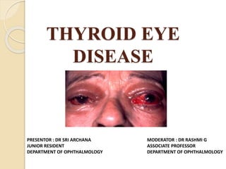
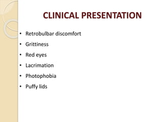
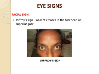
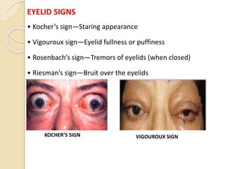
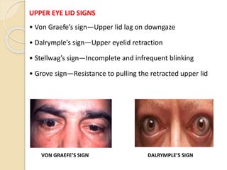
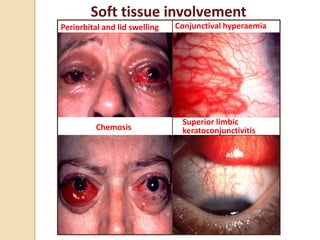
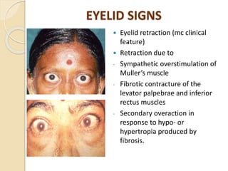
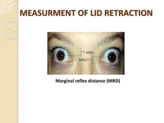
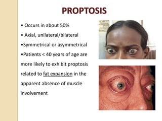
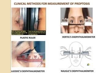
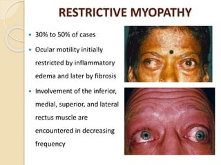
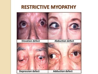
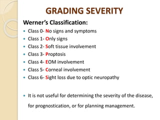
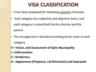
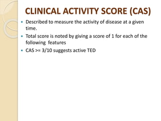
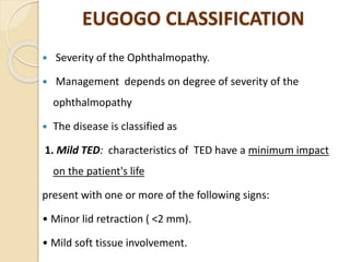
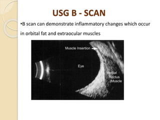
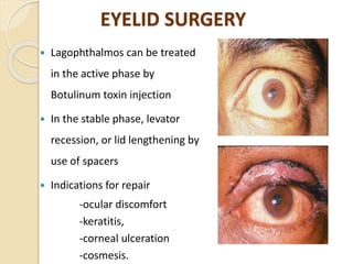
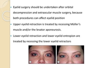
• Thyroid eye disease (TED) is an autoimmune inflammatory
disorder .
• It is also called as Thyroid orbitopathy, Thyroid associated
ophthalmopathy (TAO), Thyroid ophthalmopathy
• It is the commonest cause of both unilateral and bilateral
proptosis in adults
RISK FACTORS
 Fourth to fifth decade
 Female to Maleratio4:1
 Cigarette smoking ( modifiable risk factor)
 Treatment with radioactive iodine
ETIOLOGY
 Graves’ disease (90%)
 Euthyroidism (6%)
 Hyperthyroidism – other causes
(Toxic nodular goiter, Hashimoto’s thyroiditis (3%),
Factitious hyperthyroidism, Thyroid malignancy)
 Hypothyroidism (1%)
PATHOGENISIS
 Thyroid eye disease is an organ specific autoimmune-
mediated inflammation of the extraocular muscle and
periorbital connective tissue.
 Target antigen is shared between thyroid follicular
cells and orbital fibroblast
 Orbital fibroblast are the primary target of
inflammatory attack
Lymphocytic infiltration of orbital tissue
Release of cytokines (IL – 1)
Stimulation of fibroblasts, Proinflamatory genes
upregualted(IL6,IL8,PGE2)
Synthesis of Glicosaminoglycans(GAG)
Hyaluronan attracts water, GAG accumulates in fatty
connective tissue
Increase in osmotic load
Muscle odema, Proptosis, Fibrosis of muscle fibres,
Atrophy
PATHOGENISIS
GROSS PATHOLOGY
EOM
enlargement
confined to
bellies and
sparing
tendons
HISTOPATHOLOGY
The extraocular muscle fibres are separated by an
accumulation of granular material consisting of collagen
fibrils and glycosaminoglycans, among which hyaluronan
predominates
Focal and perivascular inflammatory
mononuclear cell
Muscle replaced by fibrous connective
tissue with a scattering of mononuclear
inflammatory cells in late disease
CLINICAL PRESENTATION
• Retrobulbar discomfort
• Grittiness
• Red eyes
• Lacrimation
• Photophobia
• Puffy lids
EYE SIGNS
FACIAL SIGN:
 Joffroy’s sign—Absent creases in the forehead on
superior gaze.
JOFFROY’S SIGN
EYELID SIGNS
• Kocher’s sign—Staring appearance
• Vigouroux sign—Eyelid fullness or puffiness
• Rosenbach’s sign—Tremors of eyelids (when closed)
• Riesman’s sign—Bruit over the eyelids
KOCHER’S SIGN VIGOUROUX SIGN
UPPER EYE LID SIGNS
• Von Graefe’s sign—Upper lid lag on downgaze
• Dalrymple’s sign—Upper eyelid retraction
• Stellwag’s sign—Incomplete and infrequent blinking
• Grove sign—Resistance to pulling the retracted upper lid
VON GRAEFE’S SIGN DALRYMPLE’S SIGN
• Boston sign—Uneven, jerky movements of the upper lid on
inferior gaze
• Gellinek’s sign—Abnormal pigmentation of the upper lid
• Gifford’s sign—Difficulty in everting the upper lid
• Means sign—Increase superior scleral show on upgaze
GELLINEK’S SIGN GIFFORD’S SIGN
LOWER EYE LID SIGNS
• Enroth’s sign—Edema of the lower lid
• Griffith’s sign—Lower lid lag on upward gaze.
ENROTH’S SIGN
EXTRAOCULAR MOVEMENT SIGNS
• Moebius’ sign—Unable to converge eyes
• Ballet’s sign—Restriction of one or more extra ocular muscle
• Suker’s sign—Poor fixation on abduction
• Jendrassik’s sign—Paralysis of all EOMs.
MOEBIUS’ SIGN BALLET’S SIGN
CONJUNCTIVAL SIGNS
• Goldzeiher’s sign—Conjunctival injection.
PUPILLARY INVOLVEMENT SIGNS
• Knies’ sign—Uneven pupillary dilatation in dim light
• Cowen’s sign—Jerky contraction of pupil to light.
KNIES’ SIGN
Soft tissue involvement
Periorbital and lid swelling
Chemosis
Conjunctival hyperaemia
Superior limbic
keratoconjunctivitis
EYELID SIGNS
 Eyelid retraction (mc clinical
feature)
 Retraction due to
- Sympathetic overstimulation of
Muller’s muscle
- Fibrotic contracture of the
levator palpebrae and inferior
rectus muscles
- Secondary overaction in
response to hypo- or
hypertropia produced by
fibrosis.
Marginal reflex distance (MRD)
MEASURMENT OF LID RETRACTION
PROPTOSIS
• Occurs in about 50%
• Axial, unilateral/bilateral
•Symmetrical or asymmetrical
•Patients < 40 years of age are
more likely to exhibit proptosis
related to fat expansion in the
apparent absence of muscle
involvement
PLASTIC RULER
LUEDDE’S EXOPHTHALMOMETER
HERTEL’S EXOPHTHALMOMETER
CLINICAL METHODS FOR MEASUREMENT OF PROPTOSIS
NAUGLE’S EXOPHTHALMOMTER
RESTRICTIVE MYOPATHY
 30% to 50% of cases
 Ocular motility initially
restricted by inflammatory
edema and later by fibrosis
 Involvement of the inferior,
medial, superior, and lateral
rectus muscle are
encountered in decreasing
frequency
RESTRICTIVE MYOPATHY
Elevation defect Abduction defect
Depression defect Adduction defect
OPTIC NEUROPATHY
 5% of cases
 Compression of optic nerve or
its blood supply at the orbital
apex by congested and enlarged
recti and swollen orbital tissue
 Visual acuity is reduced
 Colour desaturation
 Visual Field defects : central and
paracentral scotoma
GRADING SEVERITY
Werner’s Classification:
 Class 0- No signs and symptoms
 Class 1- Only signs
 Class 2- Soft tissue involvement
 Class 3- Proptosis
 Class 4- EOM involvement
 Class 5- Corneal involvement
 Class 6- Sight loss due to optic neuropathy
 It is not useful for determining the severity of the disease,
for prognostication, or for planning management.
VISA CLASSIFICATION
 It has been proposed for classifying severity of disease
 Each category has subjective and objective items, and
each category is scored both by the clinician and the
patient
 The management is decided according to the score in each
category
V—Vision, and Assessment of Optic Neuropathy
I—Inflammation
S—Strabismus
A—Appearance (Proptosis, Lid Retraction) and Exposure
CLINICAL ACTIVITY SCORE (CAS)
 Described to measure the activity of disease at a given
time.
 Total score is noted by giving a score of 1 for each of the
following features
 CAS >= 3/10 suggests active TED
EUGOGO CLASSIFICATION
 Severity of the Ophthalmopathy.
 Management depends on degree of severity of the
ophthalmopathy
 The disease is classified as
1. Mild TED: characteristics of TED have a minimum impact
on the patient's life
present with one or more of the following signs:
• Minor lid retraction ( <2 mm).
• Mild soft tissue involvement.
 Exophthalmos <3 mm
 Transient or no diplopia.
 Corneal exposure responsive to lubricants.
2. Moderate-to-severe TED :
- Patients without sight-threatening TED
- Eye disease has sufficient impact on daily life to justify the
risks of immunosuppression ( if active) or surgical
intervention (if inactive).
Patients usually present one or more of the following signs:
• Lid retraction (>2mm).
• Moderate or severe soft tissue involvement.
 Exophthalmos >3 mm
• Inconstant, or constant diplopia.
3 Sight threatening TED: patients with
• Dysthyroid optic neuropathy
• Corneal breakdown due to severe exposure
• Severe forms of frozen eye
• Choroidal folds
This category warrants immediate intervention .
CLINICAL COURSE
 TED is a self-limiting disease, becoming quiescent within
3–5 years of its onset
1. Congestive or Active phase :
- Lasts for 6-18 months
- Characterized by active inflammation with marked lid
edema, conjunctival chemosis and congestion and
increasing exophthalmos
2. Static phase :
majority of cases, starts to regress and most of the signs
slowly settle down.
3. Fibrotic or Inactive phase:
- Also known as quiescent or burnt-out phase, ensues after
the regression phase.
- In this phase, eyes are white, however, a painless motility
defect may be present.
INVESTIGATION
Biochemical Profile
 Thyroid stimulating hormone
 Free and total triiodothyronine
 Free and total thyroxine
 Thyroid hormone binding ratio (THBR)
 Radioiodine uptake (RAIU)
Imaging
 Magnetic resonance imaging
 Computed tomography scanning
 Ultrasonography
CT ORBIT
 CT is helpful in assessing the relationship between the EOM
and optic nerve at the orbital apex and can aid in surgical
planning
 Allows identification of even minimally enlarged recti
 There is bilateral fusiform enlargement of the extra-ocular
muscles, with smooth borders and sparing of the tendons,
is the classic finding ( Coca-cola sign)
CT ORBIT
Coca-cola sign
MRI ORBIT
•MRI helps to identify the typical fusiform rectus muscle
enlargement and orbital fat expansion
•Helps in early diagnosis of compressive optic neuropathy
USG B - SCAN
•B scan can demonstrate inflammatory changes which occur
in orbital fat and extraocular muscles
MANAGEMENT
Management of thyroid eye disease includes :
 Supportive therapy during the active phase
 Management of sight-threatening conditions in severe
disease
 Functional and cosmetic rehabilitation during the
quiescent phase
MANAGEMENT PLAN FOR THYROID EYE
DISEASE
 Mild to moderate disease, within first year of onset:
- Stop smoking
- Lubricants
- Elevate head end of bed
- Cool compresses
 Severe sight threatening disorder at any point (optic
neuropathy, exposure keratitis):
- Systemic steroids–methylprednisolone
- Tarsorrhaphy or induction of ptosis with botulinum toxin.
 If severe disease is non-responsive to medical therapy
- Surgical decompression, orbital radiotherapy.
 Moderate or severe disease, stable, inactive is treated
with surgical correction.
- Orbital decompression
- Extraocular muscle surgery
- Lid correction.
Use of steroids:
 Benefit from intravenous methyl-prednisolone, 1 gm per
day for three days.
 A recurrent disease may need pulse doses of steroids
every three to six weeks.
 Less severe disease may respond to oral steroids(
Prednisone 1mg/kg/day then taper )
 Intra-orbital injection of long-acting steroid such as
Triamcinolone 40 mg may have some beneficial effect.
Use of immunosuppressants:
 Patients not responding to steroids, or intolerant of
steroids
- Methotrexate (7.5–15 mg one day a week)
- Azathioprine (initial dose 1.0 mg/kg/day, increased weekly
till minimum effective dose is achieved)
- Cyclophosphamide (0.1–0.2 mg/kg/day).
 These drugs have serious side effects, and need to be
used with careful monitoring under supervision
Radiotherapy:
 A dose of 2000 cGy used
 It may act by decreasing the T lymphocyte
population in the orbit.
 Radiation is contraindicated in diabetic patients, as it
can adversely affect the retinopathy.
SURGICAL MANAGEMENT
Orbital decompression
Extraocular muscle surgery
Eyelid surgery
ORBITAL DECOMPRESSION
 Expands the orbital volume to create more space for
swollen soft tissues
 Primary indications for orbital decompression
- Compressive optic neuropathy
- Excessive proptosis (can manifest as globe subluxation,
corneal ulceration, and cosmetic disfigurement)
- Steroid dependence
- Intractable pain
 Types
◦ Two – wall : floor and medial wall (3–7 mm)
◦ Three – wall : floor, medial and lateral wall (6–10 mm)
◦ Four – wall : floor, medial and lateral walls with part of
sphenoid (apex) and lateral part orbital roof (10–17 mm)
Orbital Decompression
STRABISMUS SURGERY
 Goal of strabismus treatment is to achieve single binocular
vision in primary and downgaze positions
 TED quiescent with stable eye muscle function without
corticosteroids for at least 6 months
 The most frequently performed surgical procedure
strabismus is recession of the restricted medial rectus or
inferior rectus muscle
EYELID SURGERY
 Lagophthalmos can be treated
in the active phase by
Botulinum toxin injection
 In the stable phase, levator
recession, or lid lengthening by
use of spacers
 Indications for repair
-ocular discomfort
-keratitis,
-corneal ulceration
-cosmesis.
 Eyelid surgery should be undertaken after orbital
decompression and extraocular muscle surgery, because
both procedures can affect eyelid position
 Upper eyelid retraction is treated by recessing Müller’s
muscle and/or the levator aponeurosis.
 Lower eyelid retraction and lower eyelid entropion are
treated by recessing the lower eyelid retractors
REFERENCES
 Principle and practice of ophthalmology by Albert and
Jakobie’s Volume 3 , Edition 3
 Comprehensive Ophthalmology by A K Khurana, 7th
Edition
 Kanski’s Clinical Ophthalmology 9th edition
 Postgraduate Ophthalmology by Zia Chaudhuri, First
Edition
THANK YOU

Thyroid eye disease

  • 1.
    THYROID EYE DISEASE MODERATOR :DR RASHMI G ASSOCIATE PROFESSOR DEPARTMENT OF OPHTHALMOLOGY PRESENTOR : DR SRI ARCHANA JUNIOR RESIDENT DEPARTMENT OF OPHTHALMOLOGY
  • 2.
    INTRODUCTION • Thyroid eyedisease (TED) is an autoimmune inflammatory disorder . • It is also called as Thyroid orbitopathy, Thyroid associated ophthalmopathy (TAO), Thyroid ophthalmopathy • It is the commonest cause of both unilateral and bilateral proptosis in adults
  • 3.
    RISK FACTORS  Fourthto fifth decade  Female to Maleratio4:1  Cigarette smoking ( modifiable risk factor)  Treatment with radioactive iodine
  • 4.
    ETIOLOGY  Graves’ disease(90%)  Euthyroidism (6%)  Hyperthyroidism – other causes (Toxic nodular goiter, Hashimoto’s thyroiditis (3%), Factitious hyperthyroidism, Thyroid malignancy)  Hypothyroidism (1%)
  • 5.
    PATHOGENISIS  Thyroid eyedisease is an organ specific autoimmune- mediated inflammation of the extraocular muscle and periorbital connective tissue.  Target antigen is shared between thyroid follicular cells and orbital fibroblast  Orbital fibroblast are the primary target of inflammatory attack
  • 6.
    Lymphocytic infiltration oforbital tissue Release of cytokines (IL – 1) Stimulation of fibroblasts, Proinflamatory genes upregualted(IL6,IL8,PGE2) Synthesis of Glicosaminoglycans(GAG) Hyaluronan attracts water, GAG accumulates in fatty connective tissue Increase in osmotic load Muscle odema, Proptosis, Fibrosis of muscle fibres, Atrophy
  • 7.
  • 8.
  • 9.
    HISTOPATHOLOGY The extraocular musclefibres are separated by an accumulation of granular material consisting of collagen fibrils and glycosaminoglycans, among which hyaluronan predominates Focal and perivascular inflammatory mononuclear cell Muscle replaced by fibrous connective tissue with a scattering of mononuclear inflammatory cells in late disease
  • 10.
    CLINICAL PRESENTATION • Retrobulbardiscomfort • Grittiness • Red eyes • Lacrimation • Photophobia • Puffy lids
  • 11.
    EYE SIGNS FACIAL SIGN: Joffroy’s sign—Absent creases in the forehead on superior gaze. JOFFROY’S SIGN
  • 12.
    EYELID SIGNS • Kocher’ssign—Staring appearance • Vigouroux sign—Eyelid fullness or puffiness • Rosenbach’s sign—Tremors of eyelids (when closed) • Riesman’s sign—Bruit over the eyelids KOCHER’S SIGN VIGOUROUX SIGN
  • 13.
    UPPER EYE LIDSIGNS • Von Graefe’s sign—Upper lid lag on downgaze • Dalrymple’s sign—Upper eyelid retraction • Stellwag’s sign—Incomplete and infrequent blinking • Grove sign—Resistance to pulling the retracted upper lid VON GRAEFE’S SIGN DALRYMPLE’S SIGN
  • 14.
    • Boston sign—Uneven,jerky movements of the upper lid on inferior gaze • Gellinek’s sign—Abnormal pigmentation of the upper lid • Gifford’s sign—Difficulty in everting the upper lid • Means sign—Increase superior scleral show on upgaze GELLINEK’S SIGN GIFFORD’S SIGN
  • 15.
    LOWER EYE LIDSIGNS • Enroth’s sign—Edema of the lower lid • Griffith’s sign—Lower lid lag on upward gaze. ENROTH’S SIGN
  • 16.
    EXTRAOCULAR MOVEMENT SIGNS •Moebius’ sign—Unable to converge eyes • Ballet’s sign—Restriction of one or more extra ocular muscle • Suker’s sign—Poor fixation on abduction • Jendrassik’s sign—Paralysis of all EOMs. MOEBIUS’ SIGN BALLET’S SIGN
  • 17.
    CONJUNCTIVAL SIGNS • Goldzeiher’ssign—Conjunctival injection. PUPILLARY INVOLVEMENT SIGNS • Knies’ sign—Uneven pupillary dilatation in dim light • Cowen’s sign—Jerky contraction of pupil to light. KNIES’ SIGN
  • 18.
    Soft tissue involvement Periorbitaland lid swelling Chemosis Conjunctival hyperaemia Superior limbic keratoconjunctivitis
  • 19.
    EYELID SIGNS  Eyelidretraction (mc clinical feature)  Retraction due to - Sympathetic overstimulation of Muller’s muscle - Fibrotic contracture of the levator palpebrae and inferior rectus muscles - Secondary overaction in response to hypo- or hypertropia produced by fibrosis.
  • 20.
    Marginal reflex distance(MRD) MEASURMENT OF LID RETRACTION
  • 21.
    PROPTOSIS • Occurs inabout 50% • Axial, unilateral/bilateral •Symmetrical or asymmetrical •Patients < 40 years of age are more likely to exhibit proptosis related to fat expansion in the apparent absence of muscle involvement
  • 22.
    PLASTIC RULER LUEDDE’S EXOPHTHALMOMETER HERTEL’SEXOPHTHALMOMETER CLINICAL METHODS FOR MEASUREMENT OF PROPTOSIS NAUGLE’S EXOPHTHALMOMTER
  • 23.
    RESTRICTIVE MYOPATHY  30%to 50% of cases  Ocular motility initially restricted by inflammatory edema and later by fibrosis  Involvement of the inferior, medial, superior, and lateral rectus muscle are encountered in decreasing frequency
  • 24.
    RESTRICTIVE MYOPATHY Elevation defectAbduction defect Depression defect Adduction defect
  • 25.
    OPTIC NEUROPATHY  5%of cases  Compression of optic nerve or its blood supply at the orbital apex by congested and enlarged recti and swollen orbital tissue  Visual acuity is reduced  Colour desaturation  Visual Field defects : central and paracentral scotoma
  • 26.
    GRADING SEVERITY Werner’s Classification: Class 0- No signs and symptoms  Class 1- Only signs  Class 2- Soft tissue involvement  Class 3- Proptosis  Class 4- EOM involvement  Class 5- Corneal involvement  Class 6- Sight loss due to optic neuropathy  It is not useful for determining the severity of the disease, for prognostication, or for planning management.
  • 27.
    VISA CLASSIFICATION  Ithas been proposed for classifying severity of disease  Each category has subjective and objective items, and each category is scored both by the clinician and the patient  The management is decided according to the score in each category V—Vision, and Assessment of Optic Neuropathy I—Inflammation S—Strabismus A—Appearance (Proptosis, Lid Retraction) and Exposure
  • 28.
    CLINICAL ACTIVITY SCORE(CAS)  Described to measure the activity of disease at a given time.  Total score is noted by giving a score of 1 for each of the following features  CAS >= 3/10 suggests active TED
  • 29.
    EUGOGO CLASSIFICATION  Severityof the Ophthalmopathy.  Management depends on degree of severity of the ophthalmopathy  The disease is classified as 1. Mild TED: characteristics of TED have a minimum impact on the patient's life present with one or more of the following signs: • Minor lid retraction ( <2 mm). • Mild soft tissue involvement.
  • 30.
     Exophthalmos <3mm  Transient or no diplopia.  Corneal exposure responsive to lubricants. 2. Moderate-to-severe TED : - Patients without sight-threatening TED - Eye disease has sufficient impact on daily life to justify the risks of immunosuppression ( if active) or surgical intervention (if inactive). Patients usually present one or more of the following signs: • Lid retraction (>2mm). • Moderate or severe soft tissue involvement.
  • 31.
     Exophthalmos >3mm • Inconstant, or constant diplopia. 3 Sight threatening TED: patients with • Dysthyroid optic neuropathy • Corneal breakdown due to severe exposure • Severe forms of frozen eye • Choroidal folds This category warrants immediate intervention .
  • 32.
    CLINICAL COURSE  TEDis a self-limiting disease, becoming quiescent within 3–5 years of its onset 1. Congestive or Active phase : - Lasts for 6-18 months - Characterized by active inflammation with marked lid edema, conjunctival chemosis and congestion and increasing exophthalmos 2. Static phase : majority of cases, starts to regress and most of the signs slowly settle down.
  • 33.
    3. Fibrotic orInactive phase: - Also known as quiescent or burnt-out phase, ensues after the regression phase. - In this phase, eyes are white, however, a painless motility defect may be present.
  • 34.
    INVESTIGATION Biochemical Profile  Thyroidstimulating hormone  Free and total triiodothyronine  Free and total thyroxine  Thyroid hormone binding ratio (THBR)  Radioiodine uptake (RAIU) Imaging  Magnetic resonance imaging  Computed tomography scanning  Ultrasonography
  • 35.
    CT ORBIT  CTis helpful in assessing the relationship between the EOM and optic nerve at the orbital apex and can aid in surgical planning  Allows identification of even minimally enlarged recti  There is bilateral fusiform enlargement of the extra-ocular muscles, with smooth borders and sparing of the tendons, is the classic finding ( Coca-cola sign)
  • 36.
  • 37.
    MRI ORBIT •MRI helpsto identify the typical fusiform rectus muscle enlargement and orbital fat expansion •Helps in early diagnosis of compressive optic neuropathy
  • 38.
    USG B -SCAN •B scan can demonstrate inflammatory changes which occur in orbital fat and extraocular muscles
  • 39.
    MANAGEMENT Management of thyroideye disease includes :  Supportive therapy during the active phase  Management of sight-threatening conditions in severe disease  Functional and cosmetic rehabilitation during the quiescent phase
  • 40.
    MANAGEMENT PLAN FORTHYROID EYE DISEASE  Mild to moderate disease, within first year of onset: - Stop smoking - Lubricants - Elevate head end of bed - Cool compresses  Severe sight threatening disorder at any point (optic neuropathy, exposure keratitis): - Systemic steroids–methylprednisolone - Tarsorrhaphy or induction of ptosis with botulinum toxin.
  • 41.
     If severedisease is non-responsive to medical therapy - Surgical decompression, orbital radiotherapy.  Moderate or severe disease, stable, inactive is treated with surgical correction. - Orbital decompression - Extraocular muscle surgery - Lid correction.
  • 42.
    Use of steroids: Benefit from intravenous methyl-prednisolone, 1 gm per day for three days.  A recurrent disease may need pulse doses of steroids every three to six weeks.  Less severe disease may respond to oral steroids( Prednisone 1mg/kg/day then taper )  Intra-orbital injection of long-acting steroid such as Triamcinolone 40 mg may have some beneficial effect.
  • 43.
    Use of immunosuppressants: Patients not responding to steroids, or intolerant of steroids - Methotrexate (7.5–15 mg one day a week) - Azathioprine (initial dose 1.0 mg/kg/day, increased weekly till minimum effective dose is achieved) - Cyclophosphamide (0.1–0.2 mg/kg/day).  These drugs have serious side effects, and need to be used with careful monitoring under supervision
  • 44.
    Radiotherapy:  A doseof 2000 cGy used  It may act by decreasing the T lymphocyte population in the orbit.  Radiation is contraindicated in diabetic patients, as it can adversely affect the retinopathy.
  • 45.
  • 46.
    ORBITAL DECOMPRESSION  Expandsthe orbital volume to create more space for swollen soft tissues  Primary indications for orbital decompression - Compressive optic neuropathy - Excessive proptosis (can manifest as globe subluxation, corneal ulceration, and cosmetic disfigurement) - Steroid dependence - Intractable pain
  • 47.
     Types ◦ Two– wall : floor and medial wall (3–7 mm) ◦ Three – wall : floor, medial and lateral wall (6–10 mm) ◦ Four – wall : floor, medial and lateral walls with part of sphenoid (apex) and lateral part orbital roof (10–17 mm)
  • 48.
  • 49.
    STRABISMUS SURGERY  Goalof strabismus treatment is to achieve single binocular vision in primary and downgaze positions  TED quiescent with stable eye muscle function without corticosteroids for at least 6 months  The most frequently performed surgical procedure strabismus is recession of the restricted medial rectus or inferior rectus muscle
  • 50.
    EYELID SURGERY  Lagophthalmoscan be treated in the active phase by Botulinum toxin injection  In the stable phase, levator recession, or lid lengthening by use of spacers  Indications for repair -ocular discomfort -keratitis, -corneal ulceration -cosmesis.
  • 51.
     Eyelid surgeryshould be undertaken after orbital decompression and extraocular muscle surgery, because both procedures can affect eyelid position  Upper eyelid retraction is treated by recessing Müller’s muscle and/or the levator aponeurosis.  Lower eyelid retraction and lower eyelid entropion are treated by recessing the lower eyelid retractors
  • 52.
    REFERENCES  Principle andpractice of ophthalmology by Albert and Jakobie’s Volume 3 , Edition 3  Comprehensive Ophthalmology by A K Khurana, 7th Edition  Kanski’s Clinical Ophthalmology 9th edition  Postgraduate Ophthalmology by Zia Chaudhuri, First Edition
  • 53.