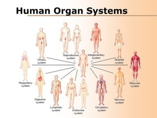
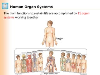
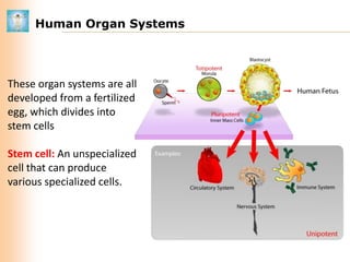
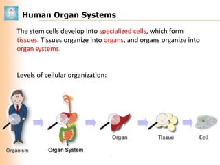
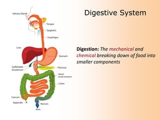
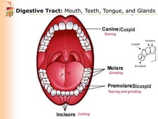
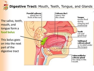
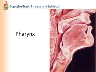
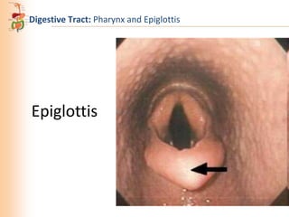
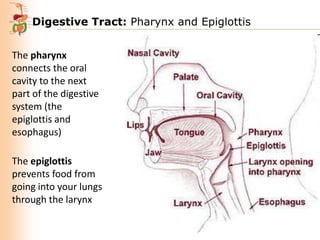
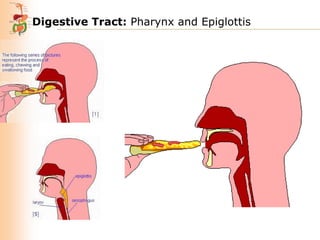
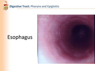
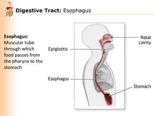
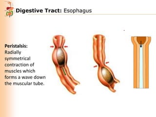
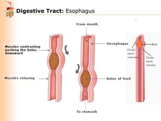
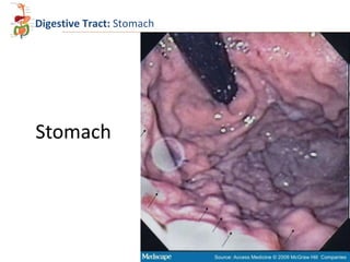
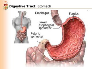
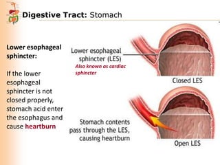
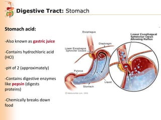
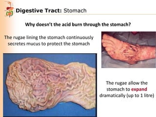
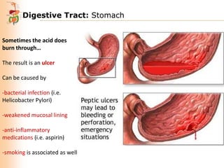
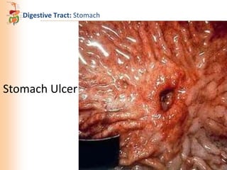
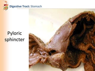
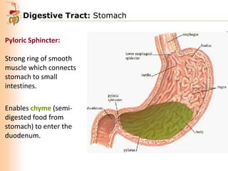
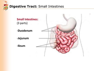
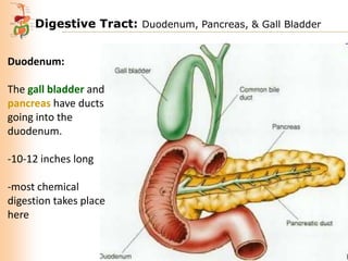
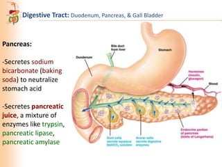
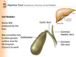
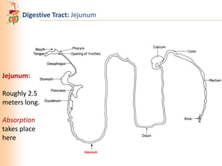
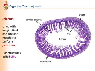
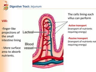
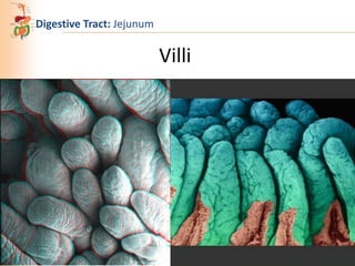
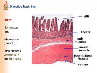
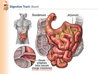
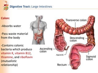
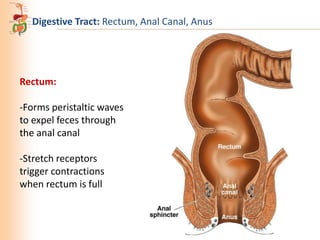
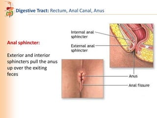
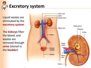
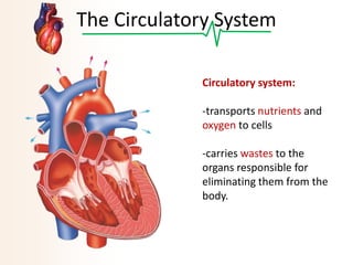
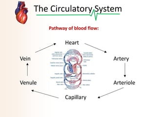
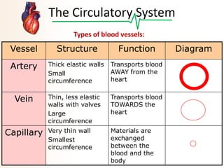
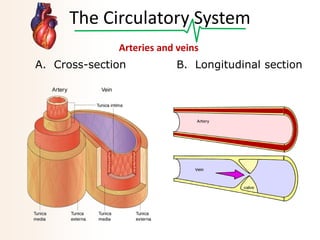
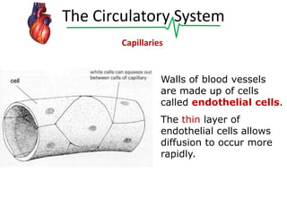
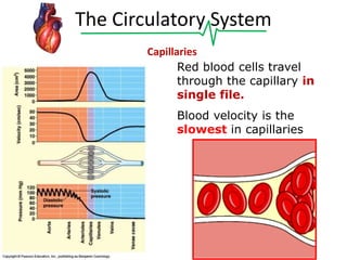
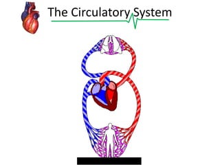
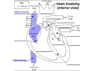
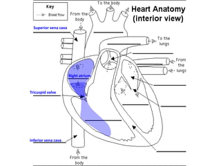
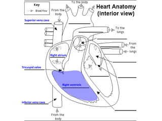
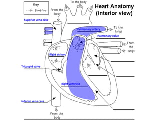
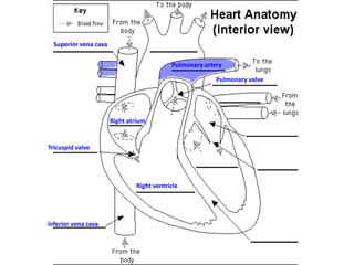
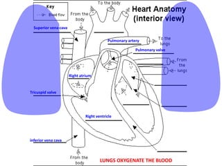
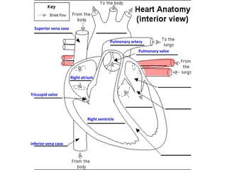
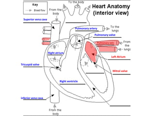
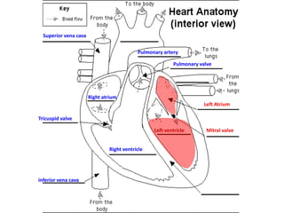
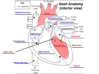
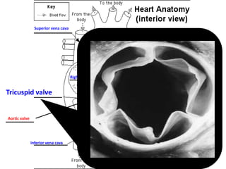
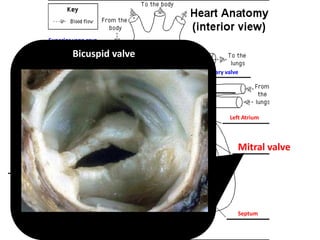
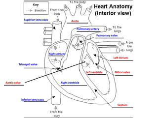
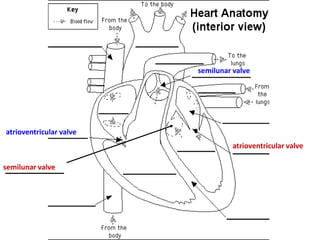
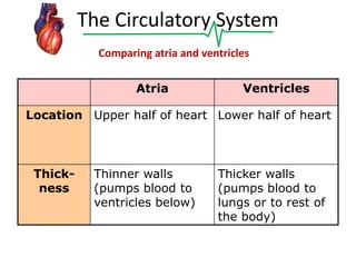
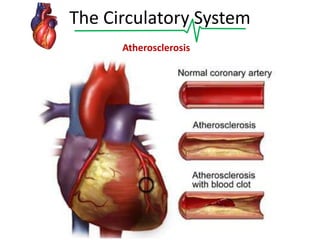
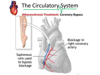
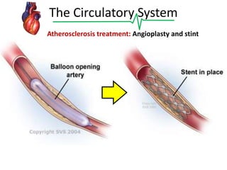
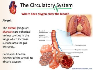
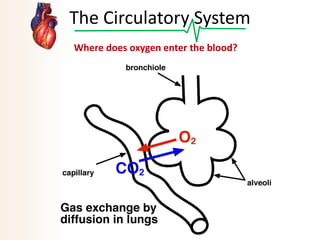
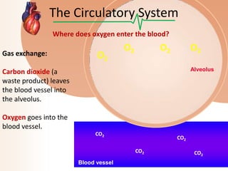
Human Organ Systems
Human Organ Systems
The main functions to sustain life are accomplished by 11 organ
systems working together
INTEGUMENTARY SYSTEM
Human Organ Systems

These organ systems are all
developed from a fertilized
egg, which divides into
stem cells
Stem cell: An unspecialized
cell that can produce
various specialized cells.
Human Organ Systems
The stem cells develop into specialized cells, which form
tissues. Tissues organize into organs, and organs organize into
organ systems.

Levels of cellular organization:
Digestive System

Digestion: The mechanical and
chemical breaking down of food into
smaller components
Digestive Tract: Mouth, Teeth, Tongue, and Glands

Tearing

/Cuspid

Grinding

/Bicuspid
Tearing and grinding

Cutting
Digestive Tract: Mouth, Teeth, Tongue, and Glands

The saliva, teeth,
mouth, and
tongue form a
food bolus
This bolus goes
on into the next
part of the
digestive tract
Digestive Tract: Pharynx and Epiglottis

Pharynx
Digestive Tract: Pharynx and Epiglottis

Epiglottis
Digestive Tract: Pharynx and Epiglottis
The pharynx
connects the oral
cavity to the next
part of the digestive
system (the
epiglottis and
esophagus)
The epiglottis
prevents food from
going into your lungs
through the larynx
Digestive Tract: Pharynx and Epiglottis

Esophagus
Digestive Tract: Esophagus

Esophagus:
Muscular tube
through which
food passes from
the pharynx to the
stomach
Digestive Tract: Esophagus

Peristalsis:
Radially
symmetrical
contraction of
muscles which
forms a wave down
the muscular tube.
Digestive Tract: Esophagus
Digestive Tract: Stomach

Stomach
Digestive Tract: Stomach

Lower esophageal
sphincter:
If the lower
esophageal
sphincter is not
closed properly,
stomach acid enter
the esophagus and
cause heartburn

Also known as cardiac
sphincter
Digestive Tract: Stomach
Stomach acid:
-Also known as gastric juice
-Contains hydrochloric acid
(HCl)
-pH of 2 (approximately)
-Contains digestive enzymes
like pepsin (digests
proteins)
-Chemically breaks down
food
Digestive Tract: Stomach
Why doesn’t the acid burn through the stomach?
The rugae lining the stomach continuously
secretes mucus to protect the stomach

The rugae allow the
stomach to expand
dramatically (up to 1 litre)
Digestive Tract: Stomach
Sometimes the acid does
burn through…
The result is an ulcer

Can be caused by
-bacterial infection (i.e.
Helicobacter Pylori)
-weakened mucosal lining
-anti-inflammatory
medications (i.e. aspirin)
-smoking is associated as well
Digestive Tract: Stomach

Stomach Ulcer
Digestive Tract: Stomach

Pyloric
sphincter
Digestive Tract: Stomach
Pyloric Sphincter:
Strong ring of smooth
muscle which connects
stomach to small
intestines.
Enables chyme (semidigested food from
stomach) to enter the
duodenum.
Digestive Tract: Stomach

Chyme
Digestive Tract: Small Intestines

Small Intestine
Digestive Tract: Small Intestines

Small Intestines:
(3 parts)
-Duodenum
-Jejunum
-Ileum
Digestive Tract: Duodenum, Pancreas, & Gall Bladder
Duodenum:
The gall bladder and
pancreas have ducts
going into the
duodenum.

-10-12 inches long
-most chemical
digestion takes place
here
Digestive Tract: Duodenum, Pancreas, & Gall Bladder

Pancreas:
-Secretes sodium
bicarbonate (baking
soda) to neutralize
stomach acid

-Secretes pancreatic
juice, a mixture of
enzymes like trypsin,
pancreatic lipase,
pancreatic amylase
Digestive Tract: Duodenum, Pancreas, & Gall Bladder

Gall Bladder:
Stores bile
produced by the
liver.
Bile emulsifies fats
to allow greater
surface area for
fat enzymes
(lipases) to work
Digestive Tract: Jejunum

Jejunum:
Roughly 2.5
meters long.

Absorption
takes place
here
Jejunum
Digestive Tract: Jejunum

Jejunum:
Lined with
longitudinal
and circular
muscles to
perform
peristalsis.
Has structures
called villi.
Digestive Tract: Jejunum

Villi:
-finger-like
projections of
the small
intestine lining
- More surface
area to absorb
nutrients.

The cells lining each
villus can perform
-Active transport
(transport of nutrients
requiring energy)

-Passive transport
(transport of nutrients not
requiring energy)
Digestive Tract: Jejunum

Villi
Digestive Tract: Ileum

Ileum:

-2.4 meters
long
-absorption
(has villi)
-also absorbs
vitamin B12
and bile salts
Digestive Tract: Ileum
Digestive Tract: Large Intestines

Colon:
-Absorbs water
-Pass waste material
from the body
-Contains colonic
bacteria which produce
vitamin K, vitamin B12,
thiamine, and riboflavin
(mutualistic
relationship)
Digestive Tract: Rectum, Anal Canal, Anus

Rectum:
-Forms peristaltic waves
to expel feces through
the anal canal
-Stretch receptors
trigger contractions
when rectum is full
Digestive Tract: Rectum, Anal Canal, Anus

Anal sphincter:
Exterior and interior
sphincters pull the anus
up over the exiting
feces
Excretory system
Liquid wastes are
eliminated by the
excretory system.
The kidneys filter
the blood, and
wastes are
removed through
urine (stored in
the bladder)
The Circulatory System
Circulatory system:
-transports nutrients and
oxygen to cells
-carries wastes to the
organs responsible for
eliminating them from the
body.
The Circulatory System
Pathway of blood flow:

Heart
Vein

Artery

Venule

Arteriole

Capillary
The Circulatory System
Types of blood vessels:

Vessel

Structure

Function

Artery

Thick elastic walls
Small
circumference

Transports blood
AWAY from the
heart

Vein

Thin, less elastic
walls with valves
Large
circumference

Transports blood
TOWARDS the
heart

Very thin wall
Smallest
circumference

Materials are
exchanged
between the
blood and the
body

Capillary

Diagram
The Circulatory System
Arteries and veins
A. Cross-section

B. Longitudinal section
The Circulatory System
Capillaries

Walls of blood vessels
are made up of cells
called endothelial cells.
The thin layer of
endothelial cells allows
diffusion to occur more
rapidly.
The Circulatory System
Capillaries
Red blood cells travel
through the capillary in
single file.
Blood velocity is the
slowest in capillaries
The Circulatory System
Superior vena cava

inferior vena cava
Superior vena cava

Right atrium

Tricuspid valve

inferior vena cava
Superior vena cava

Right atrium

Tricuspid valve

Right ventricle

inferior vena cava
Superior vena cava
Pulmonary artery
Pulmonary valve

Right atrium

Tricuspid valve

Right ventricle

inferior vena cava
Superior vena cava
Pulmonary artery
Pulmonary valve

Right atrium

Tricuspid valve

Right ventricle

inferior vena cava
Superior vena cava
Pulmonary artery
Pulmonary valve

Right atrium

Tricuspid valve

Right ventricle

inferior vena cava

LUNGS OXYGENATE THE BLOOD
Superior vena cava
Pulmonary artery
Pulmonary valve

Right atrium

Tricuspid valve

Right ventricle

inferior vena cava
Superior vena cava
Pulmonary artery
Pulmonary valve
Pulmonary vein

Right atrium
Left Atrium
Tricuspid valve
Mitral valve
Right ventricle

inferior vena cava
Superior vena cava
Pulmonary artery
Pulmonary valve

Right atrium
Left Atrium
Tricuspid valve
Left ventricle
Right ventricle

inferior vena cava

Mitral valve
Superior vena cava

Aorta
Pulmonary artery
Pulmonary valve

Right atrium
Left Atrium
Tricuspid valve
Left ventricle
Aortic valve

Mitral valve

Right ventricle

inferior vena cava
Septum
Superior vena cava

Aorta
Pulmonary artery
Pulmonary valve

Right atrium
Left Atrium

Tricuspid valve
Left ventricle
Aortic valve

Mitral valve

Right ventricle

inferior vena cava
Septum
Superior vena cava

Aorta

Bicuspid valve

Pulmonary artery
Pulmonary valve

Right atrium
Left Atrium
Tricuspid valve
Left ventricle
Aortic valve

Mitral valve

Right ventricle

inferior vena cava
Septum
Superior vena cava

Aorta
Pulmonary artery
Pulmonary valve

Right atrium
Left Atrium
Tricuspid valve
Left ventricle
Aortic valve

Mitral valve

Right ventricle

inferior vena cava
Septum
semilunar valve

atrioventricular valve

atrioventricular valve
semilunar valve
The Circulatory System
Comparing atria and ventricles
Atria

Ventricles

Location Upper half of heart Lower half of heart

Thickness

Thinner walls
(pumps blood to
ventricles below)

Thicker walls
(pumps blood to
lungs or to rest of
the body)
The Circulatory System
Atherosclerosis
The Circulatory System
Atherosclerosis Treatment: Coronary Bypass
The Circulatory System
Atherosclerosis treatment: Angioplasty and stint
The Circulatory System
Where does oxygen enter the blood?
Alveoli:
The alveoli (singular:
alveolus) are spherical
hollow cavities in the
lungs which increase
surface area for gas
exchange.
Capillaries line the
exterior of the alveoli to
absorb oxygen.
The Circulatory System
Where does oxygen enter the blood?
The Circulatory System
Where does oxygen enter the blood?
Gas exchange:

O2

O2

O2

O2
Alveolus

Carbon dioxide (a
waste product) leaves
the blood vessel into
the alveolus.
Oxygen goes into the
blood vessel.
CO2

CO2
CO2

Blood vessel

CO2

08 human organ systems

Editor's Notes

  • #49 When capillaries unite to form venules, and then veins, the area decreases and the blood flow speeds up