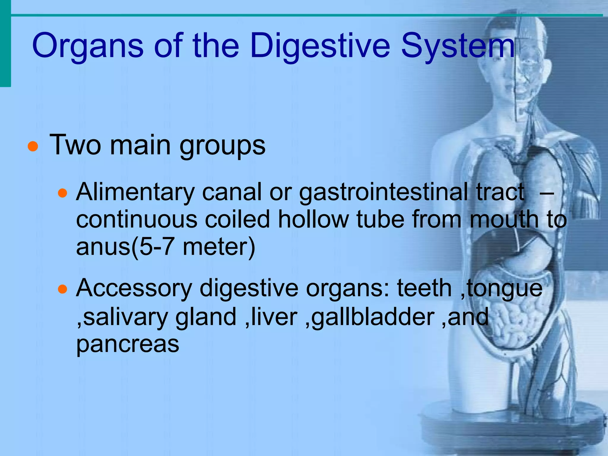
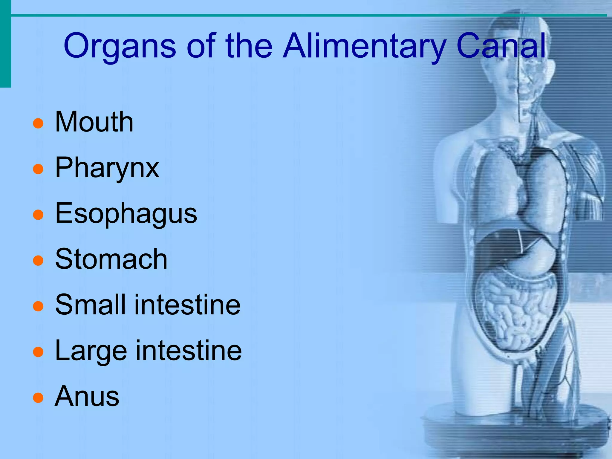
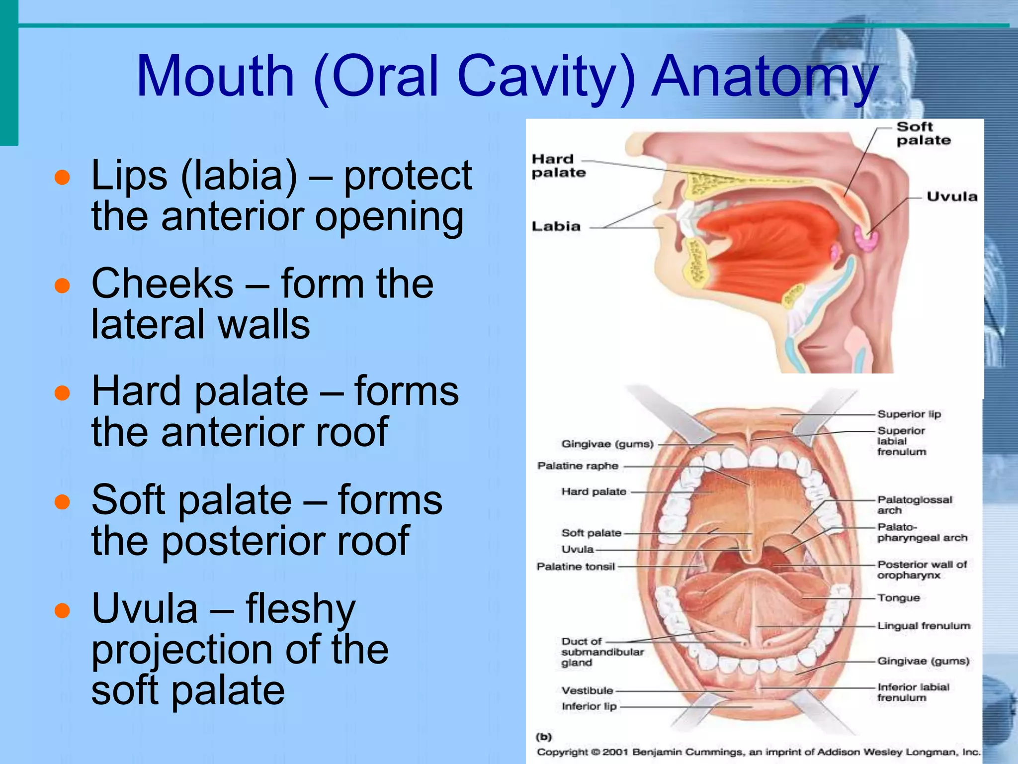
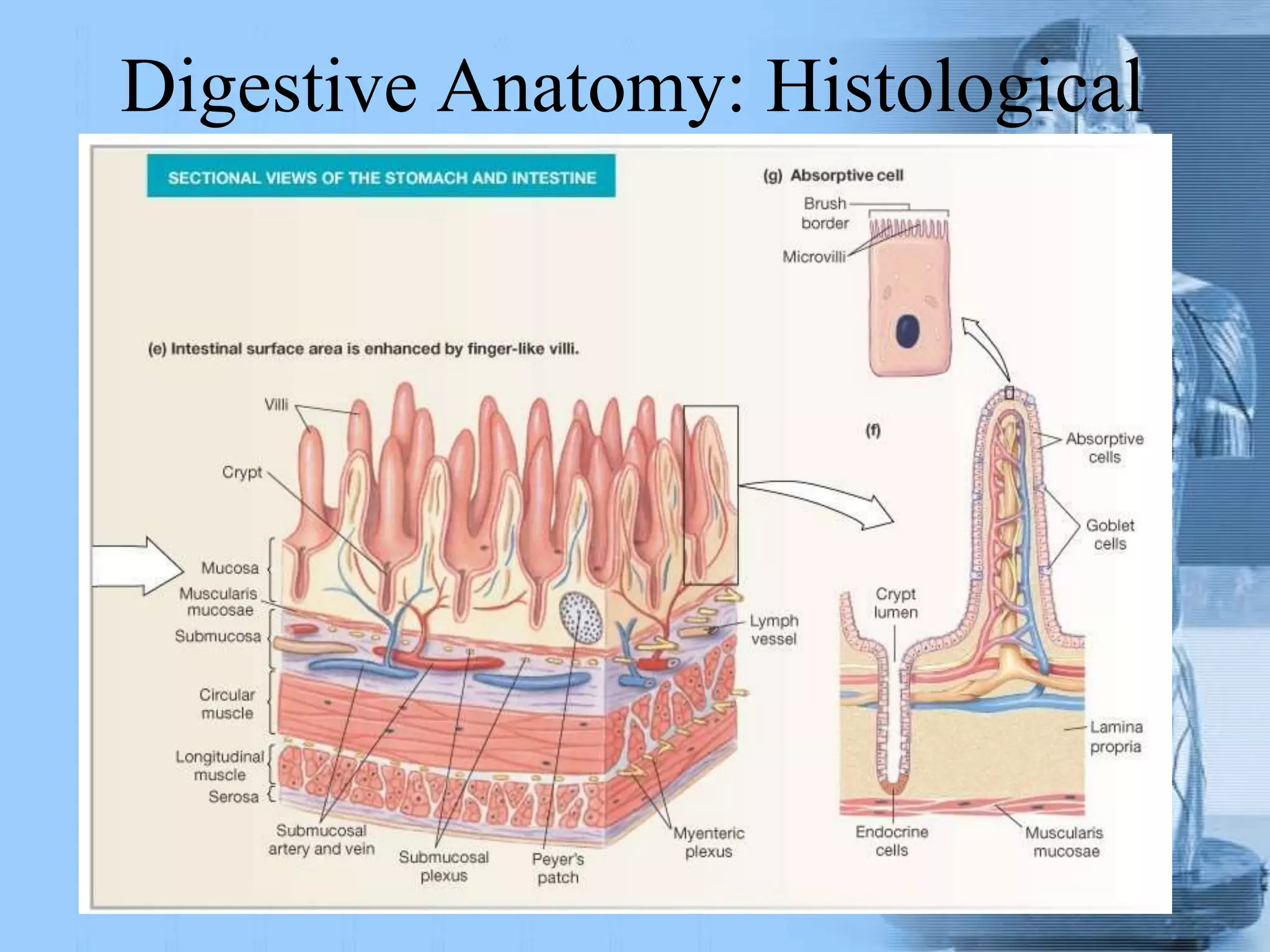
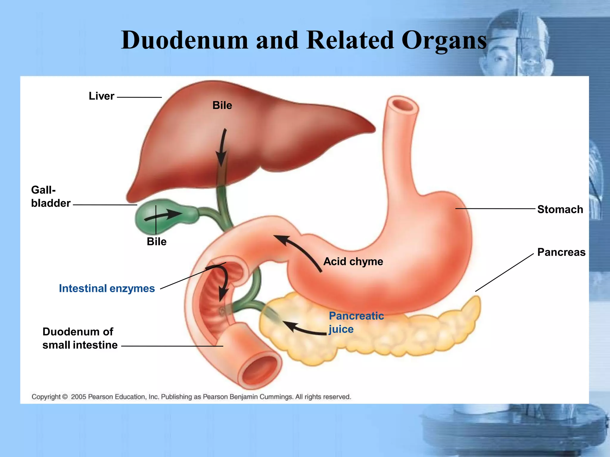
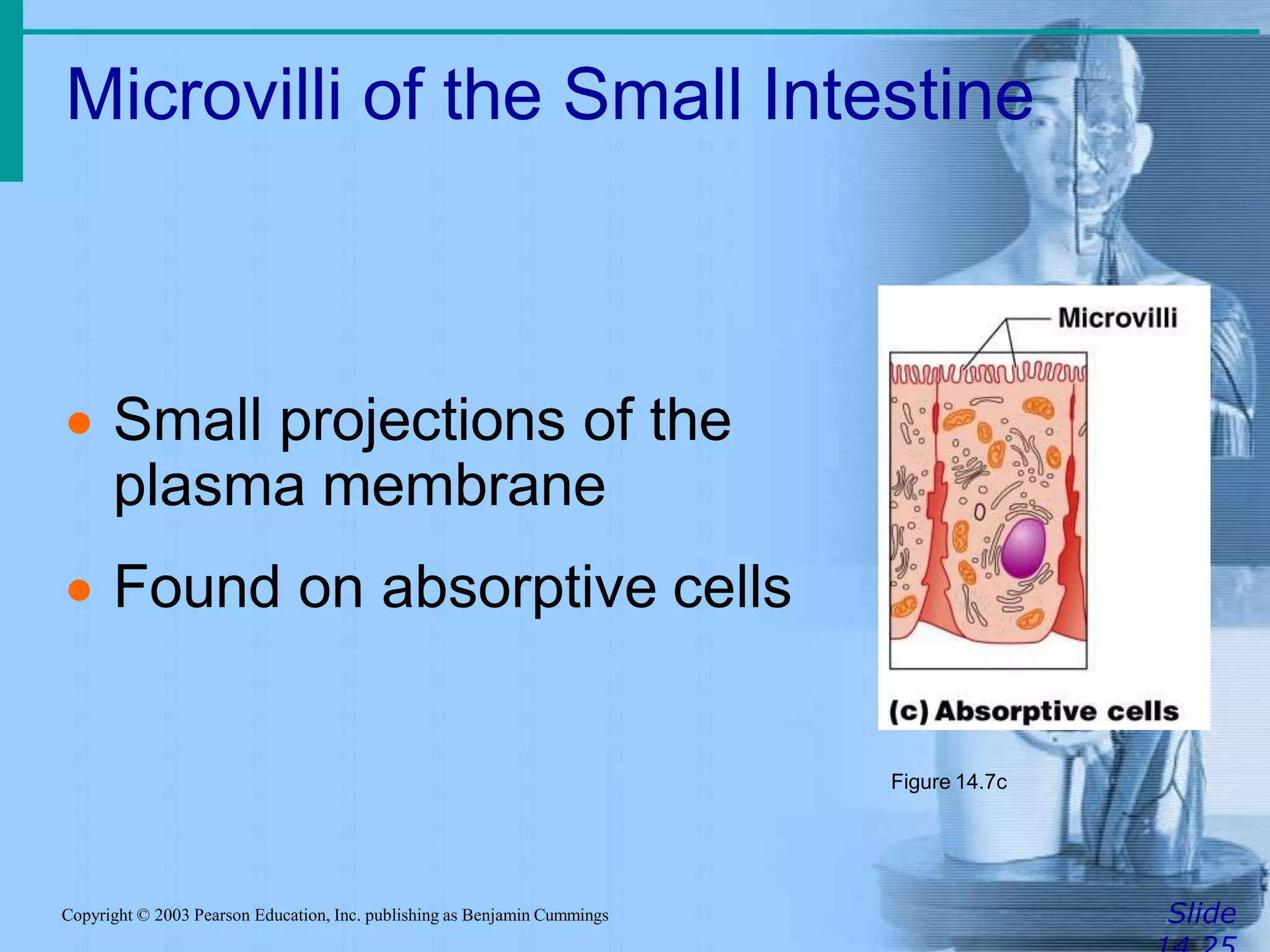
Digestive system
Overview
Function of the digestive system



 e

 ingestion: taking food and liquid into
mouth
Secretion: total about 7 liter into lumen
Mixing and propulsion: through GI
muscle and peristalsis and motility
Digestion: Breakdown of ingested food
(mechanical and chemical)
Absorption: Passage of nutrients into th
blood
 Metabolism: Production of cellular
energy (ATP)
Defecation: waste substance leave the
GI tract through anus
Organs of the Digestive System
 Two main groups
 Alimentary canal or gastrointestinal tract –
continuous coiled hollow tube from mouth to
anus(5-7 meter)
 Accessory digestive organs: teeth ,tongue
,salivary gland ,liver ,gallbladder ,and
pancreas
Organs of the Digestive System
Organs of the Alimentary Canal
 Mouth
 Pharynx
 Esophagus
 Stomach
 Small intestine
 Large intestine
 Anus
Mouth (Oral Cavity) Anatomy
 Lips (labia) – protect
the anterior opening
 Cheeks – form the
lateral walls
 Hard palate – forms
the anterior roof
 Soft palate – forms
the posterior roof
 Uvula – fleshy
projection of the
soft palate
Mouth (Oral Cavity) Anatomy
 Vestibule – space
between lips
externally and teeth
and gums internally
 Oral cavity – area
contained by the
teeth
 Tongue – attached
at hyoid bone and
styloid processes of
the skull, and by the
lingual frenulum
Tongue

 Dorsum (upper part of
tongue covered with papillae
taste receptor and buds)
filiform papillae
 fungiform papillae
 circumvallate papillae
 Paltine tonsil and
lingual tonsil
Salivary glands
-Parotid gland: In the parotid fossa, three main
structures transverse this gland – facial nerve,
external carotid artery, and retromandibular
vein. The parotid duct opens near the upper 2nd
molar tooth. The gland is completely serous.
-Submandibular gland: Sitting most
posteriorly in the submandibular triangle, it is
supplied by the facial artery and vein.
Submandibular ducts, which cross the lingual
nerves, open on both sides of the tongue
frenulum. It is mostly serous but partially
mucus,.
-Sublingual gland: The smallest salivary
gland sits beneath the oral mucosa in the floor
of the mouth. It has multiple small openings.
This gland is almost completely mucus-
secreting.
Teeth
• Teeth
(mechanical breakdown)
– Incisors used for cutting
– Canines used for stabbing
and holding
– Molars large surface area
used for grinding
• Primary or deciduous
teeth 20
• Secondary or permanent
teeth 32
Structure of Teeth
Crown - exposed surface of tooth
Neck - boundary between root and crown
Enamel - outer surface (the hardest substance in the
body 95% calcium salts)
Dentin – bone-like, but noncellular(70% calcium
salts)
Pulp cavity - hollow with blood vessels and nerves
Root canal - canal length of root
gingival sulcus - where gum and tooth meet
Processes of the Mouth
 Mastication (chewing) of food
 Mixing masticated food with saliva to
produse easy digestied food called
bolus
 Saliva contain 2 enzyme,salivary
amylase and lingual lipase
 Initiation of swallowing by the tongue
 Allowing for the sense of taste
Layers of Alimentary Canal Organs
 Submucosa
Just beneath the mucosa
Soft connective tissue with blood vessels,
nerve endings, and lymphatics also contain
submucosal plexus
Layers of Alimentary Canal Organs
 Mucosa


Innermost layer
Moist membrane
1. Surface epithelium : secretion and
absorbtion,renew every 5-7 days also
contain enteroendocrine cells
2. Small amount of connective tissue
(lamina propria): contain blood and
lymphatic vessele also contain MALT
3. Small smooth muscle layer
Layers of Alimentary Canal Organs
 Muscularis externa – smooth muscle
1.Inner circular layer
2.Outer longitudinal layer
Between them is myenteric plexus
 Serosa


Outermost layer – visceral peritoneum
Layer of serous fluid-producing cells
(mesothelium)
Layers of Alimentary Canal Organs
Digestive Anatomy: Histological
Pharynx Anatomy
 Nasopharynx –
not part of the
digestive system
 Oropharynx –
posterior to oral
cavity
 Laryngopharynx –
below the oropharynx
and connected to
the esophagus
Pharynx Function
 Serves as a passageway for air and
food
 Food is propelled to the esophagus by
two muscle layers
Longitudinal inner layer
Circular outer layer
 Food movement is by alternating
contractions of the muscle layers
(peristalsis)
Esophagus
 Runs from pharynx to stomach through
the diaphragm( 25 cm)
 Conducts food by peristalsis
(slow rhythmic squeezing): contraction
of circular layer above the food and
contraction of longitudinal below the
food
 Passageway for food only (respiratory
system branches off after the pharynx)
Esophagus
-The esophagus is posterior to the larynx
and trachea in the neck region and upper
thorax. It travels on the right side of the
descending aorta, passes through the
diaphragm, and connects with the
stomach.
-There are also inner circular and outer
longitudinal muscle layers.
-The upper third is skeletal muscle
(voluntary), middle third is mixed, and lower
third is smooth muscle (involuntary).
-esophagogastric junction is located
approximately at the level of the diaphragm.
Contractions of the diaphragm create sphincter-
like effects, preventing reflux of stomach acids
and content. The esophagogastric junction is a
functional, not anatomical, sphincter.
Peristalsis in Esophagus
Bolus of
food
Muscles relax,
allowing
passageway
to open
Stomach
Muscles
contract,
constricting
passageway
and pushing
bolus down
Muscles
relax
Muscles contract
Muscles relax
Muscles contract
Stomach Anatomy
 Located on the left side of the
abdominal cavity
 Food enters at the
cardioesophageal sphincter
Site where food is churned into chyme
Protein digestion begins
Stomach Anatomy
 Regions of the stomach
Cardiac region – near the heart
Fundus
Body
Phylorus – funnel-shaped terminal end
 Food empties into the small intestine at
the pyloric sphincter
Stomach
Stomach Anatomy
 Rugae – internal folds of the mucosa
 External regions
Lesser curvature
Greater curvature
Stomach
Stomach Anatomy
 Layers of peritoneum attached to the
stomach
Lesser omentum – attaches the liver to the
lesser curvature
Greater omentum – attaches the greater
curvature to the transverse colon which
Contains fat to insulate, cushion, and
protect abdominal organs
Stomach Anatomy
Stomach Functions
 Acts as a storage tank for food
 Site of food breakdown and mixing
 Chemical breakdown of protein begins
 Delivers chyme (processed food) to the
small intestine
Specialized Mucosa of the
Stomach
 Simple columnar epithelium




 Mucous neck cells – produce a sticky
alkaline mucus
Gastric glands – secrete gastric juice
Chief cells – produce protein-digesting
enzymes (pepsinogens)
Parietal cells – produce hydrochloric acid
and Intrinsic factor(B12 absorption)
Endocrine cells (G cell) – produce gastrin
which stimulates both parietal and chief cells)
Structure of the Stomach Mucosa
 Gastric pits
formed by
folded mucosa
 Glands and
specialized
cells are in the
gastric gland
region
Structure of the Stomach Mucosa
Peritoneum
• Is the largest serous membrane of the body consist of
mesothelium
• Divide into
1. Parietal peritoneum: lines the wall of abdominopelvic
cavity internally
2. Visceral peritoneum: cover some oh the organs in the
cavity
3. The space between them contain fluid and called
peritoneal cavity this cavity may be accumulated by
several liters of fluid state called ascites
Membranes
Mesenteries - double sheets of peritoneum, surrounding and
the digestivesuspending portions of
organs
Peritoneal folds
•
1. falciform ligament:- attach the liver to anterior abdominal
wall and diaphragm
2. Greater omentum - "fatty apron", hangs anteriorly from
stomach, double layer encloses fat
3. Lesser omentum - between stomach and liver
4. Mesentery proper - suspends and wraps the small intestine
5. Mesocolon - suspends and wraps the colon, parts are
i. transverse mesocolon
ii. sigmoid mesocolon
Ascending and descending ,pancreas, first 2 parts of the
duodenum and kidneys are Retroperitoneal structure
peritoneum
Mesenteries
• Greater omentum
and transverse colon
reflected
Mesenteries
• Superficial view
of the abdominal
organs
Small Intestine
 The body’s major digestive organ
 Site of nutrient absorption into the blood
 Muscular tube extending form the
pyloric sphincter to the ileocecal valve
 Suspended from the posterior
abdominal wall by the mesentery
Subdivisions of the Small Intestine
 Duodenum(25cm)
Attached to the stomach
Curves around the head of the pancreas
Fixed retroperitoneal structure
 Jejunum (2.5m)
Attaches anteriorly to the duodenum
 Ileum (3.5m)
Extends from jejunum to large intestine
Regions of Small Intestine
Small intestine
Duodenum and Related Organs
Liver
Bile
Gall-
bladder
Bile
Duodenum of
small intestine
Acid chyme
Pancreatic
juice
Intestinal enzymes
Stomach
Pancreas
Chemical Digestion in the Small
Intestine
SlideCopyright © 2003 Pearson Education, Inc. publishing as Benjamin Cummings
 Source of enzymes that are mixed with
chyme
Intestinal cells
Pancreas
 Bile enters from the gall bladder
Villi of the Small Intestine
SlideCopyright © 2003 Pearson Education, Inc. publishing as Benjamin Cummings
 Fingerlike
structures formed
by the mucosa
 Give the small
intestine more
surface area
Figure 14.7a
Microvilli of the Small Intestine
SlideCopyright © 2003 Pearson Education, Inc. publishing as Benjamin Cummings
 Small projections of the
plasma membrane
 Found on absorptive cells
Figure 14.7c
Structures Involved in Absorption
of Nutrients
SlideCopyright © 2003 Pearson Education, Inc. publishing as Benjamin Cummings
 Absorptive cells
 Blood capillaries
 Lacteals (specialized
lymphatic capillaries)
Figure 14.7b
Folds of the Small Intestine
SlideCopyright © 2003 Pearson Education, Inc. publishing as Benjamin Cummings
 Called circular folds or plicae circulares
 Deep folds of the mucosa and
submucosa
 Do not disappear when filled with food
 The submucosa has Peyer’s patches
(collections of lymphatic tissue)
Digestion in the Small Intestine
Slide
 Enzymes from the brush border
Break double sugars into simple sugars
Complete some protein digestion
 Pancreatic enzymes play the major
digestive function
Help complete digestion of starch
(pancreatic amylase)
Carry out about half of all protein digestion
(trypsin, etc.)
Copyright © 2003 Pearson Education, Inc. publishing as Benjamin Cummings
Chemical Digestion in the Small
Intestine
SlideCopyright © 2003 Pearson Education, Inc. publishing as Benjamin Cummings
Figure 14.6
Digestion in the Small Intestine
SlideCopyright © 2003 Pearson Education, Inc. publishing as Benjamin Cummings
 Pancreatic enzymes play the major
digestive function (continued)
Responsible for fat digestion (lipase)
Digest nucleic acids (nucleases)
Alkaline content neutralizes acidic chyme
Absorption in the Small Intestine
SlideCopyright © 2003 Pearson Education, Inc. publishing as Benjamin Cummings
 Water is absorbed along the length of
the small intestine
 End products of digestion
Most substances are absorbed by active
transport through cell membranes
Lipids are absorbed by diffusion
 Substances are transported to the liver
by the hepatic portal vein or lymph
Propulsion in the Small Intestine
SlideCopyright © 2003 Pearson Education, Inc. publishing as Benjamin Cummings
 Peristalsis is the major means of
moving food
 Segmental movements
Mix chyme with digestive juices
Aid in propelling food
Digestive Secretions:
(7 L / Day From Tissues into
Lumen)• Salivary glands
• Pancreas
• Water
• Enzymes
• Mucus
• Ions: H+, K+, Na+
• HCO3
-, Cl-
• Mass Balance (H2O)
Large Intestine
SlideCopyright © 2003 Pearson Education, Inc. publishing as Benjamin Cummings
 Larger in diameter, but shorter than the
small intestine
 Frames the internal abdomen
Large Intestine
SlideCopyright © 2003 Pearson Education, Inc. publishing as Benjamin Cummings
Figure 14.8
Regions of Large Intestine
Cecum – pocket at proximal end with
Appendix
Colon
Ascending colon - on right, between
cecum and right colic flexure
Transverse colon - horizontal portion
Descending colon - left side, between
left colic flexure and
Sigmoid colon - S bend near terminal
end
Rectum – terminal end is anal canal - ending at the anus -
which has internal involuntary sphincter and external voluntary
sphincter
1. Mucosa - abundant goblet cells, stratified
squamous epithelium near anal canal
2. No villi
3. Longitudinal muscle layer incomplete, forms
three bands or taenia coli
4. Circular muscle - forms pockets or haustra
between bands
Histology of Large Intestine
Functions of the Large Intestine
SlideCopyright © 2003 Pearson Education, Inc. publishing as Benjamin Cummings
 Absorption of water
 Eliminates indigestible food from the
body as feces
 Does not participate in digestion of food
 Goblet cells produce mucus to act as a
lubricant
Structures of the Large Intestine
SlideCopyright © 2003 Pearson Education, Inc. publishing as Benjamin Cummings
 Cecum – saclike first part of the large
intestine
 Appendix
Accumulation of lymphatic tissue that
sometimes becomes inflamed
(appendicitis)
Hangs from the cecum
Structures of the Large Intestine
SlideCopyright © 2003 Pearson Education, Inc. publishing as Benjamin Cummings
 Colon
Ascending
Transverse
Descending
S-shaped sigmoidal
 Rectum
 Anus – external body opening
Food Breakdown and Absorption in
the Large Intestine
SlideCopyright © 2003 Pearson Education, Inc. publishing as Benjamin Cummings
 No digestive enzymes are produced
 Resident bacteria digest remaining
nutrients
Produce some vitamin K and B
Release gases
 Water and vitamins K and B are absorbed
 Remaining materials are eliminated via
feces
Propulsion in the Large Intestine
SlideCopyright © 2003 Pearson Education, Inc. publishing as Benjamin Cummings
 Sluggish peristalsis
 Mass movements
Slow, powerful movements
Occur three to four times per day
 Presence of feces in the rectum causes
a defecation reflex
Internal anal sphincter is relaxed
Defecation occurs with relaxation of the
voluntary (external) anal sphincter
Saliva
SlideCopyright © 2003 Pearson Education, Inc. publishing as Benjamin Cummings
 Mixture of mucus and serous fluids
 Helps to form a food bolus
 Contains salivary amylase to begin
starch digestion
 Dissolves chemicals so they can be
tasted
Chemistry of Digestion:
Carbohydrates
Enzymes in Small Intestine
Pancreas
Slide
 Produces a wide spectrum of digestive
enzymes that break down all categories of food
 Enzymes are secreted into the duodenum
 Alkaline fluid introduced with enzymes
neutralizes acidic chyme
 Endocrine products of pancreas (langerhans
island)
Insulin
Glucagons
Somatostatin
Exocrine Pancreas: Histology
Composition and Function of
Pancreatic Juice
• Examples include
• Trypsinogen is activated to trypsin
• Procarboxypeptidase is activated to
carboxypeptidase
• Active enzymes secreted
• Amylase, lipases, and nucleases
• These enzymes require ions or bile for
optimal activity
• Retroperitoneal :compose
of head, body and tail
• Endocrine and exocrine
gland
• Common bile duct and
major pancreatic duct lead
to ampulla of vater then to
second part of duodenum
through sphincter of oddi
Pancreas
Liver
SlideCopyright © 2003 Pearson Education, Inc. publishing as Benjamin Cummings
 Largest gland in the body
 Located on the right side of the body
under the diaphragm
 Consists of four lobes suspended from
the diaphragm and abdominal wall by
the falciform ligament
 Connected to the gall bladder via the
common hepatic duct
Liver
e
,
On right under diaphragm,
largest organ made up of 4
lobes (left and right, caudat
and quadrate)
Hilus (porta hepatis) –
underside "entry" point
Gall bladder
Microscopic anatomy: Liver lobules and triads
Microscopic Anatomy of
Liver
Visceral Surface of the Liver
Role of the Liver in Metabolism
SlideCopyright © 2003 Pearson Education, Inc. publishing as Benjamin Cummings
 Several roles in digestion
 Detoxifies drugs and alcohol
 Degrades hormones
 Produce cholesterol, blood proteins
(albumin and clotting proteins)
 Plays a central role in metabolism
Bile
SlideCopyright © 2003 Pearson Education, Inc. publishing as Benjamin Cummings
 Produced by cells in the liver
 Composition
Bile salts
Bile pigment (mostly bilirubin from the
breakdown of hemoglobin)
Cholesterol
Phospholipids
Electrolytes
Gall Bladder
SlideCopyright © 2003 Pearson Education, Inc. publishing as Benjamin Cummings
 Sac found in hollow fossa of liver
 Stores bile from the liver by way of the
cystic duct
 Bile is introduced into the duodenum in
the presence of fatty food
 Gallstones can cause blockages
Chemical Digestion in the Small
Intestine
SlideCopyright © 2003 Pearson Education, Inc. publishing as Benjamin Cummings
Figure 14.6
Gallbladder
• Stores and concentrates bile to ten folds
• Expels bile into duodenum
– Bile emulsifies fats
Processes of the Digestive System
SlideCopyright © 2003 Pearson Education, Inc. publishing as Benjamin Cummings
 Ingestion – getting food into the mouth
 Propulsion – moving foods from one
region of the digestive system to
another
Processes of the Digestive System
SlideCopyright © 2003 Pearson Education, Inc. publishing as Benjamin Cummings
Peristalsis – alternating
waves of contraction
Segmentation – moving
materials back and forth
to aid in mixing
Figure 14.12
Processes of the Digestive System
SlideCopyright © 2003 Pearson Education, Inc. publishing as Benjamin Cummings
 Mechanical digestion
Mixing of food in the mouth by the tongue
Churning of food in the stomach
Segmentation in the small intestine
Processes of the Digestive System
SlideCopyright © 2003 Pearson Education, Inc. publishing as Benjamin Cummings
 Chemical Digestion
Enzymes break down food molecules into
their building blocks
Each major food group uses different
enzymes
Carbohydrates are broken to simple sugars
Proteins are broken to amino acids
Fats are broken to fatty acids and alcohols
Processes of the Digestive System
SlideCopyright © 2003 Pearson Education, Inc. publishing as Benjamin Cummings
 Absorption
End products of digestion are absorbed in
the blood or lymph
Food must enter mucosal cells and then
into blood or lymph capillaries
 Defecation
Elimination of indigestible substances as
feces
Processes of the Digestive System
Slide
Control of Digestive Activity
SlideCopyright © 2003 Pearson Education, Inc. publishing as Benjamin Cummings
 Mostly controlled by reflexes via the
parasympathetic division
 Chemical and mechanical receptors are
located in organ walls that trigger
reflexes
Nutrition
Slide
 Nutrient – substance used by the
body for growth, maintenance, and
repair
 Categories of nutrients
Carbohydrates: simple sugars, starches,
fiber
Lipids: triglycerides, phospholipids, fatty
acids
Proteins: amino acids
Vitamins
Mineral
Water

Digestive System Anatomy

  • 1.
  • 2.
    Function of thedigestive system     e   ingestion: taking food and liquid into mouth Secretion: total about 7 liter into lumen Mixing and propulsion: through GI muscle and peristalsis and motility Digestion: Breakdown of ingested food (mechanical and chemical) Absorption: Passage of nutrients into th blood  Metabolism: Production of cellular energy (ATP) Defecation: waste substance leave the GI tract through anus
  • 3.
    Organs of theDigestive System  Two main groups  Alimentary canal or gastrointestinal tract – continuous coiled hollow tube from mouth to anus(5-7 meter)  Accessory digestive organs: teeth ,tongue ,salivary gland ,liver ,gallbladder ,and pancreas
  • 4.
    Organs of theDigestive System
  • 5.
    Organs of theAlimentary Canal  Mouth  Pharynx  Esophagus  Stomach  Small intestine  Large intestine  Anus
  • 6.
    Mouth (Oral Cavity)Anatomy  Lips (labia) – protect the anterior opening  Cheeks – form the lateral walls  Hard palate – forms the anterior roof  Soft palate – forms the posterior roof  Uvula – fleshy projection of the soft palate
  • 7.
    Mouth (Oral Cavity)Anatomy  Vestibule – space between lips externally and teeth and gums internally  Oral cavity – area contained by the teeth  Tongue – attached at hyoid bone and styloid processes of the skull, and by the lingual frenulum
  • 8.
    Tongue   Dorsum (upperpart of tongue covered with papillae taste receptor and buds) filiform papillae  fungiform papillae  circumvallate papillae  Paltine tonsil and lingual tonsil
  • 9.
    Salivary glands -Parotid gland:In the parotid fossa, three main structures transverse this gland – facial nerve, external carotid artery, and retromandibular vein. The parotid duct opens near the upper 2nd molar tooth. The gland is completely serous. -Submandibular gland: Sitting most posteriorly in the submandibular triangle, it is supplied by the facial artery and vein. Submandibular ducts, which cross the lingual nerves, open on both sides of the tongue frenulum. It is mostly serous but partially mucus,. -Sublingual gland: The smallest salivary gland sits beneath the oral mucosa in the floor of the mouth. It has multiple small openings. This gland is almost completely mucus- secreting.
  • 10.
    Teeth • Teeth (mechanical breakdown) –Incisors used for cutting – Canines used for stabbing and holding – Molars large surface area used for grinding • Primary or deciduous teeth 20 • Secondary or permanent teeth 32
  • 11.
    Structure of Teeth Crown- exposed surface of tooth Neck - boundary between root and crown Enamel - outer surface (the hardest substance in the body 95% calcium salts) Dentin – bone-like, but noncellular(70% calcium salts) Pulp cavity - hollow with blood vessels and nerves Root canal - canal length of root gingival sulcus - where gum and tooth meet
  • 12.
    Processes of theMouth  Mastication (chewing) of food  Mixing masticated food with saliva to produse easy digestied food called bolus  Saliva contain 2 enzyme,salivary amylase and lingual lipase  Initiation of swallowing by the tongue  Allowing for the sense of taste
  • 13.
    Layers of AlimentaryCanal Organs  Submucosa Just beneath the mucosa Soft connective tissue with blood vessels, nerve endings, and lymphatics also contain submucosal plexus
  • 14.
    Layers of AlimentaryCanal Organs  Mucosa   Innermost layer Moist membrane 1. Surface epithelium : secretion and absorbtion,renew every 5-7 days also contain enteroendocrine cells 2. Small amount of connective tissue (lamina propria): contain blood and lymphatic vessele also contain MALT 3. Small smooth muscle layer
  • 15.
    Layers of AlimentaryCanal Organs  Muscularis externa – smooth muscle 1.Inner circular layer 2.Outer longitudinal layer Between them is myenteric plexus  Serosa   Outermost layer – visceral peritoneum Layer of serous fluid-producing cells (mesothelium)
  • 16.
  • 17.
  • 18.
    Pharynx Anatomy  Nasopharynx– not part of the digestive system  Oropharynx – posterior to oral cavity  Laryngopharynx – below the oropharynx and connected to the esophagus
  • 19.
    Pharynx Function  Servesas a passageway for air and food  Food is propelled to the esophagus by two muscle layers Longitudinal inner layer Circular outer layer  Food movement is by alternating contractions of the muscle layers (peristalsis)
  • 20.
    Esophagus  Runs frompharynx to stomach through the diaphragm( 25 cm)  Conducts food by peristalsis (slow rhythmic squeezing): contraction of circular layer above the food and contraction of longitudinal below the food  Passageway for food only (respiratory system branches off after the pharynx)
  • 21.
    Esophagus -The esophagus isposterior to the larynx and trachea in the neck region and upper thorax. It travels on the right side of the descending aorta, passes through the diaphragm, and connects with the stomach. -There are also inner circular and outer longitudinal muscle layers. -The upper third is skeletal muscle (voluntary), middle third is mixed, and lower third is smooth muscle (involuntary). -esophagogastric junction is located approximately at the level of the diaphragm. Contractions of the diaphragm create sphincter- like effects, preventing reflux of stomach acids and content. The esophagogastric junction is a functional, not anatomical, sphincter.
  • 22.
    Peristalsis in Esophagus Bolusof food Muscles relax, allowing passageway to open Stomach Muscles contract, constricting passageway and pushing bolus down Muscles relax Muscles contract Muscles relax Muscles contract
  • 23.
    Stomach Anatomy  Locatedon the left side of the abdominal cavity  Food enters at the cardioesophageal sphincter Site where food is churned into chyme Protein digestion begins
  • 24.
    Stomach Anatomy  Regionsof the stomach Cardiac region – near the heart Fundus Body Phylorus – funnel-shaped terminal end  Food empties into the small intestine at the pyloric sphincter
  • 25.
  • 26.
    Stomach Anatomy  Rugae– internal folds of the mucosa  External regions Lesser curvature Greater curvature
  • 27.
  • 28.
    Stomach Anatomy  Layersof peritoneum attached to the stomach Lesser omentum – attaches the liver to the lesser curvature Greater omentum – attaches the greater curvature to the transverse colon which Contains fat to insulate, cushion, and protect abdominal organs
  • 29.
  • 31.
    Stomach Functions  Actsas a storage tank for food  Site of food breakdown and mixing  Chemical breakdown of protein begins  Delivers chyme (processed food) to the small intestine
  • 32.
    Specialized Mucosa ofthe Stomach  Simple columnar epithelium      Mucous neck cells – produce a sticky alkaline mucus Gastric glands – secrete gastric juice Chief cells – produce protein-digesting enzymes (pepsinogens) Parietal cells – produce hydrochloric acid and Intrinsic factor(B12 absorption) Endocrine cells (G cell) – produce gastrin which stimulates both parietal and chief cells)
  • 33.
    Structure of theStomach Mucosa  Gastric pits formed by folded mucosa  Glands and specialized cells are in the gastric gland region
  • 34.
    Structure of theStomach Mucosa
  • 35.
    Peritoneum • Is thelargest serous membrane of the body consist of mesothelium • Divide into 1. Parietal peritoneum: lines the wall of abdominopelvic cavity internally 2. Visceral peritoneum: cover some oh the organs in the cavity 3. The space between them contain fluid and called peritoneal cavity this cavity may be accumulated by several liters of fluid state called ascites
  • 36.
    Membranes Mesenteries - doublesheets of peritoneum, surrounding and the digestivesuspending portions of organs Peritoneal folds • 1. falciform ligament:- attach the liver to anterior abdominal wall and diaphragm 2. Greater omentum - "fatty apron", hangs anteriorly from stomach, double layer encloses fat 3. Lesser omentum - between stomach and liver 4. Mesentery proper - suspends and wraps the small intestine 5. Mesocolon - suspends and wraps the colon, parts are i. transverse mesocolon ii. sigmoid mesocolon Ascending and descending ,pancreas, first 2 parts of the duodenum and kidneys are Retroperitoneal structure
  • 37.
  • 38.
    Mesenteries • Greater omentum andtransverse colon reflected
  • 39.
  • 40.
    Small Intestine  Thebody’s major digestive organ  Site of nutrient absorption into the blood  Muscular tube extending form the pyloric sphincter to the ileocecal valve  Suspended from the posterior abdominal wall by the mesentery
  • 41.
    Subdivisions of theSmall Intestine  Duodenum(25cm) Attached to the stomach Curves around the head of the pancreas Fixed retroperitoneal structure  Jejunum (2.5m) Attaches anteriorly to the duodenum  Ileum (3.5m) Extends from jejunum to large intestine
  • 42.
  • 43.
  • 44.
    Duodenum and RelatedOrgans Liver Bile Gall- bladder Bile Duodenum of small intestine Acid chyme Pancreatic juice Intestinal enzymes Stomach Pancreas
  • 45.
    Chemical Digestion inthe Small Intestine SlideCopyright © 2003 Pearson Education, Inc. publishing as Benjamin Cummings  Source of enzymes that are mixed with chyme Intestinal cells Pancreas  Bile enters from the gall bladder
  • 46.
    Villi of theSmall Intestine SlideCopyright © 2003 Pearson Education, Inc. publishing as Benjamin Cummings  Fingerlike structures formed by the mucosa  Give the small intestine more surface area Figure 14.7a
  • 47.
    Microvilli of theSmall Intestine SlideCopyright © 2003 Pearson Education, Inc. publishing as Benjamin Cummings  Small projections of the plasma membrane  Found on absorptive cells Figure 14.7c
  • 48.
    Structures Involved inAbsorption of Nutrients SlideCopyright © 2003 Pearson Education, Inc. publishing as Benjamin Cummings  Absorptive cells  Blood capillaries  Lacteals (specialized lymphatic capillaries) Figure 14.7b
  • 49.
    Folds of theSmall Intestine SlideCopyright © 2003 Pearson Education, Inc. publishing as Benjamin Cummings  Called circular folds or plicae circulares  Deep folds of the mucosa and submucosa  Do not disappear when filled with food  The submucosa has Peyer’s patches (collections of lymphatic tissue)
  • 50.
    Digestion in theSmall Intestine Slide  Enzymes from the brush border Break double sugars into simple sugars Complete some protein digestion  Pancreatic enzymes play the major digestive function Help complete digestion of starch (pancreatic amylase) Carry out about half of all protein digestion (trypsin, etc.) Copyright © 2003 Pearson Education, Inc. publishing as Benjamin Cummings
  • 51.
    Chemical Digestion inthe Small Intestine SlideCopyright © 2003 Pearson Education, Inc. publishing as Benjamin Cummings Figure 14.6
  • 52.
    Digestion in theSmall Intestine SlideCopyright © 2003 Pearson Education, Inc. publishing as Benjamin Cummings  Pancreatic enzymes play the major digestive function (continued) Responsible for fat digestion (lipase) Digest nucleic acids (nucleases) Alkaline content neutralizes acidic chyme
  • 53.
    Absorption in theSmall Intestine SlideCopyright © 2003 Pearson Education, Inc. publishing as Benjamin Cummings  Water is absorbed along the length of the small intestine  End products of digestion Most substances are absorbed by active transport through cell membranes Lipids are absorbed by diffusion  Substances are transported to the liver by the hepatic portal vein or lymph
  • 54.
    Propulsion in theSmall Intestine SlideCopyright © 2003 Pearson Education, Inc. publishing as Benjamin Cummings  Peristalsis is the major means of moving food  Segmental movements Mix chyme with digestive juices Aid in propelling food
  • 55.
    Digestive Secretions: (7 L/ Day From Tissues into Lumen)• Salivary glands • Pancreas • Water • Enzymes • Mucus • Ions: H+, K+, Na+ • HCO3 -, Cl- • Mass Balance (H2O)
  • 56.
    Large Intestine SlideCopyright ©2003 Pearson Education, Inc. publishing as Benjamin Cummings  Larger in diameter, but shorter than the small intestine  Frames the internal abdomen
  • 57.
    Large Intestine SlideCopyright ©2003 Pearson Education, Inc. publishing as Benjamin Cummings Figure 14.8
  • 58.
    Regions of LargeIntestine Cecum – pocket at proximal end with Appendix Colon Ascending colon - on right, between cecum and right colic flexure Transverse colon - horizontal portion Descending colon - left side, between left colic flexure and Sigmoid colon - S bend near terminal end Rectum – terminal end is anal canal - ending at the anus - which has internal involuntary sphincter and external voluntary sphincter
  • 59.
    1. Mucosa -abundant goblet cells, stratified squamous epithelium near anal canal 2. No villi 3. Longitudinal muscle layer incomplete, forms three bands or taenia coli 4. Circular muscle - forms pockets or haustra between bands Histology of Large Intestine
  • 60.
    Functions of theLarge Intestine SlideCopyright © 2003 Pearson Education, Inc. publishing as Benjamin Cummings  Absorption of water  Eliminates indigestible food from the body as feces  Does not participate in digestion of food  Goblet cells produce mucus to act as a lubricant
  • 61.
    Structures of theLarge Intestine SlideCopyright © 2003 Pearson Education, Inc. publishing as Benjamin Cummings  Cecum – saclike first part of the large intestine  Appendix Accumulation of lymphatic tissue that sometimes becomes inflamed (appendicitis) Hangs from the cecum
  • 62.
    Structures of theLarge Intestine SlideCopyright © 2003 Pearson Education, Inc. publishing as Benjamin Cummings  Colon Ascending Transverse Descending S-shaped sigmoidal  Rectum  Anus – external body opening
  • 63.
    Food Breakdown andAbsorption in the Large Intestine SlideCopyright © 2003 Pearson Education, Inc. publishing as Benjamin Cummings  No digestive enzymes are produced  Resident bacteria digest remaining nutrients Produce some vitamin K and B Release gases  Water and vitamins K and B are absorbed  Remaining materials are eliminated via feces
  • 64.
    Propulsion in theLarge Intestine SlideCopyright © 2003 Pearson Education, Inc. publishing as Benjamin Cummings  Sluggish peristalsis  Mass movements Slow, powerful movements Occur three to four times per day  Presence of feces in the rectum causes a defecation reflex Internal anal sphincter is relaxed Defecation occurs with relaxation of the voluntary (external) anal sphincter
  • 65.
    Saliva SlideCopyright © 2003Pearson Education, Inc. publishing as Benjamin Cummings  Mixture of mucus and serous fluids  Helps to form a food bolus  Contains salivary amylase to begin starch digestion  Dissolves chemicals so they can be tasted
  • 66.
  • 67.
  • 68.
    Pancreas Slide  Produces awide spectrum of digestive enzymes that break down all categories of food  Enzymes are secreted into the duodenum  Alkaline fluid introduced with enzymes neutralizes acidic chyme  Endocrine products of pancreas (langerhans island) Insulin Glucagons Somatostatin
  • 69.
  • 71.
    Composition and Functionof Pancreatic Juice • Examples include • Trypsinogen is activated to trypsin • Procarboxypeptidase is activated to carboxypeptidase • Active enzymes secreted • Amylase, lipases, and nucleases • These enzymes require ions or bile for optimal activity
  • 72.
    • Retroperitoneal :compose ofhead, body and tail • Endocrine and exocrine gland • Common bile duct and major pancreatic duct lead to ampulla of vater then to second part of duodenum through sphincter of oddi Pancreas
  • 73.
    Liver SlideCopyright © 2003Pearson Education, Inc. publishing as Benjamin Cummings  Largest gland in the body  Located on the right side of the body under the diaphragm  Consists of four lobes suspended from the diaphragm and abdominal wall by the falciform ligament  Connected to the gall bladder via the common hepatic duct
  • 74.
    Liver e , On right underdiaphragm, largest organ made up of 4 lobes (left and right, caudat and quadrate) Hilus (porta hepatis) – underside "entry" point Gall bladder Microscopic anatomy: Liver lobules and triads
  • 75.
  • 76.
  • 77.
    Role of theLiver in Metabolism SlideCopyright © 2003 Pearson Education, Inc. publishing as Benjamin Cummings  Several roles in digestion  Detoxifies drugs and alcohol  Degrades hormones  Produce cholesterol, blood proteins (albumin and clotting proteins)  Plays a central role in metabolism
  • 78.
    Bile SlideCopyright © 2003Pearson Education, Inc. publishing as Benjamin Cummings  Produced by cells in the liver  Composition Bile salts Bile pigment (mostly bilirubin from the breakdown of hemoglobin) Cholesterol Phospholipids Electrolytes
  • 79.
    Gall Bladder SlideCopyright ©2003 Pearson Education, Inc. publishing as Benjamin Cummings  Sac found in hollow fossa of liver  Stores bile from the liver by way of the cystic duct  Bile is introduced into the duodenum in the presence of fatty food  Gallstones can cause blockages
  • 80.
    Chemical Digestion inthe Small Intestine SlideCopyright © 2003 Pearson Education, Inc. publishing as Benjamin Cummings Figure 14.6
  • 81.
    Gallbladder • Stores andconcentrates bile to ten folds • Expels bile into duodenum – Bile emulsifies fats
  • 82.
    Processes of theDigestive System SlideCopyright © 2003 Pearson Education, Inc. publishing as Benjamin Cummings  Ingestion – getting food into the mouth  Propulsion – moving foods from one region of the digestive system to another
  • 83.
    Processes of theDigestive System SlideCopyright © 2003 Pearson Education, Inc. publishing as Benjamin Cummings Peristalsis – alternating waves of contraction Segmentation – moving materials back and forth to aid in mixing Figure 14.12
  • 84.
    Processes of theDigestive System SlideCopyright © 2003 Pearson Education, Inc. publishing as Benjamin Cummings  Mechanical digestion Mixing of food in the mouth by the tongue Churning of food in the stomach Segmentation in the small intestine
  • 85.
    Processes of theDigestive System SlideCopyright © 2003 Pearson Education, Inc. publishing as Benjamin Cummings  Chemical Digestion Enzymes break down food molecules into their building blocks Each major food group uses different enzymes Carbohydrates are broken to simple sugars Proteins are broken to amino acids Fats are broken to fatty acids and alcohols
  • 86.
    Processes of theDigestive System SlideCopyright © 2003 Pearson Education, Inc. publishing as Benjamin Cummings  Absorption End products of digestion are absorbed in the blood or lymph Food must enter mucosal cells and then into blood or lymph capillaries  Defecation Elimination of indigestible substances as feces
  • 87.
    Processes of theDigestive System Slide
  • 88.
    Control of DigestiveActivity SlideCopyright © 2003 Pearson Education, Inc. publishing as Benjamin Cummings  Mostly controlled by reflexes via the parasympathetic division  Chemical and mechanical receptors are located in organ walls that trigger reflexes
  • 89.
    Nutrition Slide  Nutrient –substance used by the body for growth, maintenance, and repair  Categories of nutrients Carbohydrates: simple sugars, starches, fiber Lipids: triglycerides, phospholipids, fatty acids Proteins: amino acids Vitamins Mineral Water