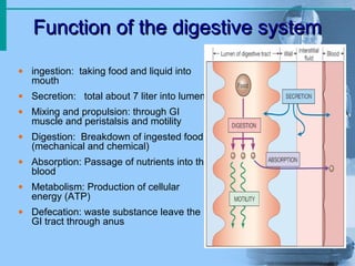
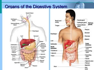
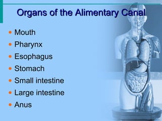
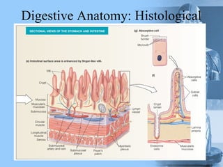
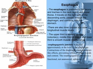
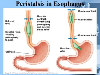
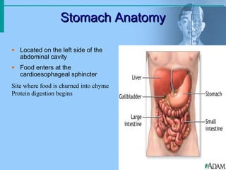
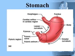
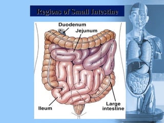
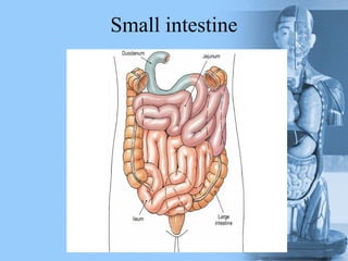
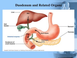
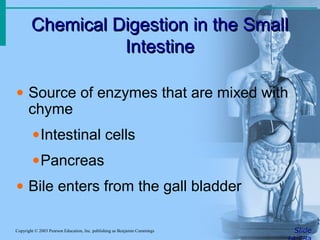
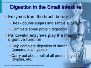
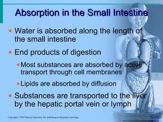
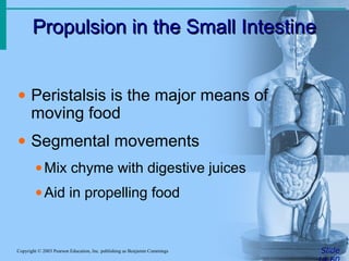
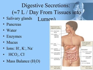
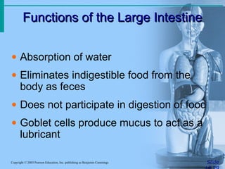
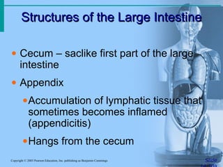
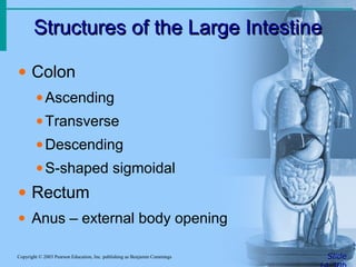
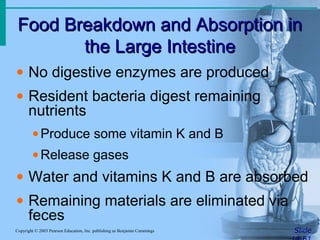
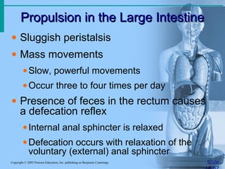
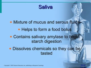
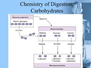
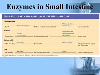
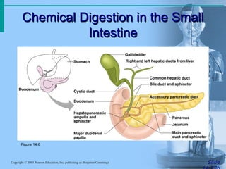
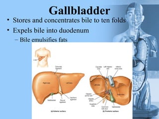
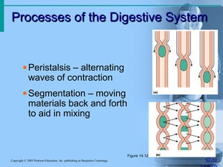
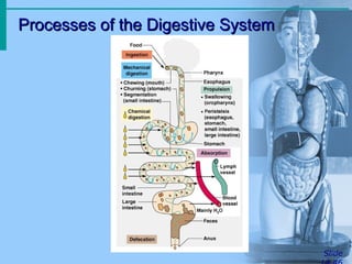
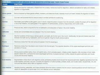
The digestive system breaks down ingested food into nutrients that can be absorbed and used by the body. The main organs include the mouth, esophagus, stomach, small intestine, large intestine, liver, pancreas, and gallbladder. In the mouth, food is chewed and mixed with saliva. The esophagus transports food to the stomach through peristalsis. The stomach mixes food with acids and enzymes to form chyme. The small intestine further digests food with enzymes from the pancreas and bile from the liver. Nutrients are absorbed through the intestinal walls. Undigested waste passes to the large intestine, where water is absorbed before waste is excreted as feces through the anus