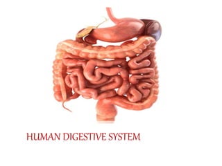
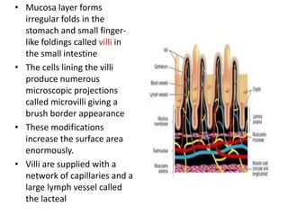
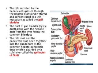
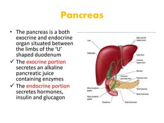
HUMAN DIGESTIVE SYSTEM
CONTENT
• Digestive system
• Digestion of food
• Absorption of digested food
• Disorders of digestive system
The human digestive system consists of
Alimentary canal
Digestive glands
Alimentary Canal
• The alimentary canal begins with an anterior
opening – the mouth and it opens out posteriorly
through the anus
• The mouth leads to the buccal cavity or oral
cavity
• The oral cavity has a number of teeth and a
muscular tongue
• The oral cavity leads into a short pharynx which
serves as a common passage for food and air
• The oesophagus and the trachea (wind pipe)
open into the pharynx
• A cartilaginous flap called epiglottis prevents
the entry of food into the glottis – opening of
the wind pipe – during swallowing
• The oesophagus is a thin, long tube which
extends posteriorly passing through the neck,
thorax and diaphragm and leads to a ‘J’
shaped bag like structure called stomach
• A muscular sphincter
(gastro-oesophageal)
regulates the opening of
oesophagus into the
stomach
• Stomach has three major
parts
 a cardiac portion into which
the oesophagus opens
 fundic region
 pyloric portion which opens
into the first part of small
intestine
• The opening of the stomach
into the duodenum is
guarded by the pyloric
sphincter
Small intestine is
distinguishable into
three regions
 U shaped duodenum
 long coiled middle
portion jejunum
 highly coiled ileum
• Ileum opens into the large intestine
• It consists of caecum, colon and rectum
 Caecum is a small blind sac which hosts some symbiotic micro-
organisms.
• A narrow finger-like tubular projection, the vermiform appendix
which is a vestigial organ, arises from the caecum.
• The caecum opens into the colon
 The colon is divided into three parts –
 an ascending part
 a transverse part
 a descending part
• The descending part opens into the rectum which opens out
through the anus
The wall of alimentary canal from oesophagus
to rectum possesses four layers
serosa
muscularis
sub-mucosa
mucosa
• Serosa is the outermost
layer made up of a thin
mesothelium (epithelium of
visceral organs) with some
connective tissues
• Muscularis is formed by
smooth muscles usually
arranged into an inner
circular and an outer
longitudinal layer
• The submucosal layer is
formed of loose connective
tissues containing nerves,
blood and lymph vessels
• The innermost layer lining
the lumen of the alimentary
canal is the mucosa
• Mucosa layer forms
irregular folds in the
stomach and small finger-
like foldings called villi in
the small intestine
• The cells lining the villi
produce numerous
microscopic projections
called microvilli giving a
brush border appearance
• These modifications
increase the surface area
enormously.
• Villi are supplied with a
network of capillaries and a
large lymph vessel called
the lacteal
• Mucosal epithelium has
goblet cells which secrete
mucus that help in
lubrication.
• Mucosa also forms glands
in the stomach (gastric
glands) and crypts in
between the bases of villi in
the intestine (crypts of
Lieberkuhn)
• All the four layers show
modifications in different
parts of the alimentary
canal
DIGESTIVE GLANDS
The digestive glands associated with the
alimentary canal include
salivary glands
liver
pancreas
Salivary glands
• Saliva is mainly produced
by three pairs of salivary
glands
 parotids (cheek),
 sub-maxillary/sub-
mandibular (lower jaw)
 sublinguals (below the
tongue).
• These glands situated just
outside the buccal cavity
secrete salivary juice into
the buccal cavity
Liver
• The largest gland of the
body weighing about 1.2 to
1.5 kg in an adult human
• Situated in the abdominal
cavity, just below the
diaphragm and has two
lobes
• The hepatic lobules are the
structural and functional
units of liver containing
hepatic cells
• Each lobule is covered by a
thin connective tissue
sheath called the Glisson’s
capsule
• The bile secreted by the
hepatic cells passes through
the hepatic ducts and is stored
and concentrated in a thin
muscular sac called the gall
bladder
• The duct of gall bladder (cystic
duct) along with the hepatic
duct from the liver forms the
common bile duct
• The bile duct and the
pancreatic duct open together
into the duodenum as the
common hepato-pancreatic
duct which is guarded by a
sphincter called the sphincter
of Oddi
Pancreas
• The pancreas is a both
exocrine and endocrine
organ situated between
the limbs of the ‘U’
shaped duodenum
 The exocrine portion
secretes an alkaline
pancreatic juice
containing enzymes
 The endocrine portion
secretes hormones,
insulin and glucagon
DIGESTION OF FOOD
• The process of digestion is accomplished by
mechanical and chemical processes
• The buccal cavity performs two major functions,
mastication of food and facilitation of swallowing
• Mucus in saliva helps in lubricating and adhering
the masticated food particles into a bolus
• The bolus is then conveyed into the pharynx and
then into the oesophagus by swallowing
• The bolus further passes down through the
oesophagus by successive waves of muscular
contractions called peristalsis
• The saliva secreted into the oral cavity
contains electrolytes (Na+ , K+ , Cl– , HCO– )
and enzymes, salivary amylase and lysozyme
• The chemical process of digestion is initiated
in the oral cavity by the hydrolytic action of
the carbohydrate splitting enzyme, the
salivary amylase
• Lysozyme present in saliva acts as an
antibacterial agent that prevents infections
The mucosa of stomach has gastric glands
which have three major types of cells
mucus neck cells secrete mucus
peptic or chief cells secrete proenzyme and
pepsinogen
parietal or oxyntic cells secrete HCl and
intrinsic factor (factor essential for absorption
of vitamin B12).
• The stomach stores the food for 4-5 hours.
• The food mixes thoroughly with the acidic gastric juice of the
stomach by the churning movements of its muscular wall and
is called the chyme
• The proenzyme pepsinogen, on exposure to hydrochloric acid
gets converted into the active enzyme pepsin
• Pepsin converts proteins into proteoses and peptones
(peptides)
• The mucus and bicarbonates present in the gastric juice play
an important role in lubrication and protection of the mucosal
epithelium from excoriation by the highly concentrated
hydrochloric acid
• HCl provides the acidic pH (pH 1.8) optimal for pepsins
• Rennin is a proteolytic enzyme found in gastric juice of infants
which helps in the digestion of milk proteins.
• Small amounts of lipases are also secreted by gastric glands
• The bile, pancreatic juice and the intestinal juice are the
secretions released into the small intestine
• The pancreatic juice contains inactive enzymes –
 trypsinogen
 chymotrypsinogen
 procarboxypeptidases
 amylases
 lipases
 nucleases
• Trypsinogen is activated by an enzyme,
enterokinase, secreted by the intestinal mucosa
into active trypsin, which in turn activates the
other enzymes in the pancreatic juice
• The bile released into the duodenum contains
bile pigments (bilirubin and bili-verdin), bile salts,
cholesterol and phospholipids but no enzymes
• Bile helps in emulsification of fats, i.e., breaking
down of the fats into very small micelles
• Bile also activates lipases
• The intestinal mucosal epithelium has goblet cells which
secrete mucus
• The secretions of the brush border cells of the mucosa
alongwith the secretions of the goblet cells constitute the
intestinal juice or succus entericus
• This juice contains a variety of enzymes like
 disaccharidases
 dipeptidases
 lipases
 nucleosidases
• The mucus along with the bicarbonates from the pancreas
protects the intestinal mucosa from acid as well as provide
an alkaline medium (pH 7.8) for enzymatic activities
• Sub-mucosal glands (Brunner’s glands) also help in this
Proteins/peptones/
proteoses
Trypsin/chemotrypsin/
carboxypeptidase
dipeptides
starch
amylase
disaccharides
fats
lipase
diglycerides
monoglyceri
des
Nucleic acids
nucleases
nucleotides nucleotides
dipeptides diptidases Amino acids
maltose
maltase
glucose
Di/ mono-
glycerides lipase Fatty acid
glycerols
ABSORPTION OF DIGESTED
PRODUCTS
• Absorption is the process by which the end products of
digestion pass through the intestinal mucosa into the blood
or lymph
• It is carried out by passive, active or facilitated transport
mechanisms.
• Small amounts of monosacharides like glucose, amino
acids and some of electrolytes like chloride ions are
generally absorbed by simple diffusion
• The passage of these substances into the blood depends
upon the concentration gradients
• Some of the substances like fructose and some amino acids
are absorbed with the help of the carrier ions like Na+
• This mechanism is called the facilitated transport
• Fatty acids and glycerol being insoluble,
cannot be absorbed into the blood
• They are first incorporated into small droplets
called micelles which move into the intestinal
mucosa
• They are re-formed into very small protein
coated fat globules called the chylomicrons
which are transported into the lymph vessels
(lacteals) in the villi
• These lymph vessels ultimately release the
absorbed substances into the blood stream
DISORDERS OF DIGESTIVE SYSTEM
• Jaundice: The liver is affected, skin and eyes turn yellow
due to the deposit of bile pigments.
• Vomiting: It is the ejection of stomach contents through
the mouth. This reflex action is controlled by the vomit
centre in the medulla. A feeling of nausea precedes
vomiting.
• Diarrhoea: The abnormal frequency of bowel movement
and increased liquidity of the faecal discharge is known as
diarrhoea. It reduces the absorption of food.
• Constipation: In constipation, the faeces are retained
within the rectum as the bowel movements occur
irregularly.
• Indigestion: In this condition, the food is not properly
digested leading to a feeling of fullness. The causes of
indigestion are inadequate enzyme secretion, anxiety, food
poisoning, over eating, and spicy food.

Human digestive system

  • 1.
  • 2.
    CONTENT • Digestive system •Digestion of food • Absorption of digested food • Disorders of digestive system
  • 4.
    The human digestivesystem consists of Alimentary canal Digestive glands
  • 5.
    Alimentary Canal • Thealimentary canal begins with an anterior opening – the mouth and it opens out posteriorly through the anus • The mouth leads to the buccal cavity or oral cavity • The oral cavity has a number of teeth and a muscular tongue • The oral cavity leads into a short pharynx which serves as a common passage for food and air
  • 6.
    • The oesophagusand the trachea (wind pipe) open into the pharynx • A cartilaginous flap called epiglottis prevents the entry of food into the glottis – opening of the wind pipe – during swallowing • The oesophagus is a thin, long tube which extends posteriorly passing through the neck, thorax and diaphragm and leads to a ‘J’ shaped bag like structure called stomach
  • 8.
    • A muscularsphincter (gastro-oesophageal) regulates the opening of oesophagus into the stomach • Stomach has three major parts  a cardiac portion into which the oesophagus opens  fundic region  pyloric portion which opens into the first part of small intestine • The opening of the stomach into the duodenum is guarded by the pyloric sphincter
  • 9.
    Small intestine is distinguishableinto three regions  U shaped duodenum  long coiled middle portion jejunum  highly coiled ileum
  • 10.
    • Ileum opensinto the large intestine • It consists of caecum, colon and rectum  Caecum is a small blind sac which hosts some symbiotic micro- organisms. • A narrow finger-like tubular projection, the vermiform appendix which is a vestigial organ, arises from the caecum. • The caecum opens into the colon  The colon is divided into three parts –  an ascending part  a transverse part  a descending part • The descending part opens into the rectum which opens out through the anus
  • 12.
    The wall ofalimentary canal from oesophagus to rectum possesses four layers serosa muscularis sub-mucosa mucosa
  • 13.
    • Serosa isthe outermost layer made up of a thin mesothelium (epithelium of visceral organs) with some connective tissues • Muscularis is formed by smooth muscles usually arranged into an inner circular and an outer longitudinal layer • The submucosal layer is formed of loose connective tissues containing nerves, blood and lymph vessels • The innermost layer lining the lumen of the alimentary canal is the mucosa
  • 14.
    • Mucosa layerforms irregular folds in the stomach and small finger- like foldings called villi in the small intestine • The cells lining the villi produce numerous microscopic projections called microvilli giving a brush border appearance • These modifications increase the surface area enormously. • Villi are supplied with a network of capillaries and a large lymph vessel called the lacteal
  • 15.
    • Mucosal epitheliumhas goblet cells which secrete mucus that help in lubrication. • Mucosa also forms glands in the stomach (gastric glands) and crypts in between the bases of villi in the intestine (crypts of Lieberkuhn) • All the four layers show modifications in different parts of the alimentary canal
  • 16.
    DIGESTIVE GLANDS The digestiveglands associated with the alimentary canal include salivary glands liver pancreas
  • 17.
    Salivary glands • Salivais mainly produced by three pairs of salivary glands  parotids (cheek),  sub-maxillary/sub- mandibular (lower jaw)  sublinguals (below the tongue). • These glands situated just outside the buccal cavity secrete salivary juice into the buccal cavity
  • 18.
    Liver • The largestgland of the body weighing about 1.2 to 1.5 kg in an adult human • Situated in the abdominal cavity, just below the diaphragm and has two lobes • The hepatic lobules are the structural and functional units of liver containing hepatic cells • Each lobule is covered by a thin connective tissue sheath called the Glisson’s capsule
  • 19.
    • The bilesecreted by the hepatic cells passes through the hepatic ducts and is stored and concentrated in a thin muscular sac called the gall bladder • The duct of gall bladder (cystic duct) along with the hepatic duct from the liver forms the common bile duct • The bile duct and the pancreatic duct open together into the duodenum as the common hepato-pancreatic duct which is guarded by a sphincter called the sphincter of Oddi
  • 20.
    Pancreas • The pancreasis a both exocrine and endocrine organ situated between the limbs of the ‘U’ shaped duodenum  The exocrine portion secretes an alkaline pancreatic juice containing enzymes  The endocrine portion secretes hormones, insulin and glucagon
  • 21.
  • 22.
    • The processof digestion is accomplished by mechanical and chemical processes • The buccal cavity performs two major functions, mastication of food and facilitation of swallowing • Mucus in saliva helps in lubricating and adhering the masticated food particles into a bolus • The bolus is then conveyed into the pharynx and then into the oesophagus by swallowing • The bolus further passes down through the oesophagus by successive waves of muscular contractions called peristalsis
  • 23.
    • The salivasecreted into the oral cavity contains electrolytes (Na+ , K+ , Cl– , HCO– ) and enzymes, salivary amylase and lysozyme • The chemical process of digestion is initiated in the oral cavity by the hydrolytic action of the carbohydrate splitting enzyme, the salivary amylase • Lysozyme present in saliva acts as an antibacterial agent that prevents infections
  • 24.
    The mucosa ofstomach has gastric glands which have three major types of cells mucus neck cells secrete mucus peptic or chief cells secrete proenzyme and pepsinogen parietal or oxyntic cells secrete HCl and intrinsic factor (factor essential for absorption of vitamin B12).
  • 25.
    • The stomachstores the food for 4-5 hours. • The food mixes thoroughly with the acidic gastric juice of the stomach by the churning movements of its muscular wall and is called the chyme • The proenzyme pepsinogen, on exposure to hydrochloric acid gets converted into the active enzyme pepsin • Pepsin converts proteins into proteoses and peptones (peptides) • The mucus and bicarbonates present in the gastric juice play an important role in lubrication and protection of the mucosal epithelium from excoriation by the highly concentrated hydrochloric acid • HCl provides the acidic pH (pH 1.8) optimal for pepsins • Rennin is a proteolytic enzyme found in gastric juice of infants which helps in the digestion of milk proteins. • Small amounts of lipases are also secreted by gastric glands
  • 26.
    • The bile,pancreatic juice and the intestinal juice are the secretions released into the small intestine • The pancreatic juice contains inactive enzymes –  trypsinogen  chymotrypsinogen  procarboxypeptidases  amylases  lipases  nucleases
  • 27.
    • Trypsinogen isactivated by an enzyme, enterokinase, secreted by the intestinal mucosa into active trypsin, which in turn activates the other enzymes in the pancreatic juice • The bile released into the duodenum contains bile pigments (bilirubin and bili-verdin), bile salts, cholesterol and phospholipids but no enzymes • Bile helps in emulsification of fats, i.e., breaking down of the fats into very small micelles • Bile also activates lipases
  • 28.
    • The intestinalmucosal epithelium has goblet cells which secrete mucus • The secretions of the brush border cells of the mucosa alongwith the secretions of the goblet cells constitute the intestinal juice or succus entericus • This juice contains a variety of enzymes like  disaccharidases  dipeptidases  lipases  nucleosidases • The mucus along with the bicarbonates from the pancreas protects the intestinal mucosa from acid as well as provide an alkaline medium (pH 7.8) for enzymatic activities • Sub-mucosal glands (Brunner’s glands) also help in this
  • 29.
  • 30.
    Nucleic acids nucleases nucleotides nucleotides dipeptidesdiptidases Amino acids maltose maltase glucose Di/ mono- glycerides lipase Fatty acid glycerols
  • 31.
  • 32.
    • Absorption isthe process by which the end products of digestion pass through the intestinal mucosa into the blood or lymph • It is carried out by passive, active or facilitated transport mechanisms. • Small amounts of monosacharides like glucose, amino acids and some of electrolytes like chloride ions are generally absorbed by simple diffusion • The passage of these substances into the blood depends upon the concentration gradients • Some of the substances like fructose and some amino acids are absorbed with the help of the carrier ions like Na+ • This mechanism is called the facilitated transport
  • 33.
    • Fatty acidsand glycerol being insoluble, cannot be absorbed into the blood • They are first incorporated into small droplets called micelles which move into the intestinal mucosa • They are re-formed into very small protein coated fat globules called the chylomicrons which are transported into the lymph vessels (lacteals) in the villi • These lymph vessels ultimately release the absorbed substances into the blood stream
  • 35.
  • 36.
    • Jaundice: Theliver is affected, skin and eyes turn yellow due to the deposit of bile pigments. • Vomiting: It is the ejection of stomach contents through the mouth. This reflex action is controlled by the vomit centre in the medulla. A feeling of nausea precedes vomiting. • Diarrhoea: The abnormal frequency of bowel movement and increased liquidity of the faecal discharge is known as diarrhoea. It reduces the absorption of food. • Constipation: In constipation, the faeces are retained within the rectum as the bowel movements occur irregularly. • Indigestion: In this condition, the food is not properly digested leading to a feeling of fullness. The causes of indigestion are inadequate enzyme secretion, anxiety, food poisoning, over eating, and spicy food.