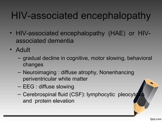
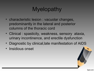
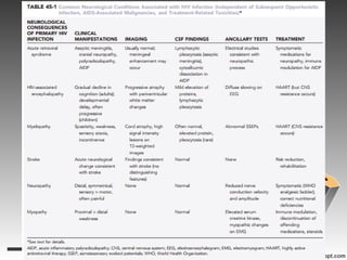
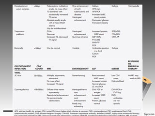
Acquired immune
deficiency syndrome
Youmans Chapter 45
Outline
• HIV infection of the nervous system
• AIDS-related malignancies
• Infection
• HIV treatment-related neurotoxicities
• Acute retroviral syndrome
• HIV-associated encephalopathy
• Myelopathy
• HIV-associated stroke
• HIV-associated neuropathy
• HIV-associated myopathy
HIV infection of the nervous system
Acute retroviral syndrome
• Acute HIV infection results in an acute retroviral
syndrome in the majority of those exposed
• aseptic meningitis 25%, facial nerve palsies,
radiculopathy, and acute demyelinating
polyneuropathy
• 2 to 6 weeks after infection
HIV-associated encephalopathy
• HIV-associated encephalopathy (HAE) or HIV-
associated dementia
• Adult
– gradual decline in cognitive, motor slowing, behavioral
changes
– Neuroimaging : diffuse atrophy, Nonenhancing
periventricular white matter
– EEG : diffuse slowing
– Cerebrospinal fluid (CSF): lymphocytic pleocytosis
and protein elevation
HIV-associated encephalopathy
• Children
– Similar to adult
– Development delay, first sign
– behavior
Myelopathy
• characteristic lesion : vacuolar changes,
predominantly in the lateral and posterior
columns of the thoracic cord
• Clinical : spasticity, weakness, sensory ataxia,
urinary incontinence, and erectile dysfunction
• Diagnostic by clinical,late manifestation of AIDS
• Insidious onset
Myelopathy
• MRI : atrophic changes of the thoracic cord with
occasional increased signal on T2-weighted and
fluid-attenuated inversion recovery (FLAIR)
sequences
• CSF : normal or mild a mild lymphocytic
pleocytosis and protein elevation
• Ddx : retroviruses (human T-cell lymphotropic
virus types I and II), cytomegalovirus (CMV),
herpes simplex virus type 2, and herpes
zoster, lymphoma, myeloma
HIV-associated stroke
• HIV infection is associated with an increased
risk for stroke
• antiphospholipid antibodies, protein S deficiency
• Vasculitis
• stroke must be considered in the differential
diagnosis of any HIV-positive individual with an
abrupt onset of neurological symptoms.
HIV-associated neuropathy
• Most common : Peripheral neuropathy
• acute inflammatory polyradiculopathy
• A painful distal symmetrical polyneuropathy
• Patients complain of numbness and burning of
the feet, which are exquisitely sensitive to touch,
hand symptom is usually late complication
• Electrical : nerve conduction velocity or
amplitude
HIV-associated myopathy
• Common : Symmetrical and proximal
myopathy
• Hip flexor , neck muscle
• Serum creatinine kinase elevated
• EMG : myopathic change
• Muscle biopsy : degeneration of myofiber
AIDS-related malignancies
• Kaposi’s sarcoma, Hodgkin’s lymphoma, non-
Hodgkin’s lymphoma (NHL), squamous cell
carcinoma, plasmacytoma, and
leiomyosarcoma
• Direct involvement of the CNS by these tumors
is rare, with one exception: primary central
nervous system lymphoma (PCNSL)
• high frequency in HIV-positive patients,
especially with the advent of HAART and
the prolongation of life associated with its use
Primary Central Nervous System
Lymphoma
• PCNSL is a rare NHL that accounts for less than
5% of primary CNS tumors.
• Clinical manifestations :
– highly variable and depend on the CNS site or sites
involved
– The tumor is often but not always multifocal and
occupies white matter spaces preferentially
Primary Central Nervous System
Lymphoma
• Symptoms range from nonspecific (i.e.,
nonlocalizing headache, mild encephalopathy,
delirium) to those associated with discrete
lesions in so-called eloquent areas of the
brain
• Radiology finding
– homogeneous contrast enhanced lesions with either
a hypodense appearance (on CT) or low signal
intensity (on T1-weighted MRI)
– Periventricular region
– Multiple but < , location : subependymal
Primary Central Nervous System
Lymphoma
Primary Central Nervous System
Lymphoma
• The entire neuraxis (eyes, brain, spinal cord,
and CSF spaces) must be evaluated for the
presence of PCNSL if suspected
– Eyes : slit-lamp examination to rule out vitreous tumor
– CSF : cytopathologic examination
– Avoiding to brain biopsy
– Wright stain,Papanicolaou stain
– Flow cytometry : predominant lymphocyte
Primary Central Nervous System
Lymphoma
• NeuroSx : call on biopsy suspected PCNSL
• Ddx : toxoplamosis, cerebral tuberculous
• Toxoplasmosis show signs of clinical
improvement within 3 days of treatment, and
improvement on neuroimaging follows within 7
to 10 days.
• In the case of suspected tuberculosis,
improvement occurs less rapidly
Primary Central Nervous System
Lymphoma
• Therefore, if this approach is adopted,
careful clinical assessment must be maintained,
and biopsy should be performed in any person
with early deterioration after the initiation of
empirical treatment of presumed infectious
disease.
• Survival after WBRT alone in PCNSL
patients is approximately 15 months and
decreases to 2 to 5 months in AIDS patients
• Methotrexate-based chemotherapy regimens
Infection
• Toxoplasmosis
• Cryptococcus
• Aspergillus
• Mycobacterium
• Treponema pallidum
• Bartonella
• JC virus
• Cytomegalovirus
Toxoplasmosis
• Toxoplasma gondii, Most common
• Exposure to cat,endemic area
• Clinical : encephalitis without meningeal
involvement
• CT,MRI : multiple nodular or ring enhancing
lesions c surrounding edema, hyperintense on
T2 , hyperintense on DWI
• Location : gray-white interface, basal ganglia,
thalamus
Toxoplasmosis
• Ddx of ring-enhancing lesions
– Lymphoma : homogenous pattern of
enhancement, DWI : uniformly restrict pattern,
periventricular and callosal involvement
– Other infection : tuberculosis and
cryptococcosis
– Small parenchymal hemorrhage in
toxoplasmosis
Toxoplasmosis
• Serum or CSF PCR for toxoplasma organism
• Empirical ATB : pyrimethamine and sulfadiazine
• Radiographic improvement within 2-4 wks
• Fail diagnosis or fail medical treatment : biopsy,
sent for Toxoplasma-specific DNA by PCR
• Reactivation form bradyzoite infection  repeat
biopsy
Cryptoccosis
• Cryptococcous neoformans
• Bird excrement,infected human via inhalation
• CD4 < 200 cell/ul
• Clinical : meningitis, Cranial neuropathies 
involve base of skull, hydrocephalus, stroke
• Radiographic : nonspecific, ring-
enhancement(ddx : toxoplasmosis), pseudocyst
in CSF space  dilated perivascular space
Cryptoccosis
• Blood C/S : positive for cryptococcosis
• CSF : india ink  budding yeast, cryptococcal
Ag, fungal culture
• Histopathologic : chronic granulomatous
changes with only a few organisms
• ATB : fluconazole or amphotericin B
• Relapse is high
• NeuroSx : place ventricular shunt for
hydrocephalus
Aspergillosis
• Aspergillus
• septate hyaline mold found in plants and soi
• severe sinopulmonary infections in
immunocompromised hosts(CD4 < 50 cell/uL)
• diffuse cerebritis, focal abscesses, or meningitis
• Vascular invasion  stroke
• Biopsy for definite diagnosis
Mycobacterial infection
• M. tuberculosis : CNS infection after pulmonary
infection
• M. avium and M. intracellulare : water, soil, and
animal host, CD4 < 50 cells/uL, MAC
• Clinical : meningitis, parenchymal brain lesion
are less common
• Radiology : leptomeningeal thickening,
tuberculoma may be single or multiple,gray-
white junction, supratentorial space
Mycobacterial infection
• MRI : isointense c low signal center on T2,target
lesion on contrast study
• Tubercular abscess : solitary, large,
multiloculated, enhance, mass effect, surround
by significant edema
• Purified protein derivative skin tests for M.
tuberculosis are unreliable in patients with HIV
• CSF culture : positive
• CSF AFB : negative
Mycobacterial infection
• AFB PCR : higher sensitivity but a negative PCR
result does not rule out M. tuberculosis in the
CNS
• CSF profile : elevated white blood cell count with
a lymphocytic predominance, glucose decrease
• NeuroSx : placement extraventruicilar drain in
tuberculous meningitis for obstructive
hydrocephalus
Mycobacterial infection
• M. tuberculosisbrain abscesses : drainage
• Treatment : multidrug regimen, duration varies
• Role of steroid is unknown
Treponema pallidum
• T.pallidum
• The tertiary stage of syphilis : diffuse systemic
involvement, including the CNS, Syphilitic
meningitis or meningovasculitis
• The classic manifestations of tertiary syphilis,
such as general paresis of the insane, tabes
dorsalis, and cerebral gummas, are less
common in the HIV/AIDS setting
Bartonella
• gram-negative bacteria Bartonella henselaeand
Bartonella Quintana
• cat-scratch disease and bacillary angiomatosis
• systemic disease with florid
meningoencephalitis is much more common
in AIDS patients
• suspected in those with typical skin lesion or
lymphadenopathy
JC virus
• JC virus
• CD4 < 100 cells/uL
• Spred via B lymphocyte to CNS  replicate in
oligodendrocytes  demyelination  focal
symptom  fatal
• Seizure
JC virus
• Laboratory finding:
– CSF : rarely pleocytosis, no decrease in glucose,
– CSF PCR : high specifity,but sensitivity vary
• MRI :
– T2/FLAIR : hyperintensity on white matter
– T1 : hypointensity
– pronounced posteriorly but can be found anywhere in
the brain, including the posterior fossa
– multifocal and have an asymmetric distribution
JC virus
JC virus
• Brain biopsy is important for diagnosis
• Histologic examination : demyelination
accompanied by “bizarre”-looking astrocytes
with pleomorphic nuclei
• No specific treatment
• HAART for immune reconstitutuion
Cytomegalovirus
• CMV
• CD 4 < 50 cells/uL
• myriad neurological syndromes
– Encephalitis : involvement periventricular tissue,
brainstem
– Myelitis
– Polyradiculopathy : most commonly affects the
lumbar spinal nerve roots, bilateral lower extremity
weakness and bowel and bladder symptoms
– Polyneuropathy : numbness and paresthesias
Cytomegalovirus
• Neurological examination : decrease in reflexes
consistent with a lower motor neuron syndrome
• MRI
– increased signal on T2/FLAIR images
– rare enhancement in the ependymal region the
periventricular white matter, and elsewhere in the
brain or brainstem
– Leptomeningeal enhancement
Cytomegalovirus
• Laboratory
– CSF : increased protein and glucose levels, as well as
white blood cells
– CS grow slowly
– CSF CMV PCR positivity is not sufficient by itself for
diagnosis because CSF CMV PCR may also be
positive in individuals with systemic CMV infection
in the absence of neurological syndrome
Cytomegalovirus
• Histology : eosinophilic intranuclear inclusions
with surrounding halos
• Specific treatment of CMV : ganciclovir or
foscarnet, or both
• HAART
• Mortality rate is high,within week
HIV treatment-related neurotoxicities
• peripheral neuropathy
– predominant neurological consequence of HAART
therapy
– The nucleoside analogue(dose dependent),
protease inhibitors
– Multifactorial : HIV infection itself, opportunistic
infection, and nutritional deficiencies.
HIV treatment-related neurotoxicities
• Immune reconstitution inflammatory syndrome
(IRIS)
– Restoration of immune competence as a result of
HAART can paradoxically cause clinical
deterioration because of innocent bystander–like
effects
– Risk for IRIS : initiating HARR, multiple infection
– neurological symptoms of IRIS : protean and largely
dependent on location.
– Onset occurs within 2 months of initiating HAART.
HIV treatment-related neurotoxicities
• Immune reconstitution inflammatory syndrome
(IRIS)
– Imagind study : contrast enhancement
– Histopatholgy : diffuse cytotoxic T-cell infiltrates
– NeuroSx : call for brain biopsy
– Management : problematic – discontinue, steroid
Thank you

045 AIDS

  • 1.
  • 2.
    Outline • HIV infectionof the nervous system • AIDS-related malignancies • Infection • HIV treatment-related neurotoxicities
  • 3.
    • Acute retroviralsyndrome • HIV-associated encephalopathy • Myelopathy • HIV-associated stroke • HIV-associated neuropathy • HIV-associated myopathy HIV infection of the nervous system
  • 4.
    Acute retroviral syndrome •Acute HIV infection results in an acute retroviral syndrome in the majority of those exposed • aseptic meningitis 25%, facial nerve palsies, radiculopathy, and acute demyelinating polyneuropathy • 2 to 6 weeks after infection
  • 5.
    HIV-associated encephalopathy • HIV-associatedencephalopathy (HAE) or HIV- associated dementia • Adult – gradual decline in cognitive, motor slowing, behavioral changes – Neuroimaging : diffuse atrophy, Nonenhancing periventricular white matter – EEG : diffuse slowing – Cerebrospinal fluid (CSF): lymphocytic pleocytosis and protein elevation
  • 6.
    HIV-associated encephalopathy • Children –Similar to adult – Development delay, first sign – behavior
  • 7.
    Myelopathy • characteristic lesion: vacuolar changes, predominantly in the lateral and posterior columns of the thoracic cord • Clinical : spasticity, weakness, sensory ataxia, urinary incontinence, and erectile dysfunction • Diagnostic by clinical,late manifestation of AIDS • Insidious onset
  • 8.
    Myelopathy • MRI :atrophic changes of the thoracic cord with occasional increased signal on T2-weighted and fluid-attenuated inversion recovery (FLAIR) sequences • CSF : normal or mild a mild lymphocytic pleocytosis and protein elevation • Ddx : retroviruses (human T-cell lymphotropic virus types I and II), cytomegalovirus (CMV), herpes simplex virus type 2, and herpes zoster, lymphoma, myeloma
  • 9.
    HIV-associated stroke • HIVinfection is associated with an increased risk for stroke • antiphospholipid antibodies, protein S deficiency • Vasculitis • stroke must be considered in the differential diagnosis of any HIV-positive individual with an abrupt onset of neurological symptoms.
  • 10.
    HIV-associated neuropathy • Mostcommon : Peripheral neuropathy • acute inflammatory polyradiculopathy • A painful distal symmetrical polyneuropathy • Patients complain of numbness and burning of the feet, which are exquisitely sensitive to touch, hand symptom is usually late complication • Electrical : nerve conduction velocity or amplitude
  • 11.
    HIV-associated myopathy • Common: Symmetrical and proximal myopathy • Hip flexor , neck muscle • Serum creatinine kinase elevated • EMG : myopathic change • Muscle biopsy : degeneration of myofiber
  • 13.
    AIDS-related malignancies • Kaposi’ssarcoma, Hodgkin’s lymphoma, non- Hodgkin’s lymphoma (NHL), squamous cell carcinoma, plasmacytoma, and leiomyosarcoma • Direct involvement of the CNS by these tumors is rare, with one exception: primary central nervous system lymphoma (PCNSL) • high frequency in HIV-positive patients, especially with the advent of HAART and the prolongation of life associated with its use
  • 14.
    Primary Central NervousSystem Lymphoma • PCNSL is a rare NHL that accounts for less than 5% of primary CNS tumors. • Clinical manifestations : – highly variable and depend on the CNS site or sites involved – The tumor is often but not always multifocal and occupies white matter spaces preferentially
  • 15.
    Primary Central NervousSystem Lymphoma • Symptoms range from nonspecific (i.e., nonlocalizing headache, mild encephalopathy, delirium) to those associated with discrete lesions in so-called eloquent areas of the brain • Radiology finding – homogeneous contrast enhanced lesions with either a hypodense appearance (on CT) or low signal intensity (on T1-weighted MRI) – Periventricular region – Multiple but < , location : subependymal
  • 16.
    Primary Central NervousSystem Lymphoma
  • 17.
    Primary Central NervousSystem Lymphoma • The entire neuraxis (eyes, brain, spinal cord, and CSF spaces) must be evaluated for the presence of PCNSL if suspected – Eyes : slit-lamp examination to rule out vitreous tumor – CSF : cytopathologic examination – Avoiding to brain biopsy – Wright stain,Papanicolaou stain – Flow cytometry : predominant lymphocyte
  • 18.
    Primary Central NervousSystem Lymphoma • NeuroSx : call on biopsy suspected PCNSL • Ddx : toxoplamosis, cerebral tuberculous • Toxoplasmosis show signs of clinical improvement within 3 days of treatment, and improvement on neuroimaging follows within 7 to 10 days. • In the case of suspected tuberculosis, improvement occurs less rapidly
  • 19.
    Primary Central NervousSystem Lymphoma • Therefore, if this approach is adopted, careful clinical assessment must be maintained, and biopsy should be performed in any person with early deterioration after the initiation of empirical treatment of presumed infectious disease. • Survival after WBRT alone in PCNSL patients is approximately 15 months and decreases to 2 to 5 months in AIDS patients • Methotrexate-based chemotherapy regimens
  • 20.
    Infection • Toxoplasmosis • Cryptococcus •Aspergillus • Mycobacterium • Treponema pallidum • Bartonella • JC virus • Cytomegalovirus
  • 21.
    Toxoplasmosis • Toxoplasma gondii,Most common • Exposure to cat,endemic area • Clinical : encephalitis without meningeal involvement • CT,MRI : multiple nodular or ring enhancing lesions c surrounding edema, hyperintense on T2 , hyperintense on DWI • Location : gray-white interface, basal ganglia, thalamus
  • 22.
    Toxoplasmosis • Ddx ofring-enhancing lesions – Lymphoma : homogenous pattern of enhancement, DWI : uniformly restrict pattern, periventricular and callosal involvement – Other infection : tuberculosis and cryptococcosis – Small parenchymal hemorrhage in toxoplasmosis
  • 23.
    Toxoplasmosis • Serum orCSF PCR for toxoplasma organism • Empirical ATB : pyrimethamine and sulfadiazine • Radiographic improvement within 2-4 wks • Fail diagnosis or fail medical treatment : biopsy, sent for Toxoplasma-specific DNA by PCR • Reactivation form bradyzoite infection  repeat biopsy
  • 24.
    Cryptoccosis • Cryptococcous neoformans •Bird excrement,infected human via inhalation • CD4 < 200 cell/ul • Clinical : meningitis, Cranial neuropathies  involve base of skull, hydrocephalus, stroke • Radiographic : nonspecific, ring- enhancement(ddx : toxoplasmosis), pseudocyst in CSF space  dilated perivascular space
  • 25.
    Cryptoccosis • Blood C/S: positive for cryptococcosis • CSF : india ink  budding yeast, cryptococcal Ag, fungal culture • Histopathologic : chronic granulomatous changes with only a few organisms • ATB : fluconazole or amphotericin B • Relapse is high • NeuroSx : place ventricular shunt for hydrocephalus
  • 26.
    Aspergillosis • Aspergillus • septatehyaline mold found in plants and soi • severe sinopulmonary infections in immunocompromised hosts(CD4 < 50 cell/uL) • diffuse cerebritis, focal abscesses, or meningitis • Vascular invasion  stroke • Biopsy for definite diagnosis
  • 27.
    Mycobacterial infection • M.tuberculosis : CNS infection after pulmonary infection • M. avium and M. intracellulare : water, soil, and animal host, CD4 < 50 cells/uL, MAC • Clinical : meningitis, parenchymal brain lesion are less common • Radiology : leptomeningeal thickening, tuberculoma may be single or multiple,gray- white junction, supratentorial space
  • 28.
    Mycobacterial infection • MRI: isointense c low signal center on T2,target lesion on contrast study • Tubercular abscess : solitary, large, multiloculated, enhance, mass effect, surround by significant edema • Purified protein derivative skin tests for M. tuberculosis are unreliable in patients with HIV • CSF culture : positive • CSF AFB : negative
  • 29.
    Mycobacterial infection • AFBPCR : higher sensitivity but a negative PCR result does not rule out M. tuberculosis in the CNS • CSF profile : elevated white blood cell count with a lymphocytic predominance, glucose decrease • NeuroSx : placement extraventruicilar drain in tuberculous meningitis for obstructive hydrocephalus
  • 30.
    Mycobacterial infection • M.tuberculosisbrain abscesses : drainage • Treatment : multidrug regimen, duration varies • Role of steroid is unknown
  • 31.
    Treponema pallidum • T.pallidum •The tertiary stage of syphilis : diffuse systemic involvement, including the CNS, Syphilitic meningitis or meningovasculitis • The classic manifestations of tertiary syphilis, such as general paresis of the insane, tabes dorsalis, and cerebral gummas, are less common in the HIV/AIDS setting
  • 32.
    Bartonella • gram-negative bacteriaBartonella henselaeand Bartonella Quintana • cat-scratch disease and bacillary angiomatosis • systemic disease with florid meningoencephalitis is much more common in AIDS patients • suspected in those with typical skin lesion or lymphadenopathy
  • 33.
    JC virus • JCvirus • CD4 < 100 cells/uL • Spred via B lymphocyte to CNS  replicate in oligodendrocytes  demyelination  focal symptom  fatal • Seizure
  • 34.
    JC virus • Laboratoryfinding: – CSF : rarely pleocytosis, no decrease in glucose, – CSF PCR : high specifity,but sensitivity vary • MRI : – T2/FLAIR : hyperintensity on white matter – T1 : hypointensity – pronounced posteriorly but can be found anywhere in the brain, including the posterior fossa – multifocal and have an asymmetric distribution
  • 35.
  • 36.
    JC virus • Brainbiopsy is important for diagnosis • Histologic examination : demyelination accompanied by “bizarre”-looking astrocytes with pleomorphic nuclei • No specific treatment • HAART for immune reconstitutuion
  • 37.
    Cytomegalovirus • CMV • CD4 < 50 cells/uL • myriad neurological syndromes – Encephalitis : involvement periventricular tissue, brainstem – Myelitis – Polyradiculopathy : most commonly affects the lumbar spinal nerve roots, bilateral lower extremity weakness and bowel and bladder symptoms – Polyneuropathy : numbness and paresthesias
  • 38.
    Cytomegalovirus • Neurological examination: decrease in reflexes consistent with a lower motor neuron syndrome • MRI – increased signal on T2/FLAIR images – rare enhancement in the ependymal region the periventricular white matter, and elsewhere in the brain or brainstem – Leptomeningeal enhancement
  • 39.
    Cytomegalovirus • Laboratory – CSF: increased protein and glucose levels, as well as white blood cells – CS grow slowly – CSF CMV PCR positivity is not sufficient by itself for diagnosis because CSF CMV PCR may also be positive in individuals with systemic CMV infection in the absence of neurological syndrome
  • 40.
    Cytomegalovirus • Histology :eosinophilic intranuclear inclusions with surrounding halos • Specific treatment of CMV : ganciclovir or foscarnet, or both • HAART • Mortality rate is high,within week
  • 43.
    HIV treatment-related neurotoxicities •peripheral neuropathy – predominant neurological consequence of HAART therapy – The nucleoside analogue(dose dependent), protease inhibitors – Multifactorial : HIV infection itself, opportunistic infection, and nutritional deficiencies.
  • 44.
    HIV treatment-related neurotoxicities •Immune reconstitution inflammatory syndrome (IRIS) – Restoration of immune competence as a result of HAART can paradoxically cause clinical deterioration because of innocent bystander–like effects – Risk for IRIS : initiating HARR, multiple infection – neurological symptoms of IRIS : protean and largely dependent on location. – Onset occurs within 2 months of initiating HAART.
  • 45.
    HIV treatment-related neurotoxicities •Immune reconstitution inflammatory syndrome (IRIS) – Imagind study : contrast enhancement – Histopatholgy : diffuse cytotoxic T-cell infiltrates – NeuroSx : call for brain biopsy – Management : problematic – discontinue, steroid
  • 46.

Editor's Notes

  • #6 เกิดอาการความจำแย่ลงก่อนและตามด้วยการเคลื่อนไหวช้าลง ความรู้สึกตัวเปลี่ยนไป
  • #9 ถ้าอาการเป็นเร็ว ปวดหลังมาก CSF ผิดปกติมาก ให้นึกถึงโรคอื่น
  • #10 ต้องสงสัยผ้ป่วยที่เป็น hiv infection ว่าเป็น stroke ถ้าเกิดอาการแบบเฉียบพลัน
  • #14 PCNSL เป้น direct involvement ของCNSพบได้มากขึ้นในคนไข้ HIV positive ร่วมกับใช้ HARRT
  • #15 พบ PCNSL ในผู้ป่วยที่ ICH ต่อมาพบมากวในผู้ป่วยที่เป็น aid .ในผู้ป่วย AIDs จะรักษาได้ยากและดื้อยามากกว่า
  • #16 อาการมีตั้งปต่ non specific ปวดหัวทั่วไปจนถงอาการ specific ตามตำแหน่ง
  • #17 T1 contrast enhanceT2 hyperintensity ตรง corpus callosum และ cerebellar hemisphere
  • #19 การทำ biops specifitivy มากที่สึด การวินิฉัยโดยการทำ biopsy มีผลข้างเคียงมาก จึงทำการรักษาโดยการใหเ ATB ไปก่อนใน toxoplasmois และ cerebral tuberculous ถ้าไม่ดีขึ้นต้องนึกถึง PCNSL
  • #21 Nervous system ถูก infection ได้จาก AIDS ลักษณะอาการอาจเหมือนกันมาก การวินิจฉัยต้องได้ histologic confirm
  • #22 Ring enhance อาจเห็นไม่ชัดในคนไข้ทีมี immune respone น้อย
  • #24 เนื่องจาก toxoplasma encephalitis พบได้บ่อย การวิจนิฉํยป็น invasive ส่วนมากจึงให้ยาไปก่อนเลย ถ้าให้ยาแล้ว ไม่ดีขึ้น ก้ให้ Neuro Sx biopsy
  • #28 -Mycobacterium avium complex (MAC). CNS involvement, when present, mimics tuberculosis and is difficult to distinguish from it
  • #37 HAART
  • #40 CMV PCR positive อย่างเดี่ยวไม่พอในการวินิจฉัย, เพราะสามารถขี้นได้ในผู้ป่วย systemic infection โดยที่ไม่มี neurological symptom
  • #44 Peripheral neuropathy IRIS เกิดจาก immune competence ดีขึ้นจากการได้ HAARt แต่อาการแย่ลง