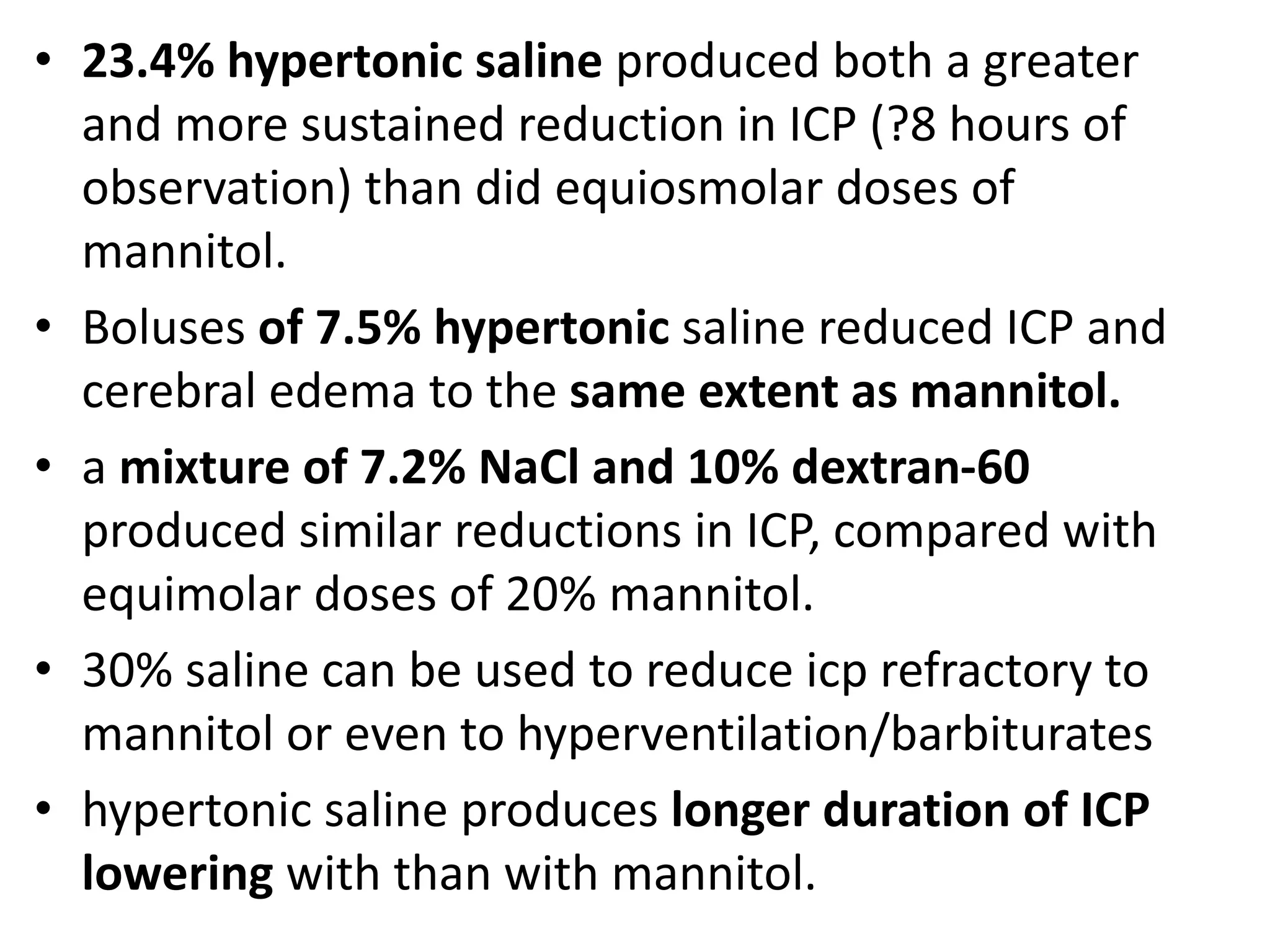
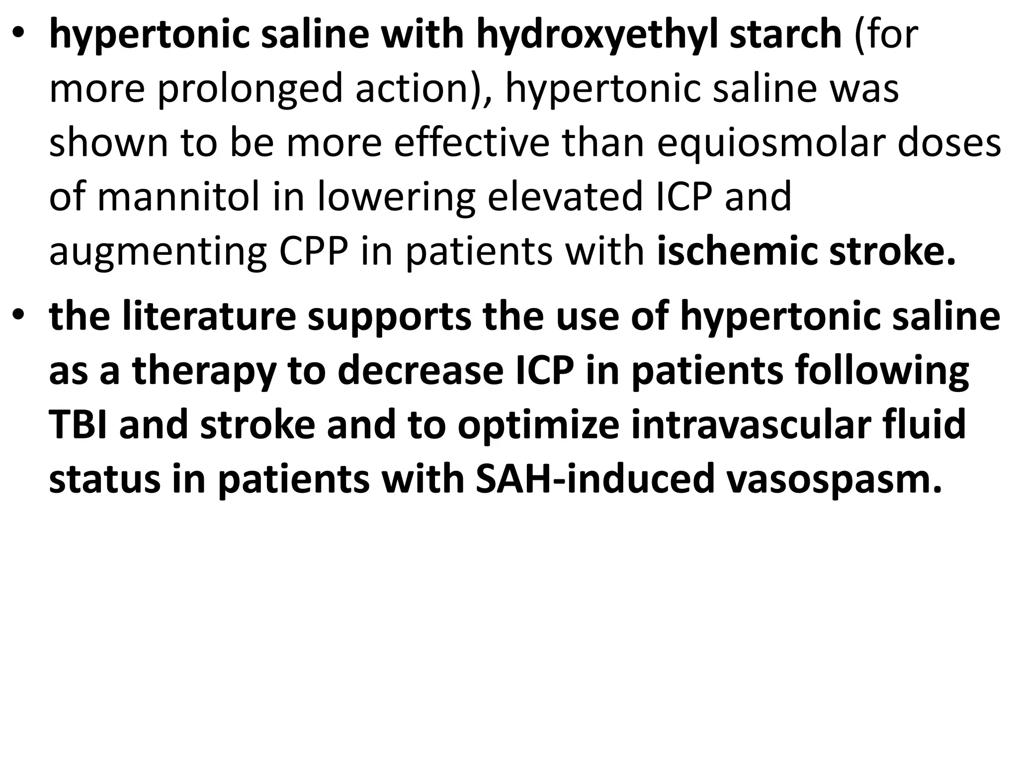
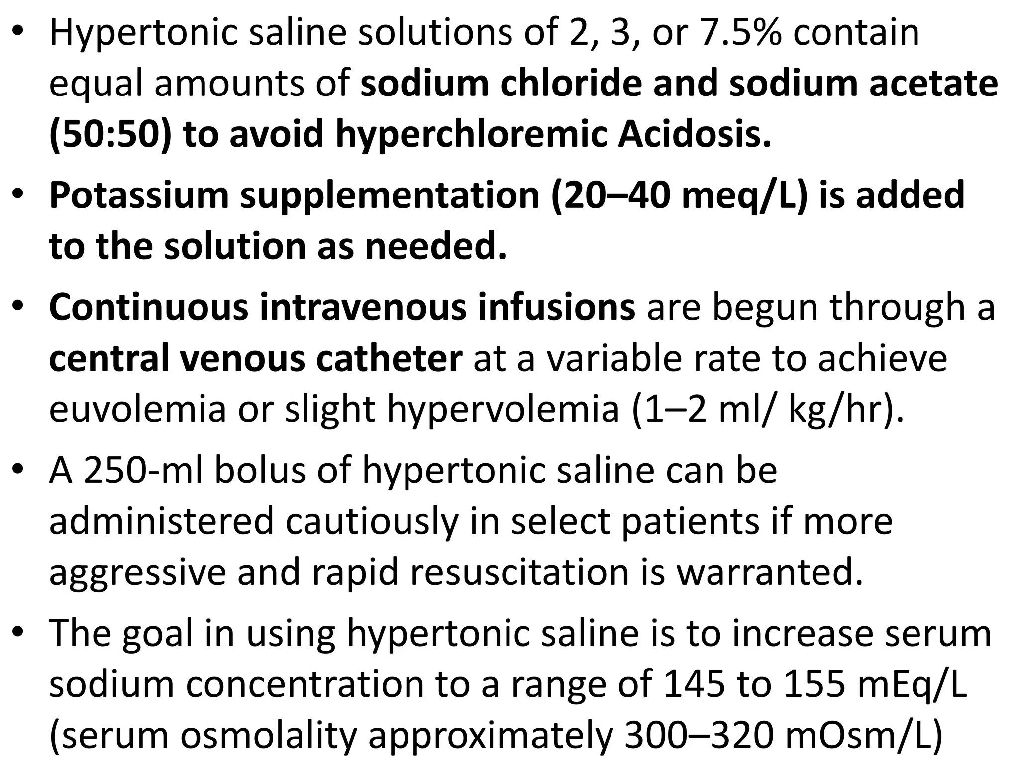
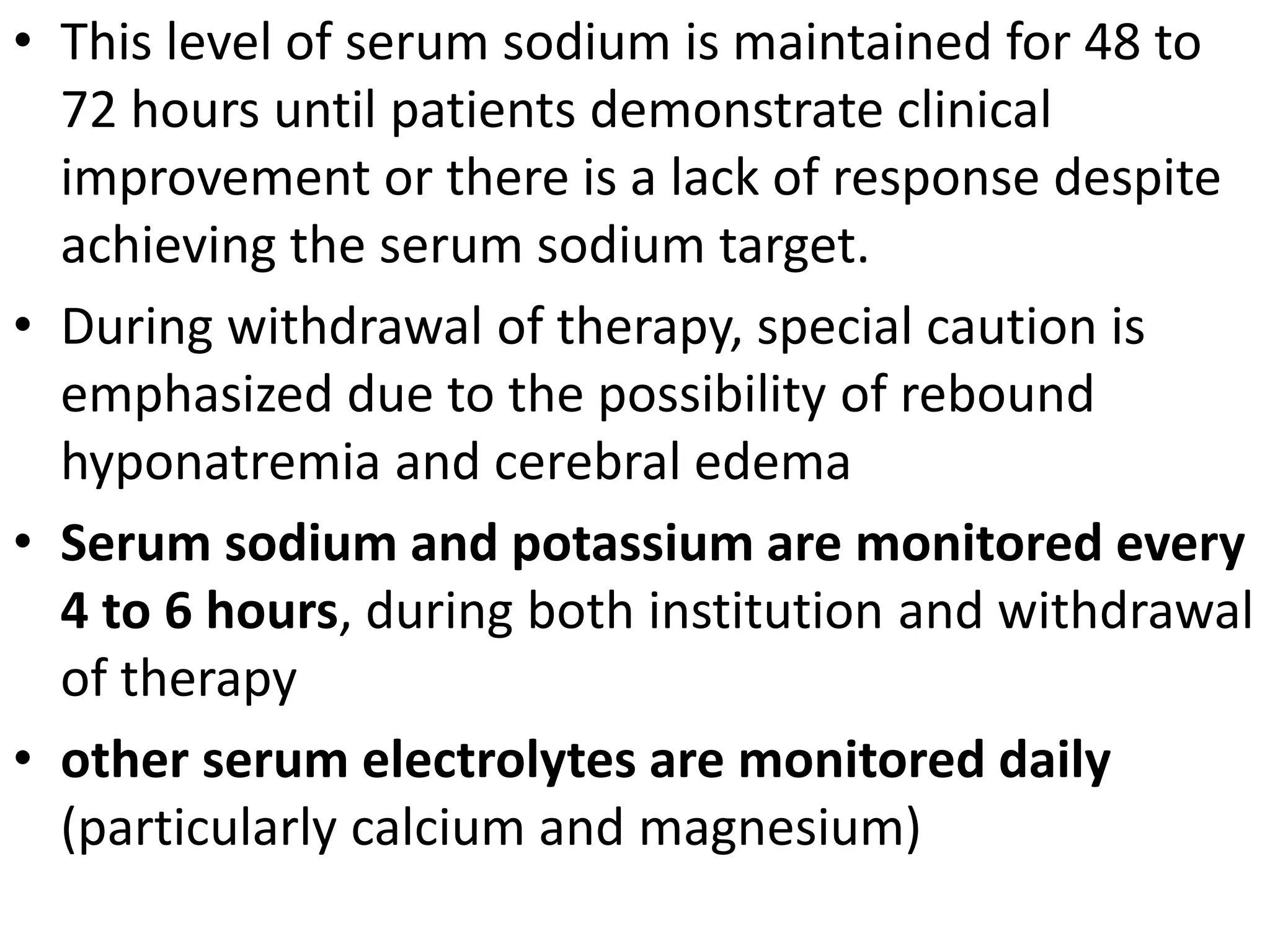
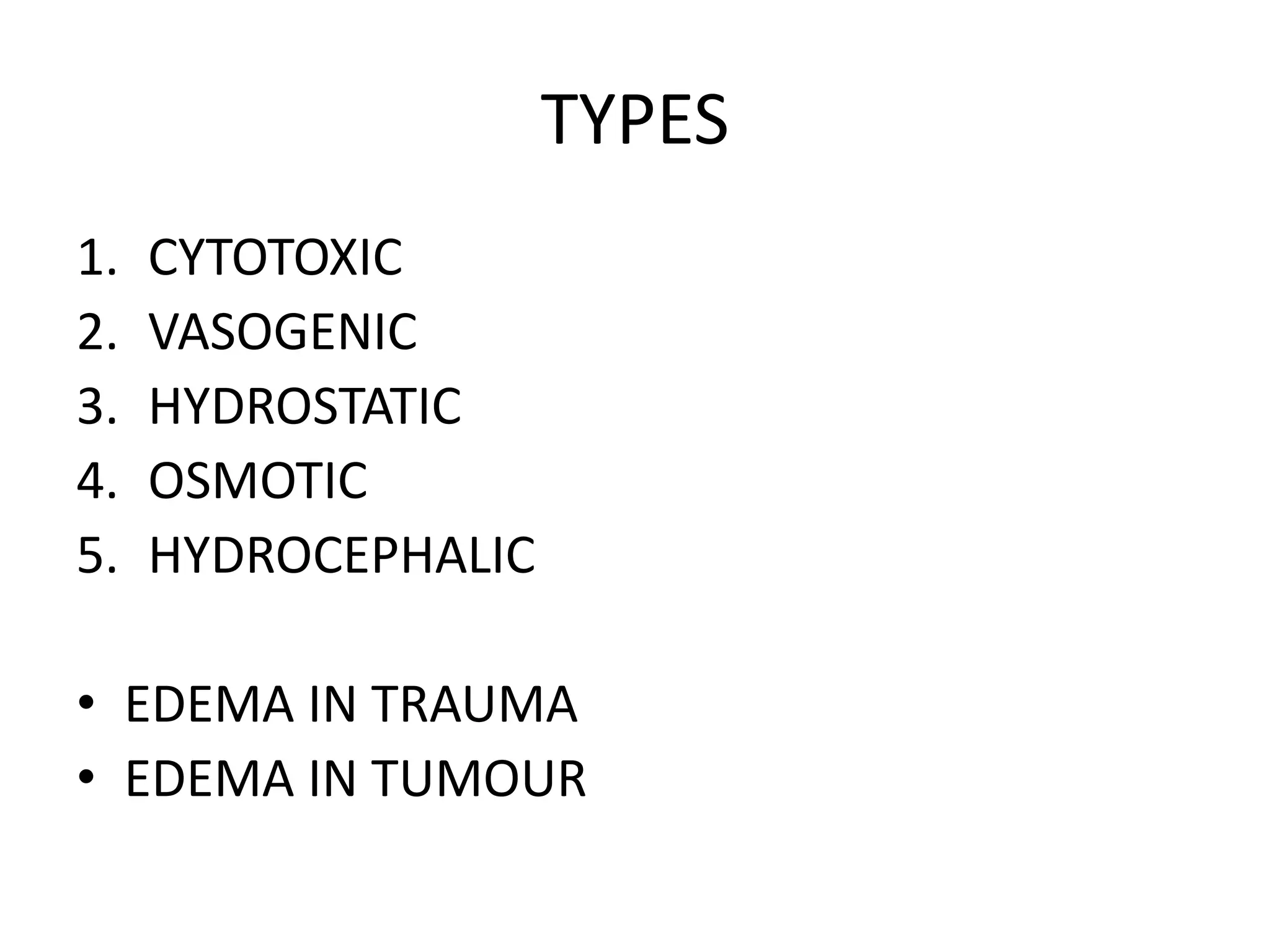
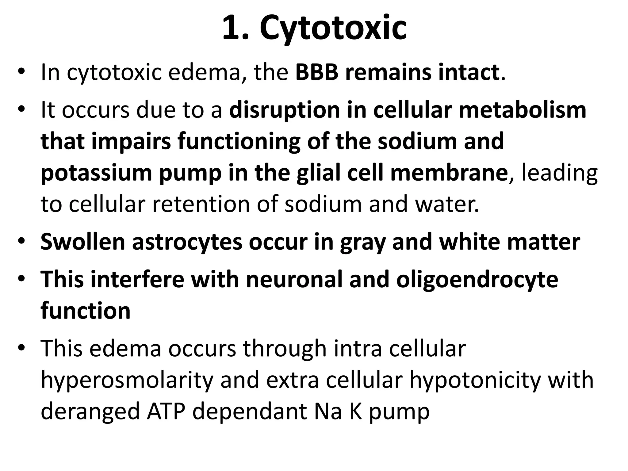
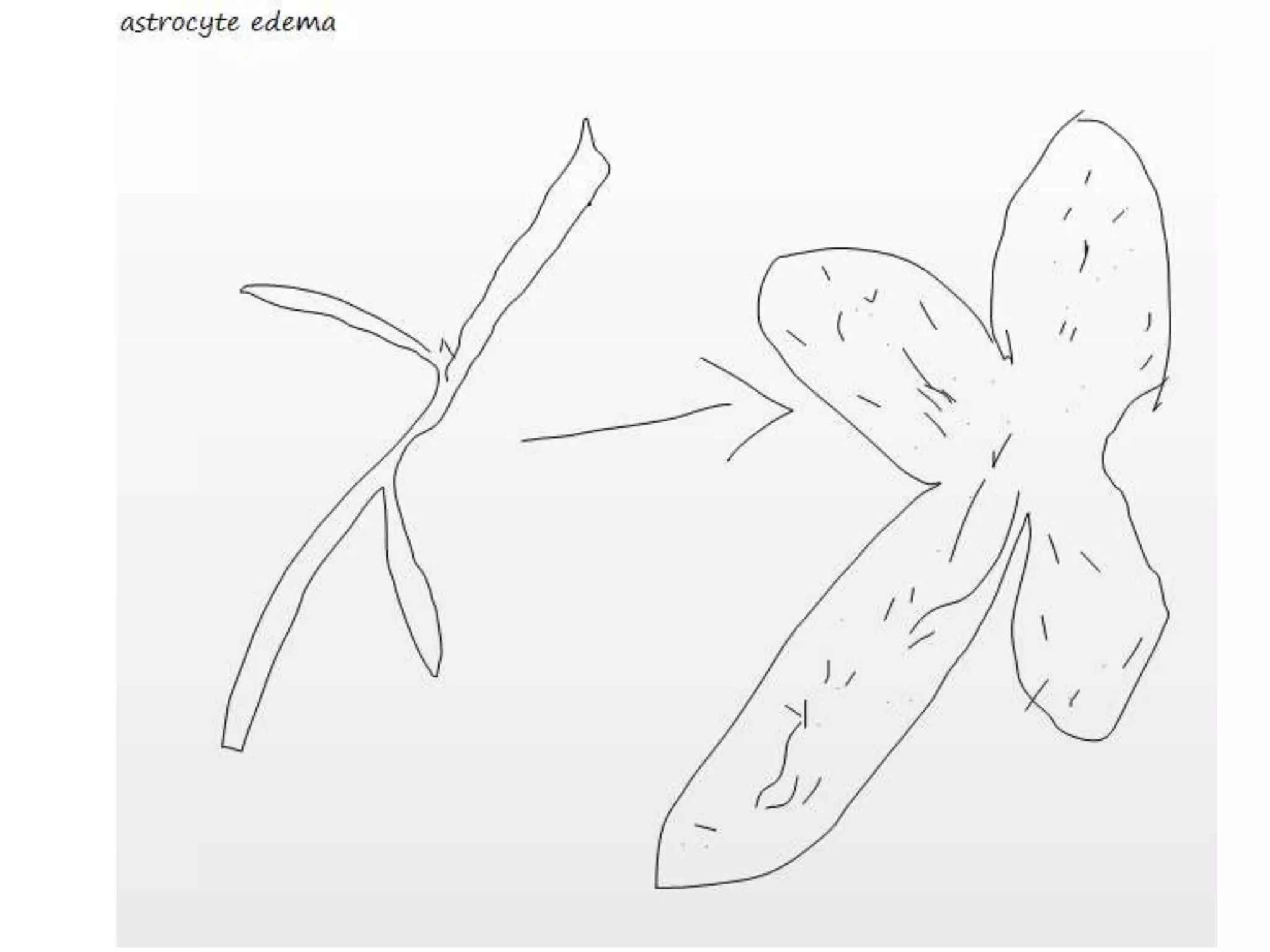
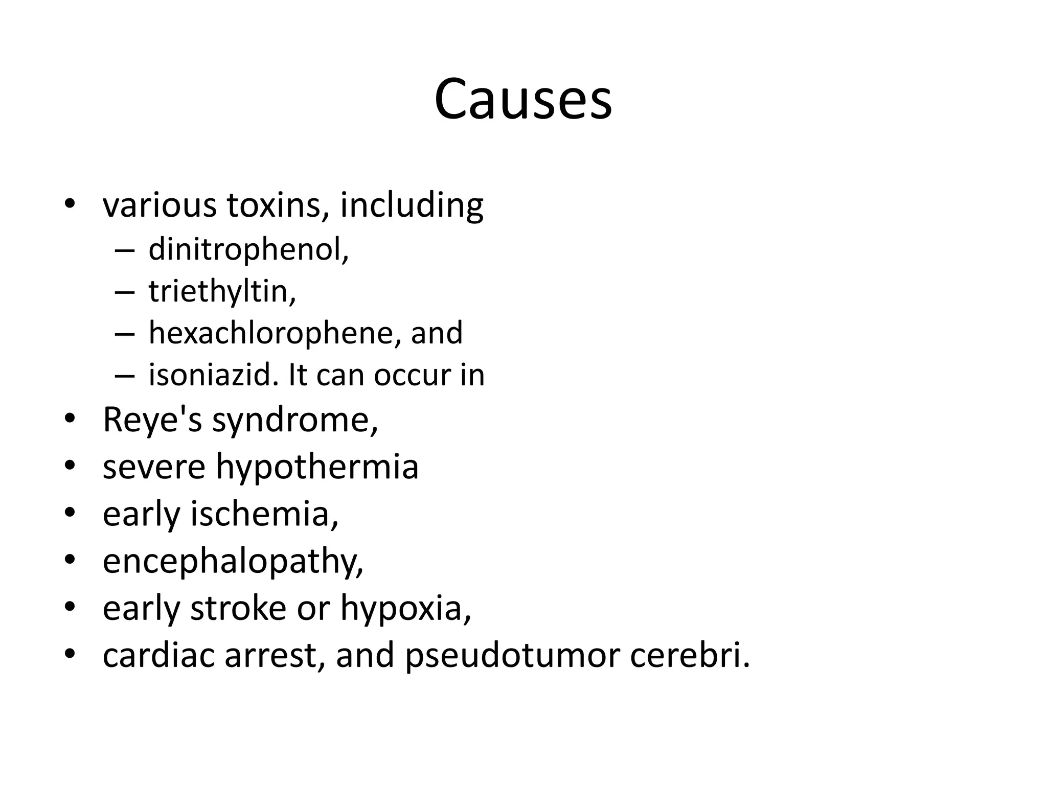
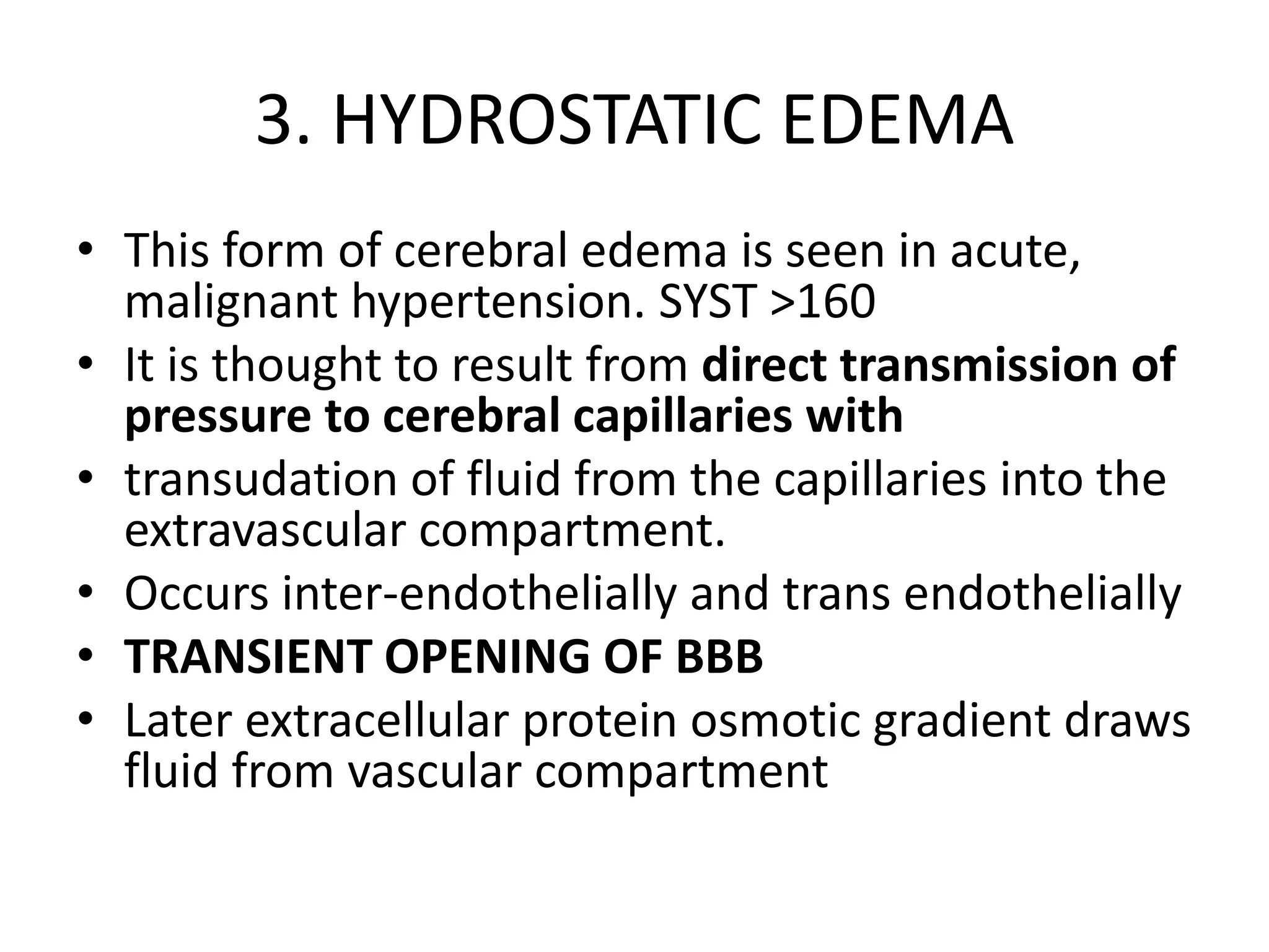
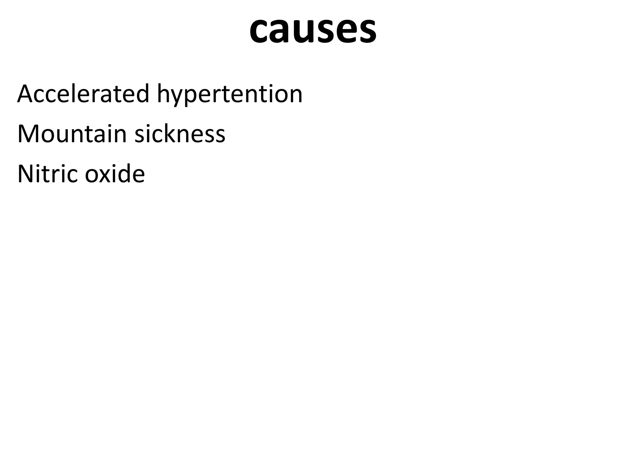
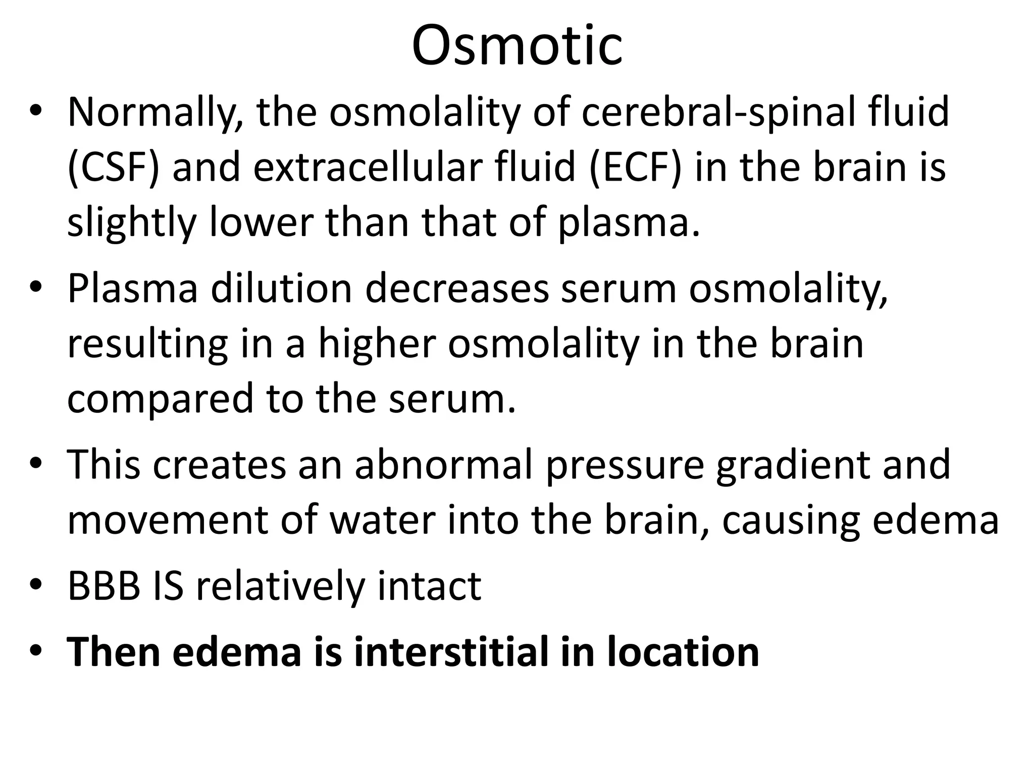
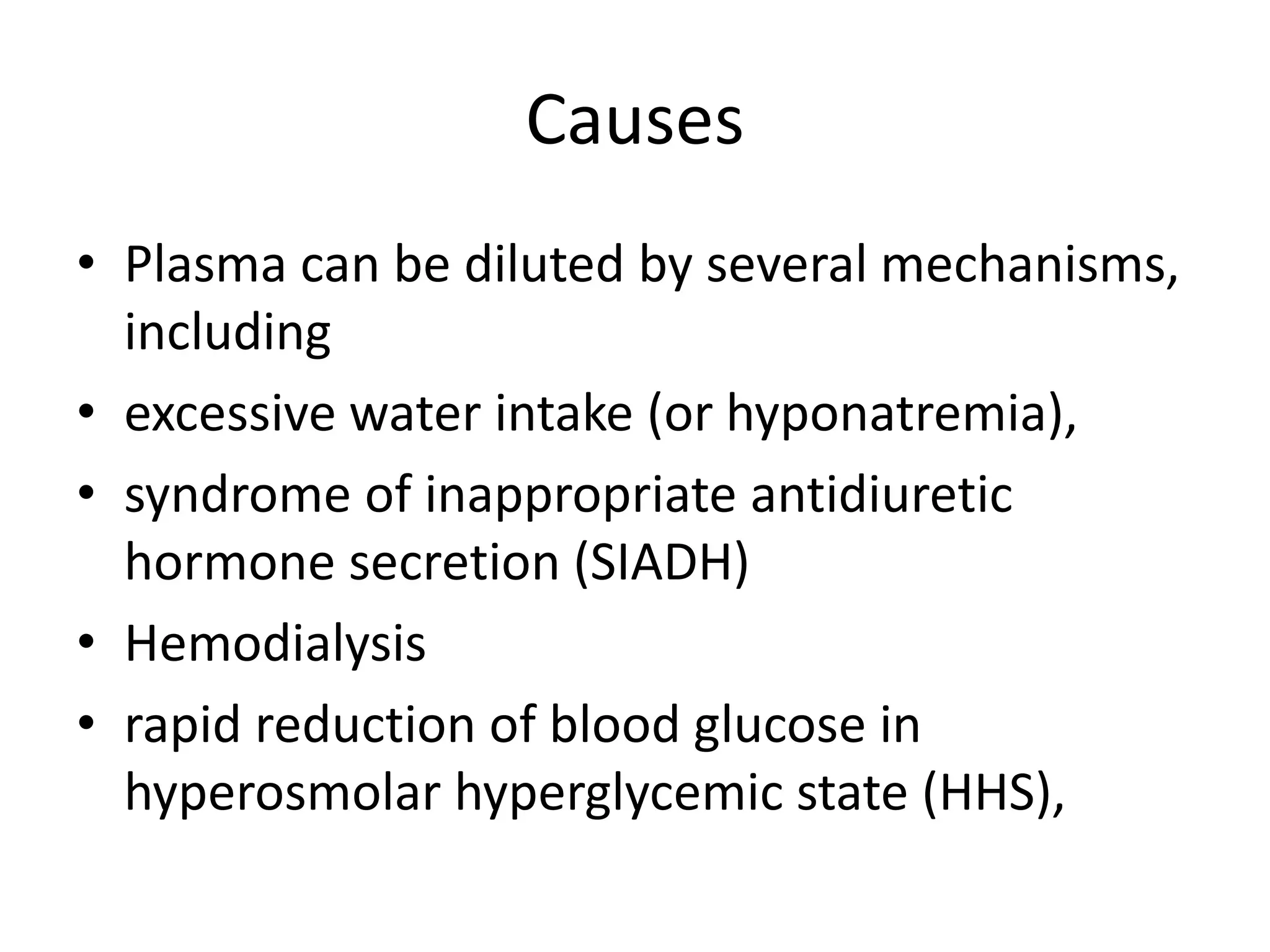
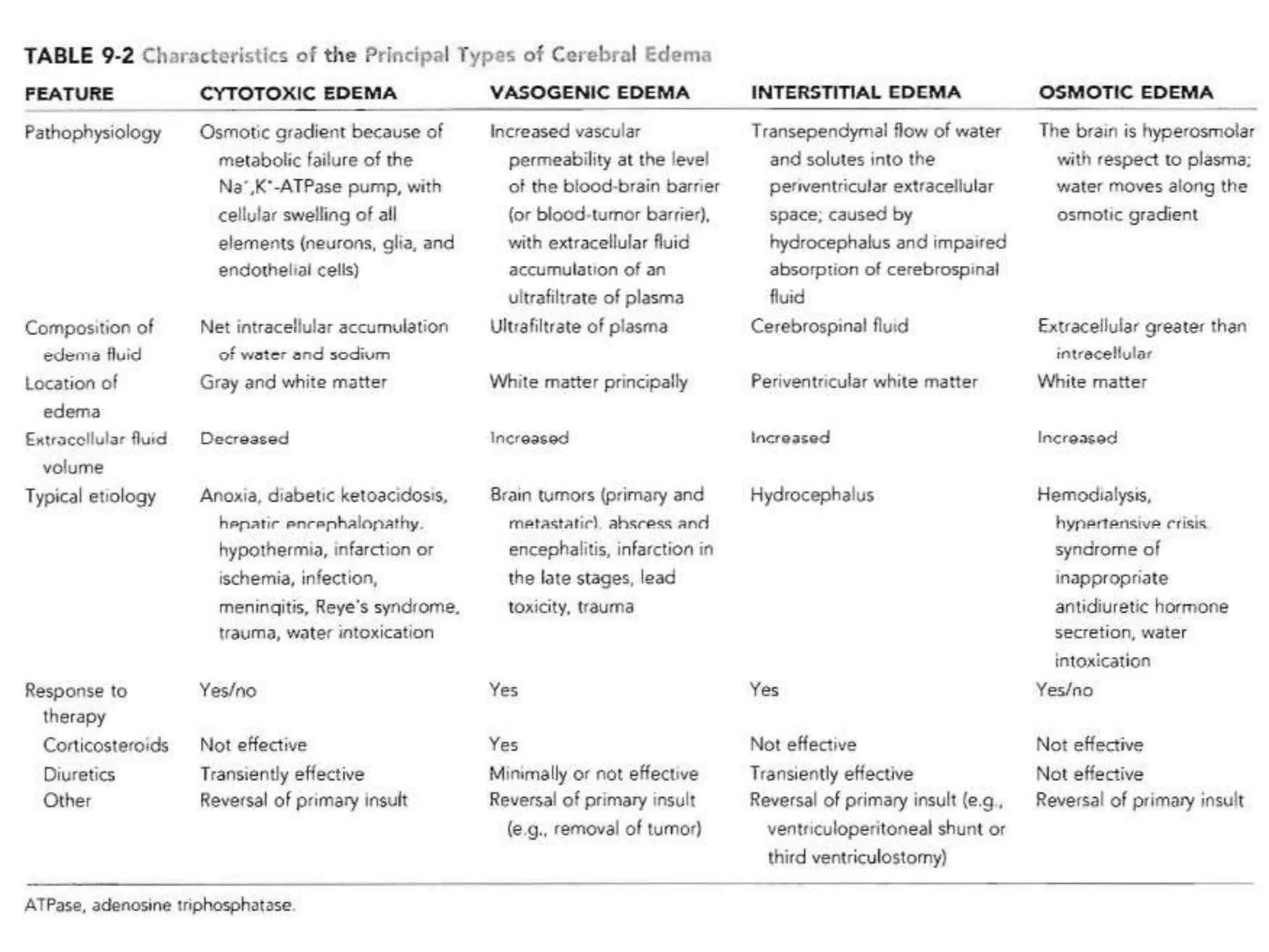
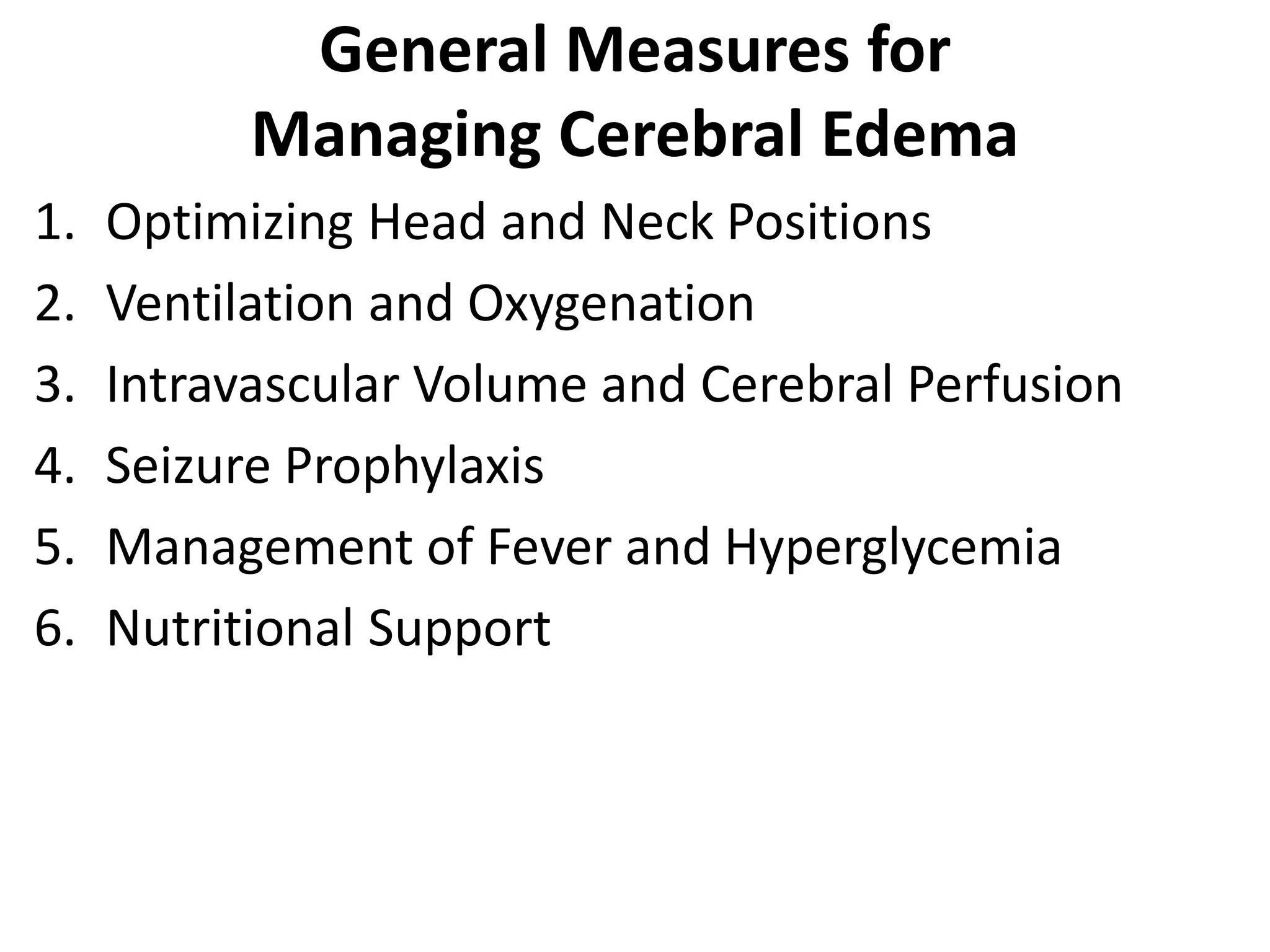
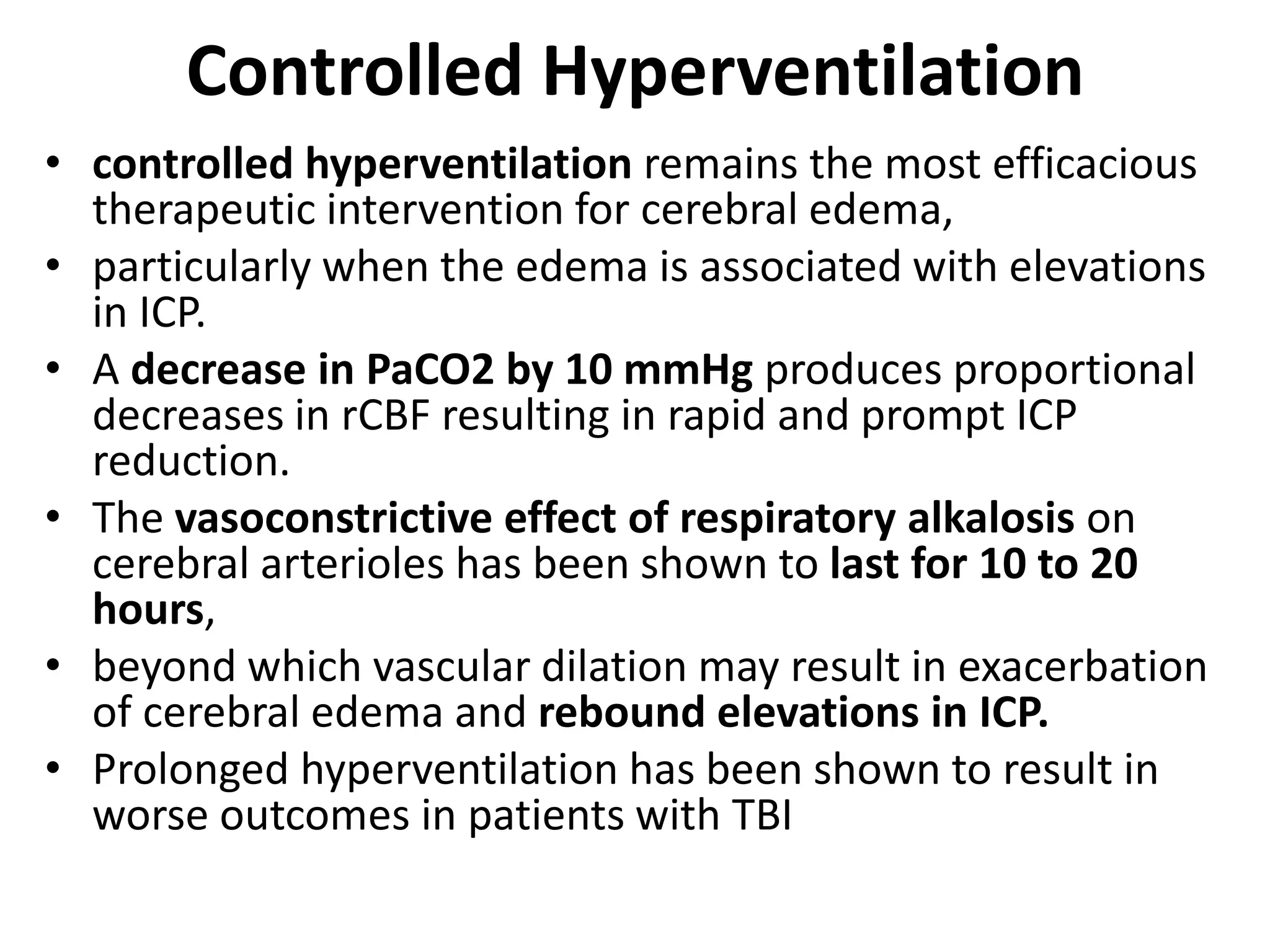
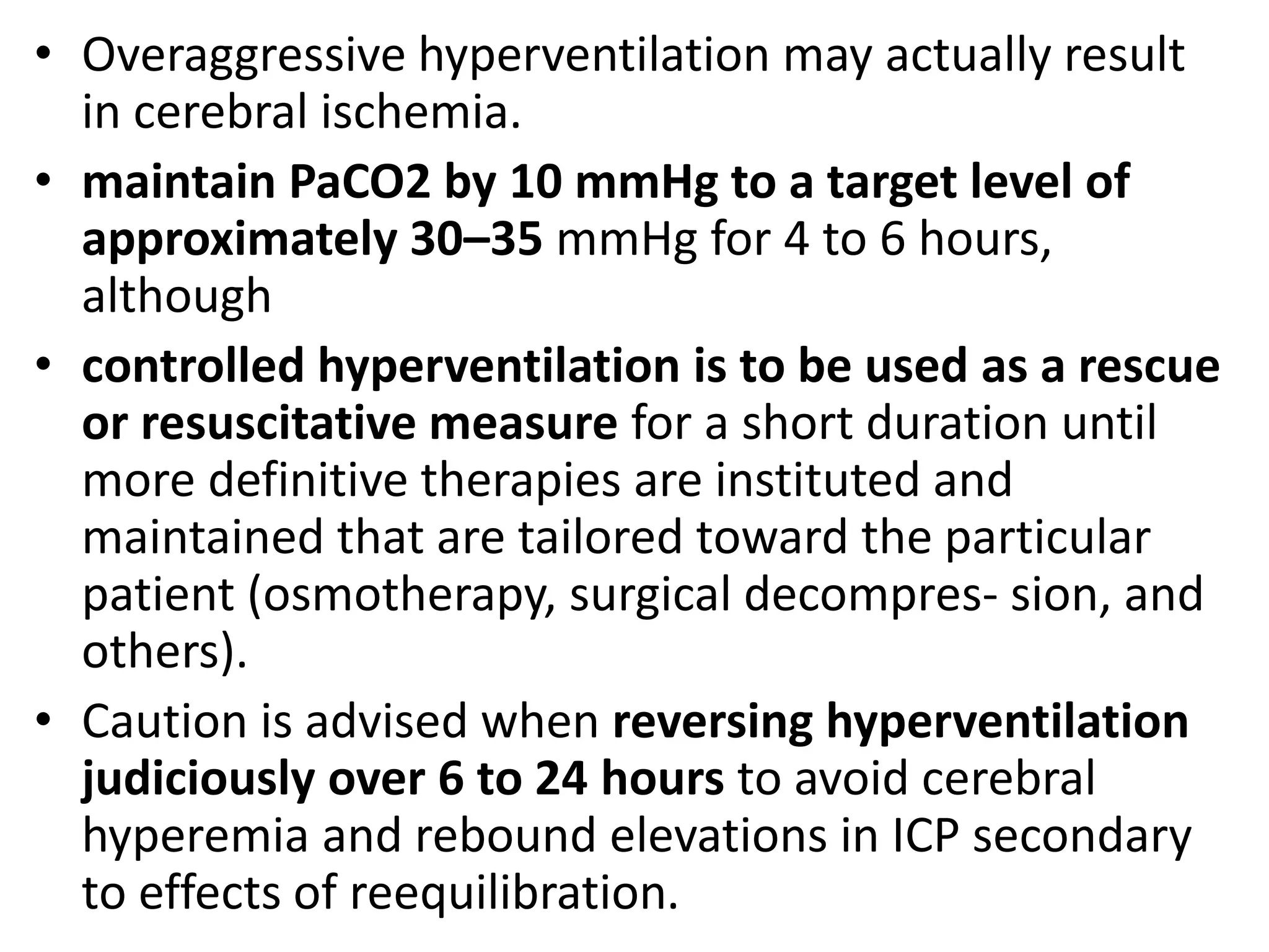
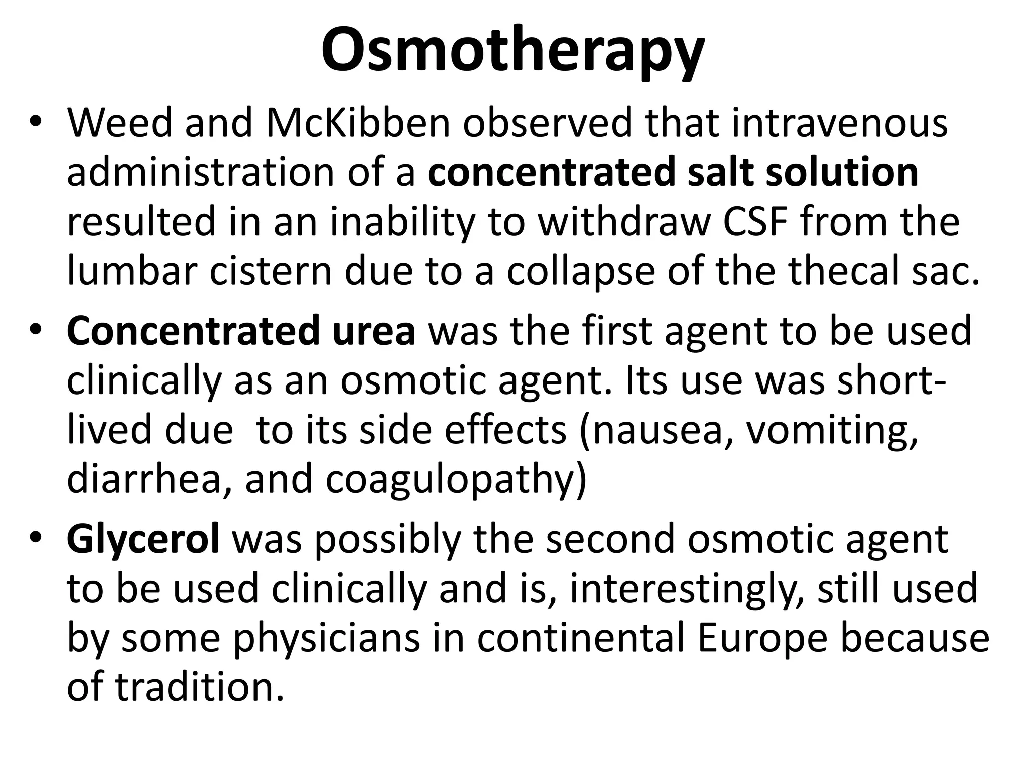
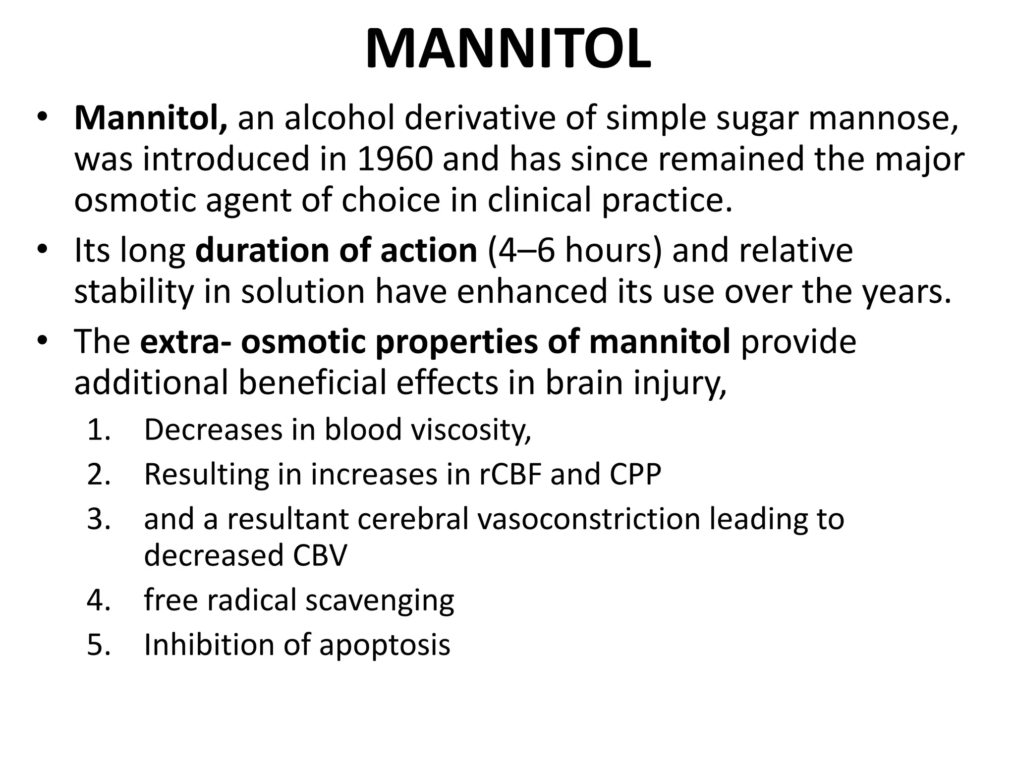
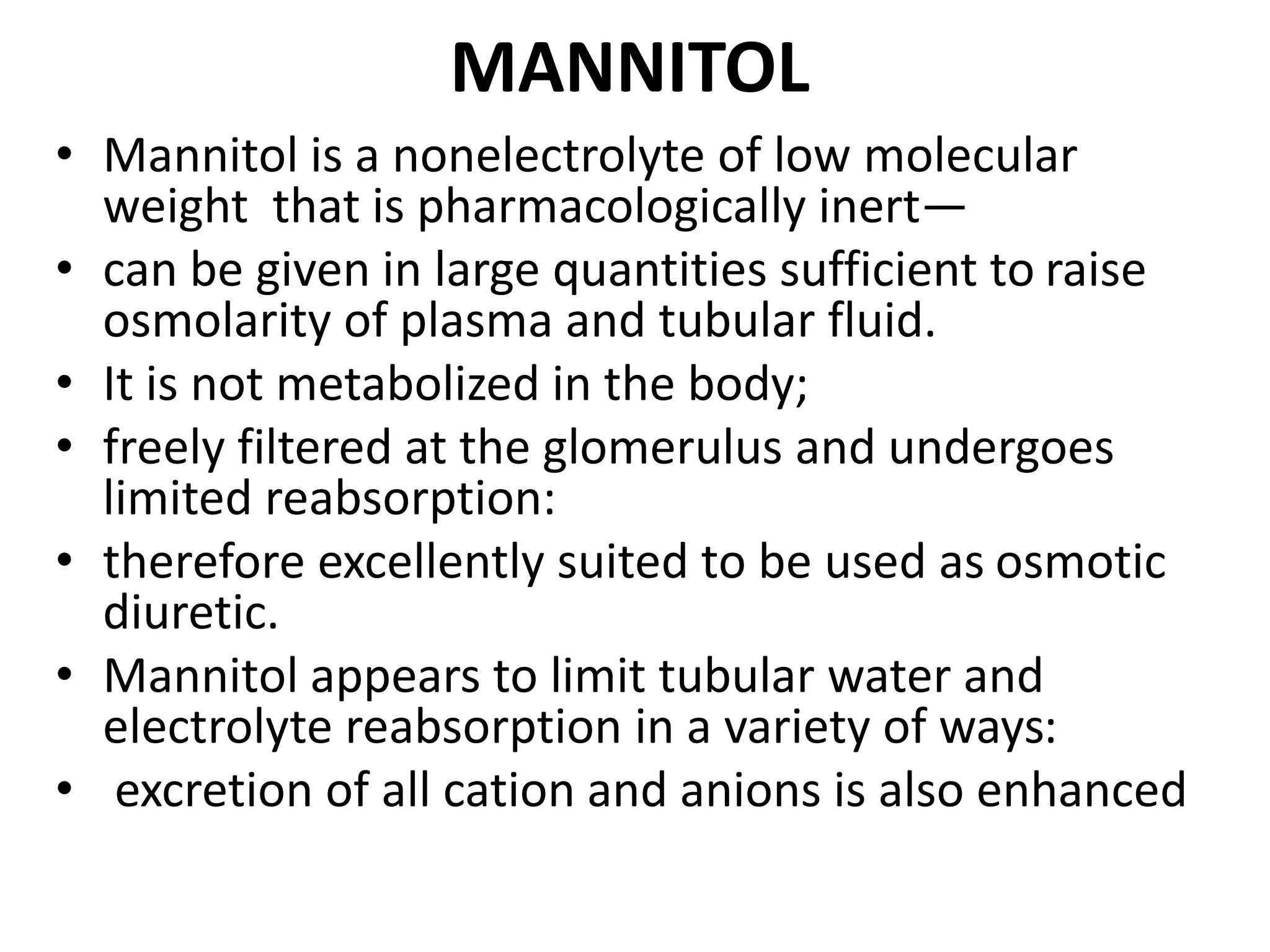
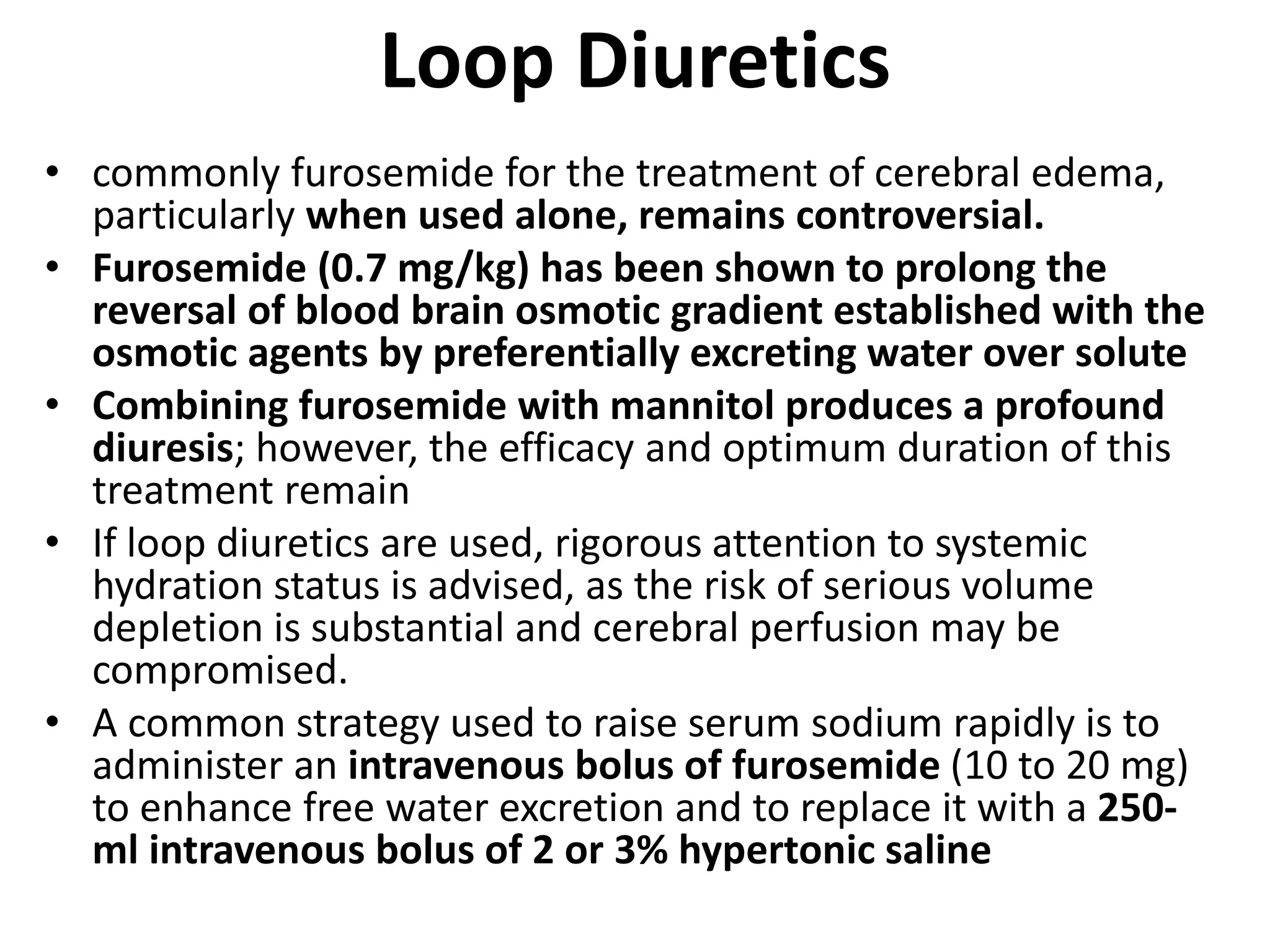
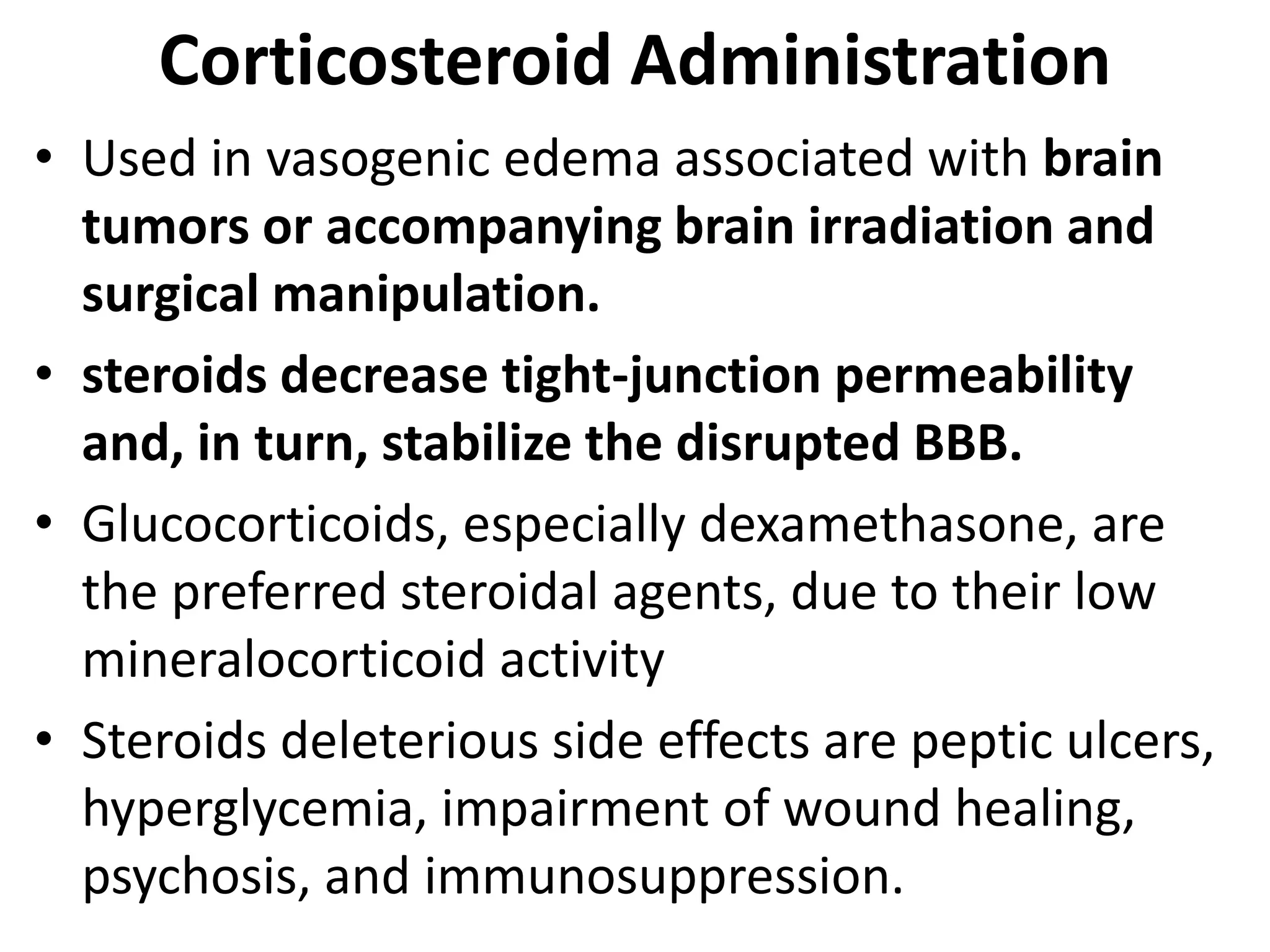
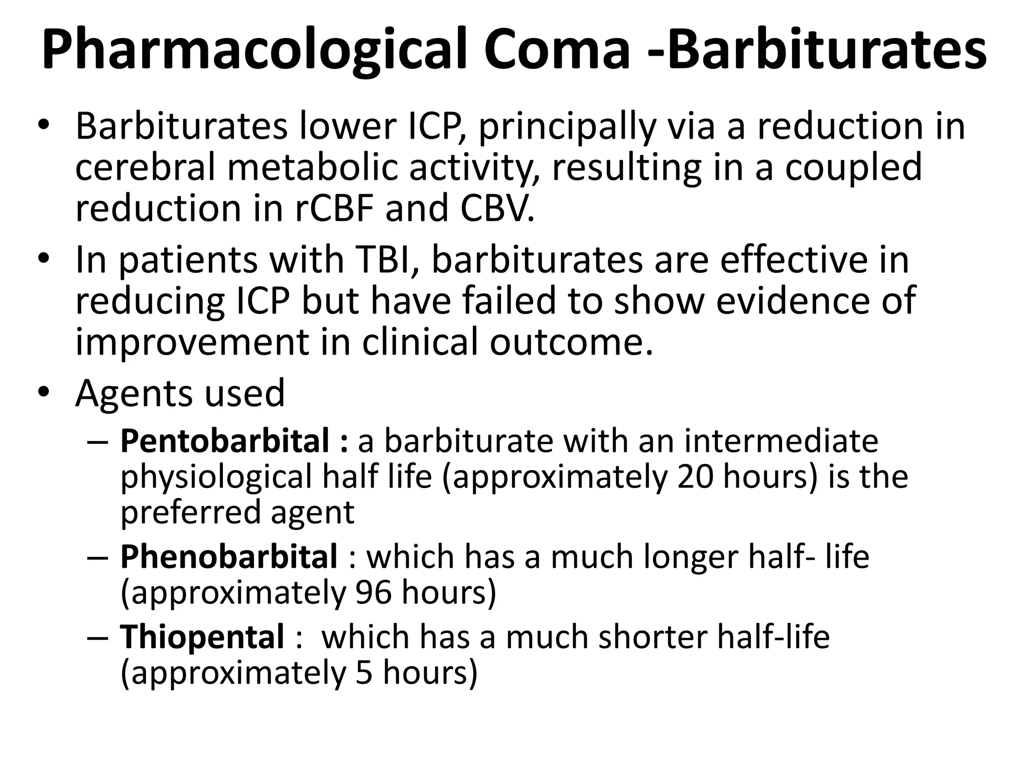
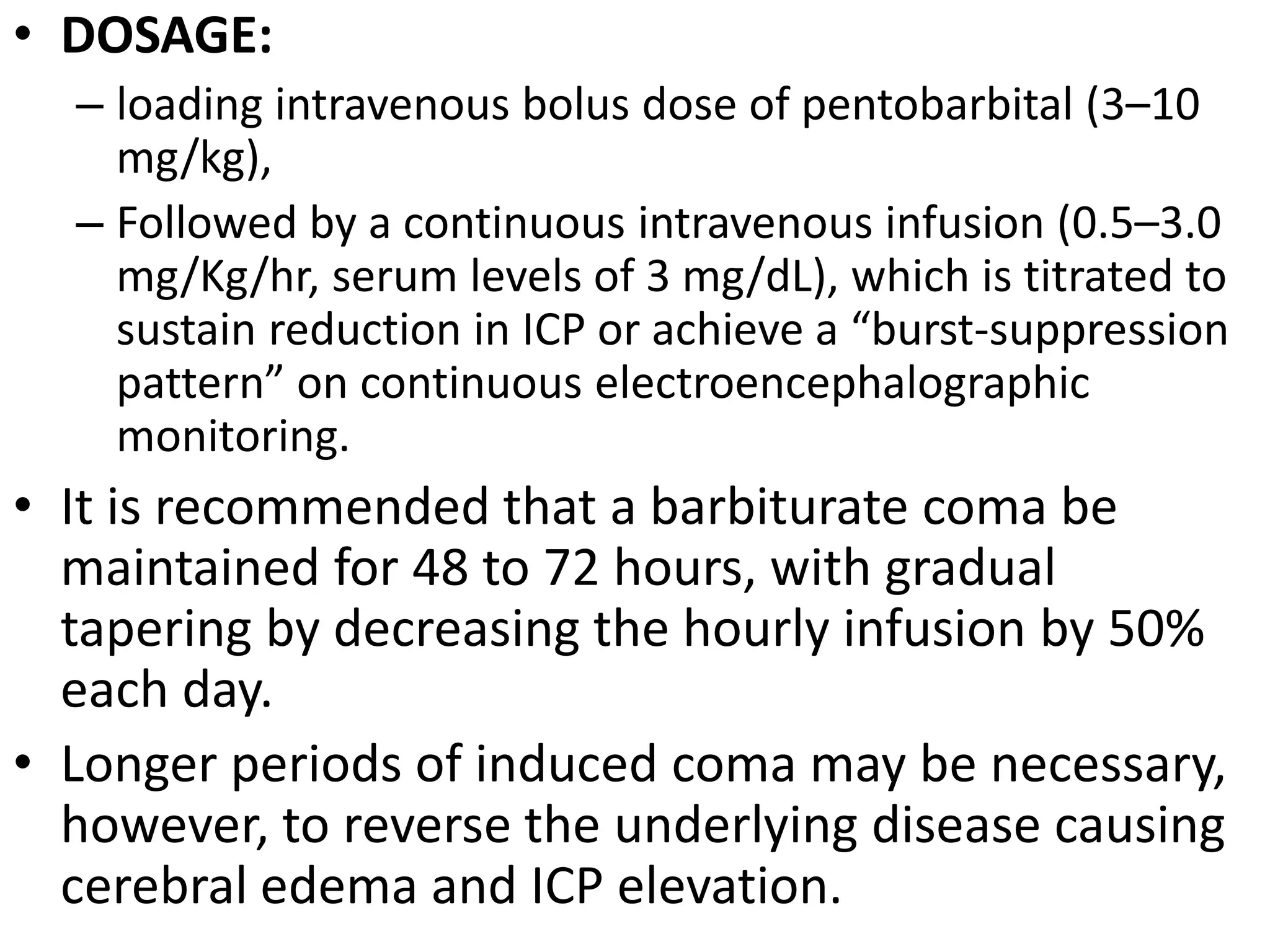
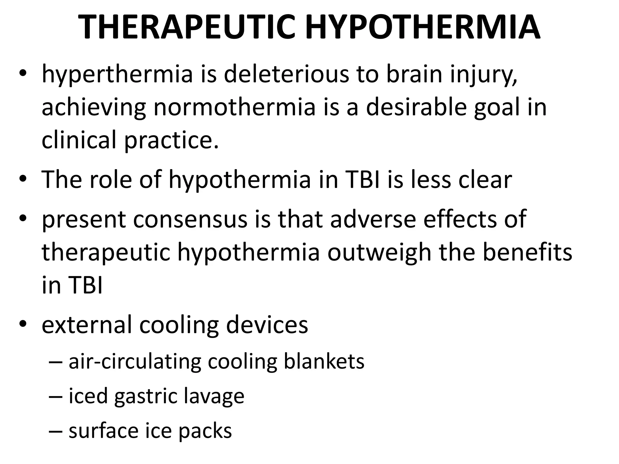
This document discusses different types of cerebral edema including cytotoxic, vasogenic, hydrostatic, osmotic, and hydrocephalic edema. It provides details on the causes, mechanisms, and management of each type. The key management strategies for cerebral edema discussed are head elevation, oxygenation, fluid management, seizure prophylaxis, fever control, nutrition, hyperventilation, osmotherapy using mannitol, and other adjunctive therapies.
Intracerebral hemorhage Diagnosis and managementRamesh Babu
About ICH - Diagnosis and management, Discussed the clinical presentation, evaluation, radiological features and management including recent guidelines
Intracerebral hemorhage Diagnosis and managementRamesh Babu
About ICH - Diagnosis and management, Discussed the clinical presentation, evaluation, radiological features and management including recent guidelines
Nursing management client with Increased intracranial pressure ( ICP)ANILKUMAR BR
The rigid cranial vault contains brain tissue (1,400 g), blood (75 ml), and CSF (75 ml)
The volume and pressure of these three components are usually in a state of equilibrium and produce the ICP.
ICP is usually measured in the lateral ventricles; normal ICP is 10 to 20 mm hg.
The Monro-kellie hypothesis states that because of the limited space for expansion within the skull, an increase in any one of the components causes a change in the volume of the others.
Increased ICP is a syndrome that affects many patients with acute neurologic conditions.
This is because pathologic conditions alter the relationship between intracranial volume and pressure.
Although an elevated ICP is most commonly associated with head injury, it also may be seen as a secondary effect in other conditions, such as brain tumors, subarachnoid hemorrhage, and toxic and viral encephalopathies.
Nursing management of the client with increased intracranial pressureANILKUMAR BR
The rigid cranial vault contains brain tissue (1,400 g), blood (75 mL), and CSF (75 mL)
The volume and pressure of these three components are usually in a state of equilibrium and produce the ICP.
ICP is usually measured in the lateral ventricles; normal ICP is 10 to 20 mm Hg. Increased ICP is a syndrome that affects many patients with acute neurologic conditions.
This is because pathologic conditions alter the relationship between intracranial volume and pressure. Although an elevated ICP is most commonly associated with head injury, it also may be seen as a secondary effect in other conditions, such as brain tumours, subarachnoid haemorrhage, and toxic and viral encephalopathies
Stroke or Cerebrovascular incident, is defined as an abrupt onset of a neurological deficit that is attributable to a focal vascular cause.
The clinical manifestations of stroke are highly variable because of the complex anatomy of the brain
Prix Galien International 2024 Forum ProgramLevi Shapiro
June 20, 2024, Prix Galien International and Jerusalem Ethics Forum in ROME. Detailed agenda including panels:
- ADVANCES IN CARDIOLOGY: A NEW PARADIGM IS COMING
- WOMEN’S HEALTH: FERTILITY PRESERVATION
- WHAT’S NEW IN THE TREATMENT OF INFECTIOUS,
ONCOLOGICAL AND INFLAMMATORY SKIN DISEASES?
- ARTIFICIAL INTELLIGENCE AND ETHICS
- GENE THERAPY
- BEYOND BORDERS: GLOBAL INITIATIVES FOR DEMOCRATIZING LIFE SCIENCE TECHNOLOGIES AND PROMOTING ACCESS TO HEALTHCARE
- ETHICAL CHALLENGES IN LIFE SCIENCES
- Prix Galien International Awards Ceremony
Flu Vaccine Alert in Bangalore Karnatakaaddon Scans
As flu season approaches, health officials in Bangalore, Karnataka, are urging residents to get their flu vaccinations. The seasonal flu, while common, can lead to severe health complications, particularly for vulnerable populations such as young children, the elderly, and those with underlying health conditions.
Dr. Vidisha Kumari, a leading epidemiologist in Bangalore, emphasizes the importance of getting vaccinated. "The flu vaccine is our best defense against the influenza virus. It not only protects individuals but also helps prevent the spread of the virus in our communities," he says.
This year, the flu season is expected to coincide with a potential increase in other respiratory illnesses. The Karnataka Health Department has launched an awareness campaign highlighting the significance of flu vaccinations. They have set up multiple vaccination centers across Bangalore, making it convenient for residents to receive their shots.
To encourage widespread vaccination, the government is also collaborating with local schools, workplaces, and community centers to facilitate vaccination drives. Special attention is being given to ensuring that the vaccine is accessible to all, including marginalized communities who may have limited access to healthcare.
Residents are reminded that the flu vaccine is safe and effective. Common side effects are mild and may include soreness at the injection site, mild fever, or muscle aches. These side effects are generally short-lived and far less severe than the flu itself.
Healthcare providers are also stressing the importance of continuing COVID-19 precautions. Wearing masks, practicing good hand hygiene, and maintaining social distancing are still crucial, especially in crowded places.
Protect yourself and your loved ones by getting vaccinated. Together, we can help keep Bangalore healthy and safe this flu season. For more information on vaccination centers and schedules, residents can visit the Karnataka Health Department’s official website or follow their social media pages.
Stay informed, stay safe, and get your flu shot today!
HOT NEW PRODUCT! BIG SALES FAST SHIPPING NOW FROM CHINA!! EU KU DB BK substit...GL Anaacs
Contact us if you are interested:
Email / Skype : kefaya1771@gmail.com
Threema: PXHY5PDH
New BATCH Ku !!! MUCH IN DEMAND FAST SALE EVERY BATCH HAPPY GOOD EFFECT BIG BATCH !
Contact me on Threema or skype to start big business!!
Hot-sale products:
NEW HOT EUTYLONE WHITE CRYSTAL!!
5cl-adba precursor (semi finished )
5cl-adba raw materials
ADBB precursor (semi finished )
ADBB raw materials
APVP powder
5fadb/4f-adb
Jwh018 / Jwh210
Eutylone crystal
Protonitazene (hydrochloride) CAS: 119276-01-6
Flubrotizolam CAS: 57801-95-3
Metonitazene CAS: 14680-51-4
Payment terms: Western Union,MoneyGram,Bitcoin or USDT.
Deliver Time: Usually 7-15days
Shipping method: FedEx, TNT, DHL,UPS etc.Our deliveries are 100% safe, fast, reliable and discreet.
Samples will be sent for your evaluation!If you are interested in, please contact me, let's talk details.
We specializes in exporting high quality Research chemical, medical intermediate, Pharmaceutical chemicals and so on. Products are exported to USA, Canada, France, Korea, Japan,Russia, Southeast Asia and other countries.
Couples presenting to the infertility clinic- Do they really have infertility...Sujoy Dasgupta
Dr Sujoy Dasgupta presented the study on "Couples presenting to the infertility clinic- Do they really have infertility? – The unexplored stories of non-consummation" in the 13th Congress of the Asia Pacific Initiative on Reproduction (ASPIRE 2024) at Manila on 24 May, 2024.
- Video recording of this lecture in English language: https://youtu.be/lK81BzxMqdo
- Video recording of this lecture in Arabic language: https://youtu.be/Ve4P0COk9OI
- Link to download the book free: https://nephrotube.blogspot.com/p/nephrotube-nephrology-books.html
- Link to NephroTube website: www.NephroTube.com
- Link to NephroTube social media accounts: https://nephrotube.blogspot.com/p/join-nephrotube-on-social-media.html
Ozempic: Preoperative Management of Patients on GLP-1 Receptor Agonists Saeid Safari
Preoperative Management of Patients on GLP-1 Receptor Agonists like Ozempic and Semiglutide
ASA GUIDELINE
NYSORA Guideline
2 Case Reports of Gastric Ultrasound
MANAGEMENT OF ATRIOVENTRICULAR CONDUCTION BLOCK.pdfJim Jacob Roy
Cardiac conduction defects can occur due to various causes.
Atrioventricular conduction blocks ( AV blocks ) are classified into 3 types.
This document describes the acute management of AV block.
ARTIFICIAL INTELLIGENCE IN HEALTHCARE.pdfAnujkumaranit
Artificial intelligence (AI) refers to the simulation of human intelligence processes by machines, especially computer systems. It encompasses tasks such as learning, reasoning, problem-solving, perception, and language understanding. AI technologies are revolutionizing various fields, from healthcare to finance, by enabling machines to perform tasks that typically require human intelligence.
Pulmonary Thromboembolism - etilogy, types, medical- Surgical and nursing man...VarunMahajani
Disruption of blood supply to lung alveoli due to blockage of one or more pulmonary blood vessels is called as Pulmonary thromboembolism. In this presentation we will discuss its causes, types and its management in depth.
Tom Selleck Health: A Comprehensive Look at the Iconic Actor’s Wellness Journeygreendigital
Tom Selleck, an enduring figure in Hollywood. has captivated audiences for decades with his rugged charm, iconic moustache. and memorable roles in television and film. From his breakout role as Thomas Magnum in Magnum P.I. to his current portrayal of Frank Reagan in Blue Bloods. Selleck's career has spanned over 50 years. But beyond his professional achievements. fans have often been curious about Tom Selleck Health. especially as he has aged in the public eye.
Follow us on: Pinterest
Introduction
Many have been interested in Tom Selleck health. not only because of his enduring presence on screen but also because of the challenges. and lifestyle choices he has faced and made over the years. This article delves into the various aspects of Tom Selleck health. exploring his fitness regimen, diet, mental health. and the challenges he has encountered as he ages. We'll look at how he maintains his well-being. the health issues he has faced, and his approach to ageing .
Early Life and Career
Childhood and Athletic Beginnings
Tom Selleck was born on January 29, 1945, in Detroit, Michigan, and grew up in Sherman Oaks, California. From an early age, he was involved in sports, particularly basketball. which played a significant role in his physical development. His athletic pursuits continued into college. where he attended the University of Southern California (USC) on a basketball scholarship. This early involvement in sports laid a strong foundation for his physical health and disciplined lifestyle.
Transition to Acting
Selleck's transition from an athlete to an actor came with its physical demands. His first significant role in "Magnum P.I." required him to perform various stunts and maintain a fit appearance. This role, which he played from 1980 to 1988. necessitated a rigorous fitness routine to meet the show's demands. setting the stage for his long-term commitment to health and wellness.
Fitness Regimen
Workout Routine
Tom Selleck health and fitness regimen has evolved. adapting to his changing roles and age. During his "Magnum, P.I." days. Selleck's workouts were intense and focused on building and maintaining muscle mass. His routine included weightlifting, cardiovascular exercises. and specific training for the stunts he performed on the show.
Selleck adjusted his fitness routine as he aged to suit his body's needs. Today, his workouts focus on maintaining flexibility, strength, and cardiovascular health. He incorporates low-impact exercises such as swimming, walking, and light weightlifting. This balanced approach helps him stay fit without putting undue strain on his joints and muscles.
Importance of Flexibility and Mobility
In recent years, Selleck has emphasized the importance of flexibility and mobility in his fitness regimen. Understanding the natural decline in muscle mass and joint flexibility with age. he includes stretching and yoga in his routine. These practices help prevent injuries, improve posture, and maintain mobilit
2. TYPES
1. CYTOTOXIC
2. VASOGENIC
3. HYDROSTATIC
4. OSMOTIC
5. HYDROCEPHALIC
• EDEMA IN TRAUMA
• EDEMA IN TUMOUR
3. 1. Cytotoxic
• In cytotoxic edema, the BBB remains intact.
• It occurs due to a disruption in cellular metabolism
that impairs functioning of the sodium and
potassium pump in the glial cell membrane, leading
to cellular retention of sodium and water.
• Swollen astrocytes occur in gray and white matter
• This interfere with neuronal and oligoendrocyte
function
• This edema occurs through intra cellular
hyperosmolarity and extra cellular hypotonicity with
deranged ATP dependant Na K pump
4.
5. Causes
• various toxins, including
– dinitrophenol,
– triethyltin,
– hexachlorophene, and
– isoniazid. It can occur in
• Reye's syndrome,
• severe hypothermia
• early ischemia,
• encephalopathy,
• early stroke or hypoxia,
• cardiac arrest, and pseudotumor cerebri.
6. • During an ischemic stroke, a lack of oxygen and glucose
leads to a breakdown of the sodium-calcium pumps
on brain cell membranes,
• which in turn results in a massive build up of sodium
and calcium intracellularly.
• This causes a rapid uptake of water and subsequent
swelling of the cells.
• It is this swelling of the individual cells of the brain
that is seen as the main distinguishing characteristic of
cytotoxic edema, as opposed to vasogenic wherein the
influx of fluid is typically seen in the interstitial space
rather than within the cells themselves
7. Vasogenic edema
• Vasogenic edema occurs due to a breakdown of the
tight endothelial junctions which make up the
blood–brain barrier (BBB).
• Swollen astrocyte foot processes reflecting initial
glial cytotoxicity( clossed barrier to open barrier
edema)
• Interendothelial fenestration widened
• Trans endothelial trafficking increased
• This allows intravascular proteins and fluid to
penetrate into the parenchymal extracellular space.
• Once plasma constituents cross the BBB, the edema
spreads; this may be quite rapid and extensive
• As water enters white matter it moves extracellularly
along fiber tracts and can also affect the gray matter.
8.
9. Causes
• trauma,
• tumors,
• focal inflammation,
• late stages of cerebral ischemia and
• hypertensive encephalopathy.
10. 3. HYDROSTATIC EDEMA
• This form of cerebral edema is seen in acute,
malignant hypertension. SYST >160
• It is thought to result from direct transmission of
pressure to cerebral capillaries with
• transudation of fluid from the capillaries into the
extravascular compartment.
• Occurs inter-endothelially and trans endothelially
• TRANSIENT OPENING OF BBB
• Later extracellular protein osmotic gradient draws
fluid from vascular compartment
13. Osmotic
• Normally, the osmolality of cerebral-spinal fluid
(CSF) and extracellular fluid (ECF) in the brain is
slightly lower than that of plasma.
• Plasma dilution decreases serum osmolality,
resulting in a higher osmolality in the brain
compared to the serum.
• This creates an abnormal pressure gradient and
movement of water into the brain, causing edema
• BBB IS relatively intact
• Then edema is interstitial in location
14. Causes
• Plasma can be diluted by several mechanisms,
including
• excessive water intake (or hyponatremia),
• syndrome of inappropriate antidiuretic
hormone secretion (SIADH)
• Hemodialysis
• rapid reduction of blood glucose in
hyperosmolar hyperglycemic state (HHS),
15.
16. Hydrocephalic edema
• Interstitial edema occurs in obstructive
hydrocephalus due to a rupture of the CSF-brain
barrier.
• This results in trans-ependymal flow of CSF,
causing CSF to penetrate the brain and spread to
the extracellular spaces and the white matter.
• Interstitial cerebral edema differs from
vasogenic edema as CSF contains almost no
protein.
17. Cerebral edema from brain tumour
• Cancerous glial cells (glioma) of the brain can
increase secretion of vascular endothelial
growth factor (VEGF),
• New and weak vessel formation
• which weakens the junctions of the blood–
brain barrier
18. Edema in trauma
• Vasogenic d/t breach in BBB
• Cytotoxic d/t inflammatory mediators
• Osmotic brain edema d/t extra vasation of
plasma products
• Hydrocephalic d/t obstruction of csf flow
19. • CEREBRAL edema, simply defined as an
increase in brain water content (above the
normal brain water content of approximately
80%).
20.
21. MONITORING ICP
• The Brain Trauma Foundation guidelines
recommend ICP monitoring in patients with TBI,
– a GCS score of less than 9, and abnormal CT scans,
– in patients with a GCS score less than 9 and normal CT
scans in the presence of two or more of the following:
1. age greater than 40 years
2. unilateral or bilateral motor posturing
3. systolic blood pressure greater than 90 mmHg.
• No such guidelines exist for ICP monitoring in other
brain injury paradigms (ischemic stroke, ICH,
cerebral neoplasm
22. General Measures for
Managing Cerebral Edema
1. Optimizing Head and Neck Positions
2. Ventilation and Oxygenation
3. Intravascular Volume and Cerebral Perfusion
4. Seizure Prophylaxis
5. Management of Fever and Hyperglycemia
6. Nutritional Support
23. Specific Measures for
Managing Cerebral Edema
1. Controlled Hyperventilation
2. Osmotherapy
3. Corticosteroid Administration
4. Pharmacological Coma
5. Therapeutic Hypothermia
6. Other Adjunct Therapies
24. Optimizing Head and Neck Positions
• 30̊elevation of the head in patients is essential for
1. avoiding jugular compression and impedance of venous
outflow from the cranium
2. for decreasing CSF hydrostatic pressure.
• to avoid the use of restricting devices and garments
around the neck (such as devices used to secure
endotracheal tubes), which may impair cerebral
venous outflow via compression of the internal
jugular veins.
• Head position elevation may be detrimental in
ischemic stroke, because it may compromise
perfusion to ischemic tissue at risk.
25. Ventilation and Oxygenation
• Hypoxia and hypercapnia are potent cerebral
vasodilator
• Pt should be intubated in:
1. GCS scores less than or equal to 8
2. Patients with poor upper airway reflexes be
intubated preemptively for airway protection.
3. aspiration pneumonitis
4. pulmonary contusion
5. acute respiratory distress syndrome.
26. • Levels of PaCO2should be maintained that support
adequate rCBF or CPP to the injured brain, and a
value of approximately 35 mmHg is a generally
accepted target in the absence of ICP elevations or
clinical herniation syndromes.
• Avoidance of hypoxemia and maintenance of PaO2
at approximately 100 mmHg are recommended.
• Delivery of PEEP greater than 10 cm H2O in
patients with severe TBI has resulted in elevated
ICP which resulted from elevations in central
venous pressures and impedance of cerebral
venous drainage.
27. • Therefore, careful monitoring of clinical
neurological status, ICP, and CPP (mean arterial
pressure – ICP) is recommended in mechanically
ventilated patients with cerebral edema with or
without elevations in ICP.
• Blunting of upper airway reflexes (coughing) with
endobronchial lidocaine before suctioning,
sedation, or, rarely, pharmacological paralysis may
be necessary for avoiding increases in ICP.
28. Intravascular Volume and Cerebral Perfusion
• Maintenance of CPP using adequate fluid
management in combination with vasopressors is
vital in patients with brain injury
• Hypotonic fluids should be avoided at all cost)
• Euvolemia or mild hypervolemia with the use of
isotonic fluids (0.9% saline) should always be
maintained through rigorous attention to daily
fluid balance, body weight, and serum electrolyte
monitoring.
29. • The recommended goal of a CPP level greater than
60 mmHg should be adhered to in patients with TBI,
and simultaneously, sharp rises in systemic blood
pressure should be avoided.
• CBF=CPP/CVR
( CBF-Cerebral blood flow, CPP-Cerebral perfusion pressure,
CVR-Cerebrovascular resistance )
• NORMAL CPP= 70 TO 90 mm of Hg
• CPP=MAP-ICP (IF ICP>JVP) (MAP-mean arterial pressure )
• CPP=MAP-JVP (IF JVP>ICP)
• MAP=DP-1/3PP (PP-Pulse pressure ,where PP= SBP-DBP)
• Normal MAP is between 70 to 110 mmHg
30. TREATING HYPERTENSION
• Judicious use of antihypertensives
1. Labetalol
2. Enalaprilate
3. Nicardipine
– is recommended for treating systemic hypertension.
• Potent vasodilators are to be avoided
– Nitroglycerine
– Nitroprusside
– as they may exacerbate cerebral edema via
accentuated cerebral hyperemia and CBV due to their
direct vasodilating effects on cerebral vasculature.
31. Seizure Prophylaxis
• Phenytoin are widely used empirically in clinical
practice in patients with acute brain injury
• Early seizures in TBI can be effectively reduced
by prophylactic administration of phenytoin for 1
or 2 weeks.
32. Management of Fever
• fever has deleterious effects of on outcome
following brain injury,
• increases in oxygen demand,
• normothermia is strongly recommended in
patients with cerebral edema, irrespective of
underlying origin.
• Acetaminophen (325–650 mg orally, or rectally
every 4–6 hours) is the most common agent
used,
• Other surface cooling devices have demonstrated
some efficacy
33. Hyperglycemia
• Hyperglycemia can also exacerbate brain injury
and cerebral edema
• current evidence suggests that rigorous glycemic
control may be beneficial in all patients with
brain injury.
34. Nutritional Support
• Unless contraindicated, the enteral route of
nutrition is preferred.
• Special attention should be given to the osmotic
content of formulations, to avoid free water
intake that may result in a hypoosmolar state and
worsen cerebral edema.
36. Controlled Hyperventilation
• controlled hyperventilation remains the most efficacious
therapeutic intervention for cerebral edema,
• particularly when the edema is associated with elevations
in ICP.
• A decrease in PaCO2 by 10 mmHg produces proportional
decreases in rCBF resulting in rapid and prompt ICP
reduction.
• The vasoconstrictive effect of respiratory alkalosis on
cerebral arterioles has been shown to last for 10 to 20
hours,
• beyond which vascular dilation may result in exacerbation
of cerebral edema and rebound elevations in ICP.
• Prolonged hyperventilation has been shown to result in
worse outcomes in patients with TBI
37. • Overaggressive hyperventilation may actually result
in cerebral ischemia.
• maintain PaCO2 by 10 mmHg to a target level of
approximately 30–35 mmHg for 4 to 6 hours,
although
• controlled hyperventilation is to be used as a rescue
or resuscitative measure for a short duration until
more definitive therapies are instituted and
maintained that are tailored toward the particular
patient (osmotherapy, surgical decompres- sion, and
others).
• Caution is advised when reversing hyperventilation
judiciously over 6 to 24 hours to avoid cerebral
hyperemia and rebound elevations in ICP secondary
to effects of reequilibration.
38. Osmotherapy
• Weed and McKibben observed that intravenous
administration of a concentrated salt solution
resulted in an inability to withdraw CSF from the
lumbar cistern due to a collapse of the thecal sac.
• Concentrated urea was the first agent to be used
clinically as an osmotic agent. Its use was short-lived
due to its side effects (nausea, vomiting,
diarrhea, and coagulopathy)
• Glycerol was possibly the second osmotic agent
to be used clinically and is, interestingly, still used
by some physicians in continental Europe because
of tradition.
39. MANNITOL
• Mannitol, an alcohol derivative of simple sugar mannose,
was introduced in 1960 and has since remained the major
osmotic agent of choice in clinical practice.
• Its long duration of action (4–6 hours) and relative
stability in solution have enhanced its use over the years.
• The extra- osmotic properties of mannitol provide
additional beneficial effects in brain injury,
1. Decreases in blood viscosity,
2. Resulting in increases in rCBF and CPP
3. and a resultant cerebral vasoconstriction leading to
decreased CBV
4. free radical scavenging
5. Inhibition of apoptosis
40. MANNITOL
• Mannitol is a nonelectrolyte of low molecular
weight that is pharmacologically inert—
• can be given in large quantities sufficient to raise
osmolarity of plasma and tubular fluid.
• It is not metabolized in the body;
• freely filtered at the glomerulus and undergoes
limited reabsorption:
• therefore excellently suited to be used as osmotic
diuretic.
• Mannitol appears to limit tubular water and
electrolyte reabsorption in a variety of ways:
• excretion of all cation and anions is also enhanced
41. • IN PROXIMAL TUBULE:
– Retains water isoosmotically in PT dilutes luminal fluid
which opposes NaCl reabsorption.
• IN LOOP OF HENLE:
– Inhibits transport processes in the thick AscLH by an
unknown mechanism.
– Major site of action is LOOP OF HENLE
• MEDULLARY OSMOTIC GRADIENT & RENAL BLOOD
FLOW:
– By extracting water from intracellular compartments,
osmotic diuretics expand the extracellular fluid volume,
decrease blood viscosity, and inhibit renin release.
– These effects increase RBF.
– and the increase in renal medullary blood flow removes
NaCl and urea from the renal medulla, thus reducing
medullary tonicity.
42. • some circumstances, prostaglandins may contribute
to the renal vasodilation and medullary washout
induced by osmotic diuretics.
• A reduction in medullary tonicity causes a decrease
in the extraction of water from the DTL, which, in
turn, limits the concentration of NaCl in the tubular
fluid entering the ATL.
• This latter effect diminishes the passive
reabsorption of NaCl in the ATL.
43. • Effects on Urinary Excretion
– Osmotic diuretics increase the urinary excretion of
nearly all electrolytes, including Na+, K+, Ca2+, Mg2+, Cl-,
HCO3 and phosphate.
• Effects on Renal Hemodynamics
– Osmotic diuretics increase RBF by a variety of
mechanisms.
– Osmotic diuretics dilate the afferent arteriole, which
increases PGC and dilute the plasma, which decreases PGC
– These effects would increase GFR were it not for the fact
that osmotic diuretics also increase PT
– In general, superficial SNGFR is increased, but total GFR
is little changed.
44.
45. PHARMACOKINETICS
• Administration Mannitol is not absorbed orally.
• has to be given i.v. as 10–20% solution.
• It is excreted with a t½ of 0.5–1.5 hour.
46. USES
• Decreases intracranial or intraocular tension
– (acute congestive glaucoma, head injury, stroke, etc.):
– by osmotic action it encourages movement of water
from brain parenchyma, CSF and aqueous humour;
– 1–1.5 g/kg is infused over 1 hour as 20% solution to
transiently raise plasma osmolarity.
– It is also used before and after ocular/brain surgery to
prevent acute rise in intraocular/intracranial pressure
47. • To maintain g.f.r. and urine flow in impending
acute renal failure,
– e.g. in shock, severe trauma, cardiac surgery, haemolytic
reactions: 500–1000 ml of the solution may be infused
over 24 hours.
– If acute renal failure has already set in, kidney is
incapable of forming urine even after an osmotic load;
mannitol is contraindicated: it will then expand plasma
volume → pulmonary edema and heart failure may
develop.
• To counteract low osmolality of plasma/e.c.f.
– due to rapid haemodialysis or peritoneal dialysis (dialysis
disequilibrium).
48. • Forced diuresis:
– Mannitol along with large volumes of saline was infused
i.v. to produce ‘forced diuresis’ in acute poisonings in the
hope of enhancing excretion of the poison.
– However, this has been found to be ineffective and to
produce electrolyte imbalances. Not recommended
now.
50. SIDE EFFECTS
• When given orally causes osmotic diarrhoea
• DEHYDRATION, HYPERKALEMIA, AND
HYPERNATREMIA
– Excessive use of mannitol without adequate water
replacement can ultimately lead to severe dehydration,
free water losses, and hypernatremia.
– As water is extracted from cells, intracellular K+
concentration rises, leading to cellular losses and
hyperkalemia.
– These complications can be avoided by careful attention
to serum ion composition and fluid balance.
51. • EXTRACELLULAR VOLUME EXPANSION
– Mannitol is rapidly distributed in the extracellular
compartment and extracts water from cells.
– Prior to the diuresis, this leads to expansion of the
extracellular volume and hyponatremia.
– This effect can complicate heart failure and may produce
florid pulmonary edema.
• Headache, nausea, and vomiting are commonly
observed in patients treated with osmotic diuretics.
52. OTHER OSMOTIC DIURETICS
• Isosorbide and glycerol
– These are orally active osmotic diuretics which
may be used to reduce intraocular or intracranial
tension.
– Intravenous glycerol can cause haemolysis.
• Dose: 0.5–1.5 g/kg as oral solution.
53. • In a prospective series of patients with elevated ICP
and diverse intracranial diseases, bolus mannitol
decreased ICP, with a mean reduction of 52% of
pretreatment values.
• in an uncontrolled series of patients with TBI,
0.25g/kg of an intra venous bolus of mannitol was
sufficient to attenuate elevated ICP.
• high-dose mannitol treatment may be preferable to
conventional doses for acute TBI.
54. • DOSE OF MANNITOL:
– The conventional osmotic agent mannitol, when
administered at a dose of 0.25 to 1.5 g/kg by
intravenous bolus injection, usually lowers ICP, with
maximal effects observed 20 to 40 minutes following its
administration.
– Repeated dosing of Mannitol may be instituted every 6
hours and should be guided by serum osmolality to a
recommended target value of approximately 320
mOsm/L; higher values result in renal tubular damage
55. HYPERTONIC SALINE
• Hypertonic saline solutions reappeared in the
1980s
– Its use resulted in lower ICP
– decreased cerebral edema
– increased rCBF
– improved oxygen delivery
• prehospital restoration of intravascular volume
improved morbidity and mortality rates and
physiological parameters (such as systemic blood
pressure, cardiac index, and tissue per- fusion) in
this subset of patients.
56. • unique extraosmotic properties of hypertonic saline
1. Modulation of CSF production resorption
2. Accentuation of tissue oxygen delivery.
3. May modulate inflammatory and neurohumoral responses
(arginine-vasopressin and atrial natriuretic peptide)
4. Following brain injury that may act together to ameliorate
cerebral edema.
• FORMULATIONS OF HYPERTONIC SALINE
– 2%
– 3% NaCl has 513 mEq/L of Na and Cl.
– 5% NaCl has 856 mEq/L of Na and Cl.
– 7% (1200 mEq/L) and
– 7.5%
– 10%
– 23.4% (approx 4000 mEq/L),
57. • 23.4% hypertonic saline produced both a greater
and more sustained reduction in ICP (?8 hours of
observation) than did equiosmolar doses of
mannitol.
• Boluses of 7.5% hypertonic saline reduced ICP and
cerebral edema to the same extent as mannitol.
• a mixture of 7.2% NaCl and 10% dextran-60
produced similar reductions in ICP, compared with
equimolar doses of 20% mannitol.
• 30% saline can be used to reduce icp refractory to
mannitol or even to hyperventilation/barbiturates
• hypertonic saline produces longer duration of ICP
lowering with than with mannitol.
58. • hypertonic saline with hydroxyethyl starch (for
more prolonged action), hypertonic saline was
shown to be more effective than equiosmolar doses
of mannitol in lowering elevated ICP and
augmenting CPP in patients with ischemic stroke.
• the literature supports the use of hypertonic saline
as a therapy to decrease ICP in patients following
TBI and stroke and to optimize intravascular fluid
status in patients with SAH-induced vasospasm.
59. • Hypertonic saline solutions of 2, 3, or 7.5% contain
equal amounts of sodium chloride and sodium acetate
(50:50) to avoid hyperchloremic Acidosis.
• Potassium supplementation (20–40 meq/L) is added
to the solution as needed.
• Continuous intravenous infusions are begun through a
central venous catheter at a variable rate to achieve
euvolemia or slight hypervolemia (1–2 ml/ kg/hr).
• A 250-ml bolus of hypertonic saline can be
administered cautiously in select patients if more
aggressive and rapid resuscitation is warranted.
• The goal in using hypertonic saline is to increase serum
sodium concentration to a range of 145 to 155 mEq/L
(serum osmolality approximately 300–320 mOsm/L)
60. • This level of serum sodium is maintained for 48 to
72 hours until patients demonstrate clinical
improvement or there is a lack of response despite
achieving the serum sodium target.
• During withdrawal of therapy, special caution is
emphasized due to the possibility of rebound
hyponatremia and cerebral edema
• Serum sodium and potassium are monitored every
4 to 6 hours, during both institution and withdrawal
of therapy
• other serum electrolytes are monitored daily
(particularly calcium and magnesium)
61. • Chest radiographs are obtained at least once every
day to try and find evidence of pulmonary edema
from congestive heart failure, especially in elderly
patients with poor cardiovascular reserve.
• Intravenous bolus injections (30 ml) of 23.4%
hypertonic saline have been used in cases of
intracranial hypertension refractory to conventional
ICP-lowering therapies;
• repeated injections of 30 ml boluses of 23.4% saline
may be given if needed to lower ICP.
• Administration of this osmotic load, to lower ICP
and maintain CPP, may allow extra time for other di-agnostic
or therapeutic interventions (such as
decompressive surgery) in critically ill patients.
62.
63. Glycerol
• Glycerol is another useful agent given in oral
• Doses:
– 30 ml every 4-6 hour or daily IV 50g in 500 ml of
2.5% saline solution
– its effectiveness appears to decrease after few
days.
– It is used in a dose of 0.5-1.0 g/kg body weight.
– In unconscious or uncooperative patients it is
given by nasogastric tube
64. Therapeutic Basis and Goal of
Osmotherapy
• fundamental goal of osmotherapy is to create an
osmotic gradient to cause egress of water from
the brain extracellular (and possibly intracellular)
compartment into the vasculature,
• thereby decreasing intracranial volume (normal
brain volume 80%, normal blood volume 10%,
and normal CSF volume 10%) and improving
intracranial elastance and compliance.
65. • In healthy individuals, serum osmolality (285–295
mOsm/L) is relatively constant, and the serum
Na+ concentration is an estimate of body water
osmolality.
• Under ideal circumstances,serum osmolality is
dependent on the major cations
– (Na+ and K+)
– plasma glucose
– blood urea nitrogen.
• Because urea is freely diffusible across cell
membranes, serum Na+ and plasma glucose are
the major molecules involved in altering serum
osmolality
66. • The goal of using osmotherapy is to maintain a
euvolemic or a slightly hypervolemic state.
• A serum osmolality in the range of 300 to 320
mOsm/L has traditionally been recommended for
patients with acute brain injury
67. An ideal osmotic agent
1. produces a favorable osmotic gradient,
2. Is inert
3. Nontoxic
4. is excluded from an intact BBB
5. has minimal systemic side effects.
• REFLECTION COEFFICIENT :
– The ability of the intact BBB to exclude a given compound has
been quantified by biophysicists.
– Very simplistically, compounds approaching 1is (completely
impermeable) are considered to be better osmotic agents
because they are completely excluded by an intact BBB, and
conversely less likely to exhibit “rebound” cerebral edema
during withdrawal of osmotherapy.
68. • With mannitol = 0.9 ,the potential for rebound
cerebral edema exists as a result of a reversal of
osmotic gradient
• glycerol (= 0.48) and urea ( = 0.59) are less than
ideal agents for osmotherapy because their
osmotic effects are transient and they are only
partly excluded by the intact BBB;
• sodium chloride has a reflection coefficient of
1.0, it has been proposed to be a potentially
more effective osmotic agent
69. Potential Complications of
Osmotherapy.
• Mannitol
– Hypotension
– Hemolysis
– hy perkalemia
– Renal insufficiency
– pulmonary edema.
• Side effect profile of hypertonic saline is superior to
mannitol, but some theoretical complications that are
possible with hypertonic saline therapy are notable (Table
2).
– Myelinolysis, the most serious complication of hypertonic
saline therapy, typically occurs when rapid corrections in
serum sodium arise from a chronic hyponatremic state to a
normonatremic or hypernatremic state.
70. Loop Diuretics
• commonly furosemide for the treatment of cerebral edema,
particularly when used alone, remains controversial.
• Furosemide (0.7 mg/kg) has been shown to prolong the
reversal of blood brain osmotic gradient established with the
osmotic agents by preferentially excreting water over solute
• Combining furosemide with mannitol produces a profound
diuresis; however, the efficacy and optimum duration of this
treatment remain
• If loop diuretics are used, rigorous attention to systemic
hydration status is advised, as the risk of serious volume
depletion is substantial and cerebral perfusion may be
compromised.
• A common strategy used to raise serum sodium rapidly is to
administer an intravenous bolus of furosemide (10 to 20 mg)
to enhance free water excretion and to replace it with a 250-
ml intravenous bolus of 2 or 3% hypertonic saline
71. Corticosteroid Administration
• Used in vasogenic edema associated with brain
tumors or accompanying brain irradiation and
surgical manipulation.
• steroids decrease tight-junction permeability
and, in turn, stabilize the disrupted BBB.
• Glucocorticoids, especially dexamethasone, are
the preferred steroidal agents, due to their low
mineralocorticoid activity
• Steroids deleterious side effects are peptic ulcers,
hyperglycemia, impairment of wound healing,
psychosis, and immunosuppression.
72. Pharmacological Coma -Barbiturates
• Barbiturates lower ICP, principally via a reduction in
cerebral metabolic activity, resulting in a coupled
reduction in rCBF and CBV.
• In patients with TBI, barbiturates are effective in
reducing ICP but have failed to show evidence of
improvement in clinical outcome.
• Agents used
– Pentobarbital : a barbiturate with an intermediate
physiological half life (approximately 20 hours) is the
preferred agent
– Phenobarbital : which has a much longer half- life
(approximately 96 hours)
– Thiopental : which has a much shorter half-life
(approximately 5 hours)
73. • DOSAGE:
– loading intravenous bolus dose of pentobarbital (3–10
mg/kg),
– Followed by a continuous intravenous infusion (0.5–3.0
mg/Kg/hr, serum levels of 3 mg/dL), which is titrated to
sustain reduction in ICP or achieve a “burst-suppression
pattern” on continuous electroencephalographic
monitoring.
• It is recommended that a barbiturate coma be
maintained for 48 to 72 hours, with gradual
tapering by decreasing the hourly infusion by 50%
each day.
• Longer periods of induced coma may be necessary,
however, to reverse the underlying disease causing
cerebral edema and ICP elevation.
74. • ADVERSE EFFECTS OF BARBITURATES:
1. Sustained vasodepressor effect (lowering of systemic
blood pressure and CPP),
2. Vasopressor support and ionotropic agent use are
frequently required
3. Cardiodepression
4. Immunosuppression leading to increased risk of
infection
5. Systemic hypothermia.
6. Inability to track subtle changes in a patient’s clinical
neurological status, which necessitates frequent serial
neuroimaging.
75. PROPOFOL
• ADVANTAGES:
– Propofol emerged as an appealing alternative, especially due to its
extremely short half-life.
– efficacy in controlling ICP in patients with TBI
– it also has ANTISEIZURE properties
– decreases cerebral metabolic rate.
• SIDE EFFECTS:
– HYPOTENSION can be the limiting factor to its use in the clinical
setting.
– HYPERTRIGLYCERIDEMIA
– Cases of “propofol infusion syndrome” that can be fatal have been
reported, particularly in children, when propofol is used over a long
period of time at high doses.
– Careful monitoring of serum triglycerides is recommended with its
use.
– Increased CO2 production due to the lipid emulsification vehicle.
76. Analgesia, Sedation and Paralysis.
• Pain and agitation can worsen cerebral edema and
raise ICP significantly, and should always be
controlled.
• Judicious intravenous doses of
– bolus morphine (2–5 mg)
– fentanyl (25– 100 ?cg)
– continuous intravenous infusion of fentanyl (25–200
cg/hour) can be used for analgesia.
• A NEUROMUSCULAR BLOCKADE:
– can be used as an adjunct to other measures when
controlling refractory ICP.
– Nondepolarizing agents should be used, because a
depolarizing agent (such as succinylcholine) can cause
elevations in ICP due to induction of muscle contraction.
77. THERAPEUTIC HYPOTHERMIA
• hyperthermia is deleterious to brain injury,
achieving normothermia is a desirable goal in
clinical practice.
• The role of hypothermia in TBI is less clear
• present consensus is that adverse effects of
therapeutic hypothermia outweigh the benefits
in TBI
• external cooling devices
– air-circulating cooling blankets
– iced gastric lavage
– surface ice packs
78. Other Adjunct Therapies
• THAM:
– a buffer (pKa ~ 7.8) introduced in the 1960s, which has been
shown to ameliorate secondary neuronal injury and cerebral
edema in experimental animal models,
– as well as in patients with TBI (presumably by ameliorating
tissue acidosis).
– Role in TBI is clearly not eatablished
• HYPERBARIC OXYGEN:
– for the treatment of cerebral edema, based on a clinical trial
(100% oxygen at 1.5 atmospheres for 1 hour every 8 hours)
that demonstrated enhanced survival in patients with TBI
• INDOMETHACIN:
– Although the mechanisms are poorly understood,
indomethacin treatment has been shown to attenuate
increases in ICP in TBI diminishing rCBF and fever prevention
have been postulated as plausible mechanisms for this
beneficial action
79.
80.
81.
82. • Adverse effects
– systemic infection
– Coagulopathy
– electrolyte derangements.
– Shivering, a common treatment accompaniment, can
be controlled with pharmacological neuromuscular
blockade or meperidine in combination with enteral
buspirone