SlideShare a Scribd company logo
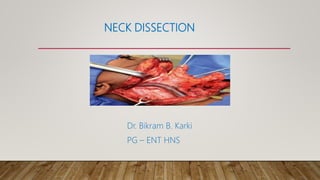
NECK DISSECTION
Dr. Bikram B. Karki
PG – ENT HNS
NECK
• Superior border
anteriorly : mandible with deep limits of FOM
posteriorly : skull base
• Inferior limits
by upper aspect of first rib and
first thoracic vertebrae.
FASCIAL LAYERS
• Superficial cervical fascia
• Deep cervical fascia
• Investing or superficial layer
• Middle layer - Muscular compartment
- Visceral compartment
• Deep layer
SUPERFICIAL NECK MUSCLES
• Platysma
• Surgical considerations
• Absent in midline of neck
• Increases blood supply to skin flaps
• Fibers run in an opposite direction to SCM
• Sternocleidomastoid
• Surgical considerations
• divides neck into
anterior and posterior triangles
• LN 2, 3, 4 lies deep to it
• Trapezius muscle
• Surgical consideration
• Posterior limit of level V neck dissection
• Denervation results in shoulder drop and winged
scapula
SUPRAHYOID AND INFRA HYOID MUSCLE
• Suprahyoid muscle
• Digastric
• Stylohyoid
• Mylohyoid
• Geniohyoid
• hyoglossus
• Digastric muscle :
• Surgical considerations
• Anterior belly :
• Landmark for identification of mylohyoid
for dissection of submandibular
triangle
• Posterior belly
• Landmark for depth of FN as it exits
stylomastoid foramen
• Infrahyoid muscle
• Sternohyoid
• Sternothyroid
• Thyrohyoid
• omohyoid
• Omohyoid muscle:
• Surgical considerations
• Absent in 10% of individuals
• Landmark demarcating level III from
IV
• 2 bellies joind by flat tendon at
point
where muscle pass superficial to IJV
TRIANGLES OF NECK
• Submental triangle
• Single midline triangle
• Contents
• Level Ia lymph nodes
• Fatty tissue
• Small veins that drain into
anterior jugular veins.
• Submandibular triangle
• Contents
• lymph nodes - level Ib
• submandibular salivary gland
• lingual nerve, hypoglossal nerve
• marginal mandibular branch of FN
• facial artery and veins
• Carotid triangle
• Contents
• Lymph nodes of jugular chain
• CCA and its branches
• IJV and its tributaries
• Vagus nerve
• Hypoglossal nerve
• Superior laryngeal nerve
• Muscular triangle:
• Contents
• Lower part of the carotid sheath
• Sternohyoid, sternothyroid,
thyrohyoid muscles
• Thyroid and parathyroid glands
• Upper aerodigestive tract
• Posterior triangle
• Divided by inferior belly of omohyoid
into
• Occipital triangle
• Supraclavicular triangles
• Posterior triangle
• Contents:
• Omohyoid muscle
• Arteries:
a) Subclavian (3rd part)
b) Transverse cervical & suprascapular
c) Occipital
• Nerves
a) cervical plexus
b) branchial plexus
c) spinal accessory nerve
• Lymphnode
MARGINAL MANDIBULAR NERVE
• Landmarks:
1 cm anterior and inferior to angle of
mandible
• Course
• Passes antero-medially across upper neck in
plane just deep to platysma muscle
• But superficial to investing layer
• Can extend as low as greater cornu of
hyoid
• Passing superficial to facial vessels and
across mandible to provide motor
supply to
a) depressor anguli oris,
b) depressor labii inferioris and
c) mentalis muscle
• Facial nerve is most at risk during
• Submandibular gland excision
• Clearance of lymph node level Ib
• Facial nerve can be protected by
• Incising investing layer to lower border of SMG or
just above hyoid bone
• Reflecting fascia superiorly along with facial vein
• Thus, retracting nerve out of surgical field
HYPOGLOSSAL NERVE
• After exiting skull via hypoglossal canal
• Nerve runs deep to IJV
• Then curves around carotid bifurcation as it heads
anteriorly
• Passing inferior to greater horn of hyoid
• Coursing superiorly, superficial to hyoglossus
to reach tongue
• Susceptible to injury
• Floor of submandibular triangle, just
deep to duct
• During surgical procedures particularly
at its lowest point near hyoid
SPINAL ACCESSORY NERVE
• Exit skull via jugular foramen
• Can be located over transverse process of atlas
• Descends obliquely in level II (forms Level IIa
and IIb)
• Enters SCM muscle
• Exits posterior border of SCM - 1 cm above
Erb’s point
• Course across posterior triangle - enter
trapezius
• Importance
• Important landmark in sub-dividing lymph node
levels IIa and IIb.
• All important structures in posterior triangle lies
caudal to SAN
• Surgical consideration
• Most commonly injured during level 5 neck
dissection
THORACIC DUCT
• Located to right and behind left CCA and vagus
nerve
• Arches upward, forward and laterally, passing
behind IJV
• In front of anterior scalene muscle and phrenic
nerve
• Opens into IJV, subclavian vein or angle formed
by their junction
CERVICAL LYMPHATICS
• Divided into superficial and deep lymphatics
• Superficial lymphatics
• Drain skin and perforate investing layer of deep
cervical fascia - drain into deep system
• Deep lymphatics
• Proximity to vessels, nerves and muscles of neck.
• Over 80% of lymph nodes - closely related to IJV
LYMPH NODE LEVELS
• Originates from memorial sloan kettering
hospital,
new york
• Was adopted by AAOHNS in 1991
• LEVEL I
• Ia
• Chin
• Lower lip
• Mandibular incisors
• Anterior floor of mouth
• Tip of tongue
• Ib
• Oral Cavity
Floor of mouth
Oral tongue
• Nasal cavity (anterior)
• Face
• LEVEL II
• First echelon lymph nodes of
oropharynx
• Oral cavity
• Nasopharynx
• Hypopharynx
• Parotid gland
• Level III
• Lower areas of Oropharynx
• Hypopharynx
• Larynx
• LEVEL IV
• Hypopharynx
• Larynx
• Thyroid
• Cervical esophagus
• LEVEL V
• From all other nodal areas
• Nasopharynx
• Posterior neck
• Cutaneous scalp primary lesions
• Level VI
• Thyroid
• Larynx (glottic and subglottic)
• Pyriform sinus apex
• Cervical esophagus
• Level VII
• Superior mediastinum node
• Harbour metastasis from
• Thyroid
• Larynx ( Subglottis )
• Trachea
• Cervical oesophagus
• STAGING : NODALCLASSIFICATION
• Nx: Regional lymph nodes cannot be assessed
• N0: No regional lymph node metastases
• N1: Single ipsilateral lymph node, < 3 cm
• N2a: Single ipsilateral lymph node more
than 3
to 6 cm
• N2b: Multiple ipsilateral lymph nodes < 6
cm
• N2c: Bilateral or contralateral nodes < 6cm
• N3a: Metastases > 6 cm
• N3b: Metastasis in single or multiple LN
with
• THYROID
• N1a – Metastasis in level VI ( pretracheal, paratracheal, prelaryngeal) or
superior mediastinum
• N1b – Metastasis in other unilateral, bilateral or contralateral cervical (
level I, II,
III, IV, V) or Retropharyngeal
• NASOPHARYNX
• NI - unilateral < 6 cm, above caudal border of cricoid cartilage
• N2 - bilateral < 6 cm, above caudal border of cricoid cartilage
• N3 - > 6 cm and/or extension below caudal border of cricoid cartilage
NECK DISSECTION
• Systemic removal of lymph nodes with their
fibrofatty tissues from various compartments of neck
• Neck dissection classification
• In 1991, published the classification
(AAO-HNS)
• Later modified in 2001
• Academy’s classification (1991)
• Radical neck dissection (RND)
• Modified radical neck dissection (MRND)
• Selective neck dissection (SND)
• Supra-omohyoid type
• Lateral type
• Posterolateral type
• Anterior compartment type
• Extended radical neck dissection
• Academy’s classification (2001)
• Radical neck dissection
• Modified radical neck dissection
• Selective neck dissection
• SND ( I to III/IV )
• SND ( II to IV )
• SND ( II to V, Postauricular, Suboccipital )
• SND ( level VI )
• Extended neck dissection
• Medina classification (1989)
• Comprehensive neck dissection
• Radical neck dissection
• Modified radical neck dissection
– Type I (XI preserved)
– Type II (XI, IJV preserved)
– Type III (XI, IJV, and SCM preserved)
• Selective neck dissection
RADICAL NECK DISSECTION
• Removes
• Lymph nodes in level I-V + fibrofatty tissue
• Non lymphatic structures :
• SAN, IJV, SCM
• Indications
• Significant operable neck disease with tumor bulk
near to or directly involving SAN and/or IJV
• Reccurent disease after previous surgery or RT
• Clinical signs of gross extranodal disease
• Contraindications
• Untreatable primary tumor
• Unresectable neck disease
• Unfit for major surgery
• Distant metastases
• Preoperative preparation
• Patient counselling
risks and possible complications
• Prophylactic antibiotic
for 24 hours
aerobic, anaerobic and Gram-negative bacteria
• Elective tracheostomy
patient undergoing bilateral neck dissection
• Position
• Supine on operating table
• Head turned to opposite side
• Sandbag under shoulders
• Upper end table elevation to 30 degree
• Draping
• Incision for neck dissection
Hockey stick Boomerang Bilateral Boomerang
McFee Apron or Bilateral
hockey
Modified apron
Schobinger Modified schobinger Utilit
y
• Four areas of special attention
• Lower end of IJV
• Junction of lateral border of clavicle with lower
edge of trapezius
• Upper end of IJV
• Submandibular triangle
• Incision
• Through skin down to platysma
• Raise flap in subplatysmal plane
• Preserve marginal mandibular and
if possible cervical branch of facial nerve.
• First area of special attention
• Lower end of IJV
• Dissection along upper border of clavicle
from trapezius to suprasternal notch.
• Divide supraclavicular nerves and vessels
• Divide SCM above clavicle
Dissection of lower end of
SCM
• IJV visualized
between sternal and clavicular heads of
SCM
• Open carotid sheath
• Expose few centimetres of IJV
• Place 3 ligating sutures (vicryl 0/0)
around vein
• Retract carotid artery and vagus nerve
medially
Ligation of internal jugular
vein.
• Extend dissection laterally towards
Chaissaignac’s triangle
• Remove scalene nodes
• Second area of special attention:
• Junction of clavicle and anterior border
trapezius
• Begin dissection at lower end of
trapezius
• Divide fatty tissues in supraclavicular
region
• Cut inferior belly of omohyoid
• Ligate transverse cervical vessels
• Continue dissection on to underlying level of
prevertebral fascia
• Identify and preserve brachial plexus and phrenic
nerve
• Continue dissection to anterior border of
trapezius
• Proceed upward direction, dissecting posterior
triangle
• Dissection of posterior triangle
• Continue dissection to uppermost point
of triangle at mastoid tip
• Clear posterior triangle
• Dissect
• Proximal attachment of SCM
• lower lobe of parotid
• Third area of special attention
• Upper end of internal jugular vein
• Clear posterior belly of digastric
• Retract superiorly to expose IJV and
accessory nerve
• Clear vein and mobilize over a few cm
• Divide IJV after ligation and transfixion
Retraction of posterior belly of
digastric muscle to show upper
end of internal jugular vein
• Mobilize specimen from
cranial and caudal direction
• Follow anterior belly of omohyoid to
insertion at hyoid bone and divide
• Fourth area of special attention:
• Submandibular triangle
• Divide fatty tissue on dissection
plane
• Dissect fascia including SMG
• Ligate facial artery and vein
• Retract mylohyoid muscle medially
and
SMG inferolaterally
Anatomy of submandibular triangle.
• Floor visible and Identify lingual and
hypoglossal nerve
• Divide submandibular duct
• Ligate facial artery – posterior inferior
border of SMG
• Remove entire specimen
• Complete haemostasis
• Wound irrigation with saline
• Neck drains
• Closure of incisions
• two layers
MODIFIED RADICAL NECK DISSECTION
• En bloc removal of lymph node–
bearing tissue from one side of neck
(levels I to V).
• Preserves
• SAN, IJV, SCM (any combination)
• Three types (Medina 1989)
• Type I: Preservation of SAN
• Type II: Preservation of SAN and IJV
• Type III: Preservation of SAN, IJV, and
SCM
• MRND Type I
• Indications
• Clinically obvious lymph node
metastases
• SAN not involved by tumor
• MRND Type II
• Indications
• Rarely planned
• Intraoperative tumor found
adherent to SCM, but not IJV and
SAN
• MRND TYPE III
• Widely accepted
• N0 neck in patients with sq. cell ca. of
hypopharynx/larynx
• Differentiated Ca. thyroid with
palpable LN metastasis in posterior triangle
SPARING OF SAN
• Careful elevation of flap in posterior triangle
• Identification of SAN
a) located 1 cm above Erb’s Point
b) Entry point in ant border of trapezius 5 cm above
clavicle
c) Cranial part of SAN:
Runs along with IJV and crosses it medially to
laterally and
Transverse process of atlas
SELECTIVE NECK DISSECTIONS
• Definition
• Procedure where one or more LN groups are preserved in addition to
non- lymphatic structures
• Four common subtypes:
• Supraomohyoid neck dissection
• Lateral neck dissection
• Posterolateral neck dissection
• Anterior neck dissection
• Selective Neck Dissection( 1-III )
• Supraomohyoid neck dissection
• En bloc removal of cervical lymph node groups I to III
• Indications
• Oral cavity carcinoma (T1 –T4 with N0 neck)
• N1 in upper neck if
postoperative radiation therapy planned
• Bilateral SOHND
• Anterior tongue Ca.
• FOM Ca. that approach the midline
• Selective Neck Dissection (I-IV)
• Extended supraomohyoid neck dissection
• Cancer of anterolateral part of tongue
• Selective Neck Dissection: Level II-IV
• Lateral neck dissection
• En bloc removal of jugular lymph nodes
(Levels II-IV)
• Indications
• N0 neck in carcinomas of
Oropharynx
Hypopharynx
Larynx
• Laryngeal Ca. with subglottic extension, Hypopharyngeal Ca, MTC
SND (II to IV and VI).
• If risk for bilateral metastasis : Bilateral SND (II to IV)
• SND: Level II-V+ suboccipital and postauricular LN
• Posterolateral neck dissection
• Indications
• Cutaneous malignancies
• Melanoma
• Squamous cell carcinoma
• Merkel cell carcinoma
• Soft tissue sarcomas of scalp and neck
• Selective Neck Dissection : Level VI
• Central compartment dissection
• En bloc removal of level VI lymph
structures
• Indications
• Thyroid carcinoma
• laryngeal cancer (Advanced glottic and
subglottic)
• Advanced pyriform sinus cancer
• Cervical esophagus cancer
• Tracheal cancer
EXTENDED NECK DISSECTION
• Definition
• Removal of one or more additional lymph node
groups and/or non-lymphatic structures relative
to RND
• Eg, level VII, Retropharygeal node,
Hypoglossal nerve,
Carotid artery, Skin of neck
SEQUELAE OF RADICAL NECK DISSECTION
• Removal of spinal accessory nerve
• Loss of trapezius function
• Decrease ability to abduct shoulder > 90
degrees
• Destabilization of scapula
• Shoulder syndrome of pain, weakness and
deformity of shoulder girdle
COMPLICATIONS OF NECK DISSECTION
• 4 types
• Intra operative
• Early post operative
• Intermediate operative
• Late complications
INTRAOPERATIVE COMPLICATIONS
• Injury to local blood vessels and nerves
• SAN
• Cervical plexus, Brachial plexus
• Marginal mandibular nerve
• Vagus nerve, Hypogloasal nerve, Phrenic nerve
• Thoracic duct injury
EARLY POST OPERATIVE COMPLICATIONS
• Haemorrhage:
• May need re-exploration.
• Airway obstruction
• Temporary elective tracheotomy to protect airway.
• Increased intracranial pressure
• If persists, head end elevation, steroids and mannitol
• Carotid sinus syndrome
May result in hypotension and bradycardia
• Pneumothorax
• Air leaks
INTERMEDIATE POST OPERATIVE COMPLICATIONS
• Carotid artery rupture
• Fatal complication resulting in immediate mortality
• Immediate finger pressure, airway management, blood transfusion
and exploration in OT
• Prevention : Use of free and pedicled flap for closure of defect
• Deep vein thrombosis
• Pulmonary complications:
• Basal collapse and bronchopneumonia
CHYLOUS LEAK
• Intraoperative
• Apparent as clear fluid - confirmed by increased
flow on valsalva manoeuvre
• Vessel - quite fragile and surrounded by fatty
tissue - prone to tearing
• Management
• Direct clamping and ligating
• If this fails,
Fibrin sealant or vicryl mesh may be used
• Muscle flaps can be used in severe cases
• Diathermy does not seal fragile lymphatic vessels
• Postoperative
• Presence of milky appearance in neck drain after starting feed
• Confirmed either by
• Identifying triglycerides >100mg/dl or
• Reduction in volume of drain fluid on stopping enteral feed
• Management
• Low output leak - close spontaneously or
Managed with aspiration, pressure dressing and low fat elemental diet
• Surgical re-exploration and ligation of the duct is advised in
a) presence of complications (e.g. flap necrosis),
b) ‘high output leak’ (300mL/ day )
• If neck exploration fails - Thoracoscopic ligation of thoracic duct
LATE POSTOPERATIVE COMPLICATIONS
• Lymph edema
If both IJV ligated , block of lymphatic drainage from head.
• Hypertrophic scars
Neck dissection

More Related Content

What's hot

Neck dissection part 1
Neck dissection part 1 Neck dissection part 1
Neck dissection part 1
Padmasree Patowary
 
Neck Dissections
Neck DissectionsNeck Dissections
Neck Dissectionsguest26910d
 
Neck dissection
Neck dissectionNeck dissection
Neck dissection
Dr-Maryam Khan
 
Closed rhinoplasty
Closed rhinoplastyClosed rhinoplasty
Closed rhinoplasty
bhakabhob mahachitsattaya
 
SELECTIVE NECK DISSECTION
SELECTIVE NECK DISSECTIONSELECTIVE NECK DISSECTION
SELECTIVE NECK DISSECTION
Ayesha Fayyaz
 
Infratemporal fossa approaches
Infratemporal fossa approachesInfratemporal fossa approaches
Infratemporal fossa approaches
Md Roohia
 
Reanimation of facial paralysis
Reanimation of facial paralysisReanimation of facial paralysis
Reanimation of facial paralysis
Dr.Amit kumar choudhary
 
surgical approaches to frontal sinus ppt
surgical approaches to frontal sinus pptsurgical approaches to frontal sinus ppt
surgical approaches to frontal sinus ppt
Vaibhav Lahane
 
Micro vascular free flaps used in head and neck reconstruction /certified fi...
Micro vascular free flaps used in head and neck reconstruction  /certified fi...Micro vascular free flaps used in head and neck reconstruction  /certified fi...
Micro vascular free flaps used in head and neck reconstruction /certified fi...
Indian dental academy
 
Overview Of Neck Dissections
Overview Of Neck DissectionsOverview Of Neck Dissections
Overview Of Neck DissectionsSaeed Al-Shomimi
 
Rhinoplasty
RhinoplastyRhinoplasty
Rhinoplasty
Disha Sharma
 
lip reconstruction
 lip reconstruction lip reconstruction
lip reconstruction
Sumer Yadav
 
Neck dissection
Neck dissectionNeck dissection
Neck dissection
Dr Safika Zaman
 
MAXILLECTOMY
MAXILLECTOMYMAXILLECTOMY
Steps in selective neck dissection
Steps in selective neck dissectionSteps in selective neck dissection
Steps in selective neck dissection
Jamil Kifayatullah
 
Local flaps in ent
Local flaps in entLocal flaps in ent
Local flaps in ent
Ajay Manickam
 
Rhinoplasty raju ppt full
Rhinoplasty raju ppt fullRhinoplasty raju ppt full
Rhinoplasty raju ppt full
Ram Raju
 
Conservative laryngeal surgery
Conservative laryngeal surgery Conservative laryngeal surgery
Conservative laryngeal surgery
Vardhaman Mahavir Medical College and SJH
 
13. neck dissection - Meghali
13. neck dissection - Meghali13. neck dissection - Meghali
13. neck dissection - Meghali
Meghali Diwaker
 
Neck Dissection.Overview
Neck Dissection.OverviewNeck Dissection.Overview
Neck Dissection.Overview
Abubakar Shah
 

What's hot (20)

Neck dissection part 1
Neck dissection part 1 Neck dissection part 1
Neck dissection part 1
 
Neck Dissections
Neck DissectionsNeck Dissections
Neck Dissections
 
Neck dissection
Neck dissectionNeck dissection
Neck dissection
 
Closed rhinoplasty
Closed rhinoplastyClosed rhinoplasty
Closed rhinoplasty
 
SELECTIVE NECK DISSECTION
SELECTIVE NECK DISSECTIONSELECTIVE NECK DISSECTION
SELECTIVE NECK DISSECTION
 
Infratemporal fossa approaches
Infratemporal fossa approachesInfratemporal fossa approaches
Infratemporal fossa approaches
 
Reanimation of facial paralysis
Reanimation of facial paralysisReanimation of facial paralysis
Reanimation of facial paralysis
 
surgical approaches to frontal sinus ppt
surgical approaches to frontal sinus pptsurgical approaches to frontal sinus ppt
surgical approaches to frontal sinus ppt
 
Micro vascular free flaps used in head and neck reconstruction /certified fi...
Micro vascular free flaps used in head and neck reconstruction  /certified fi...Micro vascular free flaps used in head and neck reconstruction  /certified fi...
Micro vascular free flaps used in head and neck reconstruction /certified fi...
 
Overview Of Neck Dissections
Overview Of Neck DissectionsOverview Of Neck Dissections
Overview Of Neck Dissections
 
Rhinoplasty
RhinoplastyRhinoplasty
Rhinoplasty
 
lip reconstruction
 lip reconstruction lip reconstruction
lip reconstruction
 
Neck dissection
Neck dissectionNeck dissection
Neck dissection
 
MAXILLECTOMY
MAXILLECTOMYMAXILLECTOMY
MAXILLECTOMY
 
Steps in selective neck dissection
Steps in selective neck dissectionSteps in selective neck dissection
Steps in selective neck dissection
 
Local flaps in ent
Local flaps in entLocal flaps in ent
Local flaps in ent
 
Rhinoplasty raju ppt full
Rhinoplasty raju ppt fullRhinoplasty raju ppt full
Rhinoplasty raju ppt full
 
Conservative laryngeal surgery
Conservative laryngeal surgery Conservative laryngeal surgery
Conservative laryngeal surgery
 
13. neck dissection - Meghali
13. neck dissection - Meghali13. neck dissection - Meghali
13. neck dissection - Meghali
 
Neck Dissection.Overview
Neck Dissection.OverviewNeck Dissection.Overview
Neck Dissection.Overview
 

Similar to Neck dissection

Neck dissections
Neck dissectionsNeck dissections
Neck dissections
Priyanka Malekar
 
TRIANGLES OF NECK
 TRIANGLES OF NECK TRIANGLES OF NECK
TRIANGLES OF NECK
ssuser7fe13d
 
Neck dissection-020116-slides
Neck dissection-020116-slidesNeck dissection-020116-slides
Neck dissection-020116-slidesmarcello ribas
 
Levels of lymph nodes in head and neck
Levels of lymph nodes in head and neckLevels of lymph nodes in head and neck
Levels of lymph nodes in head and neck
Anjali Singh
 
Neck Dissections
Neck Dissections Neck Dissections
Neck Dissections
Harmandeep Jabbal
 
Role Of Surgery In Management of Neck Nodes 2 - Copy.pptx
Role Of Surgery In Management of Neck Nodes 2 - Copy.pptxRole Of Surgery In Management of Neck Nodes 2 - Copy.pptx
Role Of Surgery In Management of Neck Nodes 2 - Copy.pptx
cheshtasharma22
 
Neck dissection
Neck dissectionNeck dissection
Neck dissection
Rehana Sultana
 
NECK DISSECTION- A COMPREHENSIVE STUDY
NECK DISSECTION- A COMPREHENSIVE STUDYNECK DISSECTION- A COMPREHENSIVE STUDY
NECK DISSECTION- A COMPREHENSIVE STUDY
Priyanko Chakraborty
 
Sch 43 surgical management of tumors of the foramen magnum
Sch 43 surgical management of tumors of the foramen magnum Sch 43 surgical management of tumors of the foramen magnum
Sch 43 surgical management of tumors of the foramen magnum
Neurosurgery Vajira
 
surgical anatomy of Triangles of neck
surgical anatomy of Triangles of neck surgical anatomy of Triangles of neck
surgical anatomy of Triangles of neck
Tasnia Mahmud
 
Neck Dissection Head Neck oncoSurgery.pptx
Neck Dissection Head Neck oncoSurgery.pptxNeck Dissection Head Neck oncoSurgery.pptx
Neck Dissection Head Neck oncoSurgery.pptx
Satishray9
 
Neck dissectionppt.pptx
Neck dissectionppt.pptxNeck dissectionppt.pptx
Neck dissectionppt.pptx
DrDhayaAnn
 
Neck swellings complete
Neck swellings completeNeck swellings complete
Neck swellings complete
Sunil Gaur
 
Surgical Anatomy of Lymph nodes
Surgical Anatomy of Lymph nodesSurgical Anatomy of Lymph nodes
Surgical Anatomy of Lymph nodes
J.Rahul Raghavender
 
Neck dissection procedure
Neck dissection procedureNeck dissection procedure
Neck dissection procedure
Jamil Kifayatullah
 
Surgical anatomy of neck and types of neck dissection
Surgical anatomy of neck and types of neck dissectionSurgical anatomy of neck and types of neck dissection
Surgical anatomy of neck and types of neck dissection
Sanika Kulkarni
 
Surgical anatomy of neck
Surgical anatomy of neck Surgical anatomy of neck
Surgical anatomy of neck
AVINAV GUPTA
 
DEEP NECK SPACE INFECTIONS by dr Priyanka FINAL-1.pptx
DEEP NECK SPACE INFECTIONS by dr Priyanka FINAL-1.pptxDEEP NECK SPACE INFECTIONS by dr Priyanka FINAL-1.pptx
DEEP NECK SPACE INFECTIONS by dr Priyanka FINAL-1.pptx
DRRamendrakumarSingh
 
Deepneck space space infections.undar.pptx
Deepneck space  space infections.undar.pptxDeepneck space  space infections.undar.pptx
Deepneck space space infections.undar.pptx
sangamadhikari9
 

Similar to Neck dissection (20)

Neck dissections
Neck dissectionsNeck dissections
Neck dissections
 
TRIANGLES OF NECK
 TRIANGLES OF NECK TRIANGLES OF NECK
TRIANGLES OF NECK
 
Neck dissection-020116-slides
Neck dissection-020116-slidesNeck dissection-020116-slides
Neck dissection-020116-slides
 
Levels of lymph nodes in head and neck
Levels of lymph nodes in head and neckLevels of lymph nodes in head and neck
Levels of lymph nodes in head and neck
 
Neck Dissections
Neck Dissections Neck Dissections
Neck Dissections
 
Role Of Surgery In Management of Neck Nodes 2 - Copy.pptx
Role Of Surgery In Management of Neck Nodes 2 - Copy.pptxRole Of Surgery In Management of Neck Nodes 2 - Copy.pptx
Role Of Surgery In Management of Neck Nodes 2 - Copy.pptx
 
Neck dissection
Neck dissectionNeck dissection
Neck dissection
 
NECK DISSECTION- A COMPREHENSIVE STUDY
NECK DISSECTION- A COMPREHENSIVE STUDYNECK DISSECTION- A COMPREHENSIVE STUDY
NECK DISSECTION- A COMPREHENSIVE STUDY
 
Sch 43 surgical management of tumors of the foramen magnum
Sch 43 surgical management of tumors of the foramen magnum Sch 43 surgical management of tumors of the foramen magnum
Sch 43 surgical management of tumors of the foramen magnum
 
surgical anatomy of Triangles of neck
surgical anatomy of Triangles of neck surgical anatomy of Triangles of neck
surgical anatomy of Triangles of neck
 
Neck Dissection Head Neck oncoSurgery.pptx
Neck Dissection Head Neck oncoSurgery.pptxNeck Dissection Head Neck oncoSurgery.pptx
Neck Dissection Head Neck oncoSurgery.pptx
 
Neck dissectionppt.pptx
Neck dissectionppt.pptxNeck dissectionppt.pptx
Neck dissectionppt.pptx
 
Neck dissections
Neck dissectionsNeck dissections
Neck dissections
 
Neck swellings complete
Neck swellings completeNeck swellings complete
Neck swellings complete
 
Surgical Anatomy of Lymph nodes
Surgical Anatomy of Lymph nodesSurgical Anatomy of Lymph nodes
Surgical Anatomy of Lymph nodes
 
Neck dissection procedure
Neck dissection procedureNeck dissection procedure
Neck dissection procedure
 
Surgical anatomy of neck and types of neck dissection
Surgical anatomy of neck and types of neck dissectionSurgical anatomy of neck and types of neck dissection
Surgical anatomy of neck and types of neck dissection
 
Surgical anatomy of neck
Surgical anatomy of neck Surgical anatomy of neck
Surgical anatomy of neck
 
DEEP NECK SPACE INFECTIONS by dr Priyanka FINAL-1.pptx
DEEP NECK SPACE INFECTIONS by dr Priyanka FINAL-1.pptxDEEP NECK SPACE INFECTIONS by dr Priyanka FINAL-1.pptx
DEEP NECK SPACE INFECTIONS by dr Priyanka FINAL-1.pptx
 
Deepneck space space infections.undar.pptx
Deepneck space  space infections.undar.pptxDeepneck space  space infections.undar.pptx
Deepneck space space infections.undar.pptx
 

Recently uploaded

Cervical & Brachial Plexus By Dr. RIG.pptx
Cervical & Brachial Plexus By Dr. RIG.pptxCervical & Brachial Plexus By Dr. RIG.pptx
Cervical & Brachial Plexus By Dr. RIG.pptx
Dr. Rabia Inam Gandapore
 
ARTHROLOGY PPT NCISM SYLLABUS AYURVEDA STUDENTS
ARTHROLOGY PPT NCISM SYLLABUS AYURVEDA STUDENTSARTHROLOGY PPT NCISM SYLLABUS AYURVEDA STUDENTS
ARTHROLOGY PPT NCISM SYLLABUS AYURVEDA STUDENTS
Dr. Vinay Pareek
 
Ophthalmology Clinical Tests for OSCE exam
Ophthalmology Clinical Tests for OSCE examOphthalmology Clinical Tests for OSCE exam
Ophthalmology Clinical Tests for OSCE exam
KafrELShiekh University
 
heat stroke and heat exhaustion in children
heat stroke and heat exhaustion in childrenheat stroke and heat exhaustion in children
heat stroke and heat exhaustion in children
SumeraAhmad5
 
ARTIFICIAL INTELLIGENCE IN HEALTHCARE.pdf
ARTIFICIAL INTELLIGENCE IN  HEALTHCARE.pdfARTIFICIAL INTELLIGENCE IN  HEALTHCARE.pdf
ARTIFICIAL INTELLIGENCE IN HEALTHCARE.pdf
Anujkumaranit
 
Pulmonary Thromboembolism - etilogy, types, medical- Surgical and nursing man...
Pulmonary Thromboembolism - etilogy, types, medical- Surgical and nursing man...Pulmonary Thromboembolism - etilogy, types, medical- Surgical and nursing man...
Pulmonary Thromboembolism - etilogy, types, medical- Surgical and nursing man...
VarunMahajani
 
How to Give Better Lectures: Some Tips for Doctors
How to Give Better Lectures: Some Tips for DoctorsHow to Give Better Lectures: Some Tips for Doctors
How to Give Better Lectures: Some Tips for Doctors
LanceCatedral
 
Hemodialysis: Chapter 3, Dialysis Water Unit - Dr.Gawad
Hemodialysis: Chapter 3, Dialysis Water Unit - Dr.GawadHemodialysis: Chapter 3, Dialysis Water Unit - Dr.Gawad
Hemodialysis: Chapter 3, Dialysis Water Unit - Dr.Gawad
NephroTube - Dr.Gawad
 
For Better Surat #ℂall #Girl Service ❤85270-49040❤ Surat #ℂall #Girls
For Better Surat #ℂall #Girl Service ❤85270-49040❤ Surat #ℂall #GirlsFor Better Surat #ℂall #Girl Service ❤85270-49040❤ Surat #ℂall #Girls
For Better Surat #ℂall #Girl Service ❤85270-49040❤ Surat #ℂall #Girls
Savita Shen $i11
 
Charaka Samhita Sutra sthana Chapter 15 Upakalpaniyaadhyaya
Charaka Samhita Sutra sthana Chapter 15 UpakalpaniyaadhyayaCharaka Samhita Sutra sthana Chapter 15 Upakalpaniyaadhyaya
Charaka Samhita Sutra sthana Chapter 15 Upakalpaniyaadhyaya
Dr KHALID B.M
 
The Normal Electrocardiogram - Part I of II
The Normal Electrocardiogram - Part I of IIThe Normal Electrocardiogram - Part I of II
The Normal Electrocardiogram - Part I of II
MedicoseAcademics
 
Ocular injury ppt Upendra pal optometrist upums saifai etawah
Ocular injury  ppt  Upendra pal  optometrist upums saifai etawahOcular injury  ppt  Upendra pal  optometrist upums saifai etawah
Ocular injury ppt Upendra pal optometrist upums saifai etawah
pal078100
 
Pharynx and Clinical Correlations BY Dr.Rabia Inam Gandapore.pptx
Pharynx and Clinical Correlations BY Dr.Rabia Inam Gandapore.pptxPharynx and Clinical Correlations BY Dr.Rabia Inam Gandapore.pptx
Pharynx and Clinical Correlations BY Dr.Rabia Inam Gandapore.pptx
Dr. Rabia Inam Gandapore
 
POST OPERATIVE OLIGURIA and its management
POST OPERATIVE OLIGURIA and its managementPOST OPERATIVE OLIGURIA and its management
POST OPERATIVE OLIGURIA and its management
touseefaziz1
 
24 Upakrama.pptx class ppt useful in all
24 Upakrama.pptx class ppt useful in all24 Upakrama.pptx class ppt useful in all
24 Upakrama.pptx class ppt useful in all
DrSathishMS1
 
Superficial & Deep Fascia of the NECK.pptx
Superficial & Deep Fascia of the NECK.pptxSuperficial & Deep Fascia of the NECK.pptx
Superficial & Deep Fascia of the NECK.pptx
Dr. Rabia Inam Gandapore
 
basicmodesofventilation2022-220313203758.pdf
basicmodesofventilation2022-220313203758.pdfbasicmodesofventilation2022-220313203758.pdf
basicmodesofventilation2022-220313203758.pdf
aljamhori teaching hospital
 
NVBDCP.pptx Nation vector borne disease control program
NVBDCP.pptx Nation vector borne disease control programNVBDCP.pptx Nation vector borne disease control program
NVBDCP.pptx Nation vector borne disease control program
Sapna Thakur
 
KDIGO 2024 guidelines for diabetologists
KDIGO 2024 guidelines for diabetologistsKDIGO 2024 guidelines for diabetologists
KDIGO 2024 guidelines for diabetologists
د.محمود نجيب
 
Ozempic: Preoperative Management of Patients on GLP-1 Receptor Agonists
Ozempic: Preoperative Management of Patients on GLP-1 Receptor Agonists  Ozempic: Preoperative Management of Patients on GLP-1 Receptor Agonists
Ozempic: Preoperative Management of Patients on GLP-1 Receptor Agonists
Saeid Safari
 

Recently uploaded (20)

Cervical & Brachial Plexus By Dr. RIG.pptx
Cervical & Brachial Plexus By Dr. RIG.pptxCervical & Brachial Plexus By Dr. RIG.pptx
Cervical & Brachial Plexus By Dr. RIG.pptx
 
ARTHROLOGY PPT NCISM SYLLABUS AYURVEDA STUDENTS
ARTHROLOGY PPT NCISM SYLLABUS AYURVEDA STUDENTSARTHROLOGY PPT NCISM SYLLABUS AYURVEDA STUDENTS
ARTHROLOGY PPT NCISM SYLLABUS AYURVEDA STUDENTS
 
Ophthalmology Clinical Tests for OSCE exam
Ophthalmology Clinical Tests for OSCE examOphthalmology Clinical Tests for OSCE exam
Ophthalmology Clinical Tests for OSCE exam
 
heat stroke and heat exhaustion in children
heat stroke and heat exhaustion in childrenheat stroke and heat exhaustion in children
heat stroke and heat exhaustion in children
 
ARTIFICIAL INTELLIGENCE IN HEALTHCARE.pdf
ARTIFICIAL INTELLIGENCE IN  HEALTHCARE.pdfARTIFICIAL INTELLIGENCE IN  HEALTHCARE.pdf
ARTIFICIAL INTELLIGENCE IN HEALTHCARE.pdf
 
Pulmonary Thromboembolism - etilogy, types, medical- Surgical and nursing man...
Pulmonary Thromboembolism - etilogy, types, medical- Surgical and nursing man...Pulmonary Thromboembolism - etilogy, types, medical- Surgical and nursing man...
Pulmonary Thromboembolism - etilogy, types, medical- Surgical and nursing man...
 
How to Give Better Lectures: Some Tips for Doctors
How to Give Better Lectures: Some Tips for DoctorsHow to Give Better Lectures: Some Tips for Doctors
How to Give Better Lectures: Some Tips for Doctors
 
Hemodialysis: Chapter 3, Dialysis Water Unit - Dr.Gawad
Hemodialysis: Chapter 3, Dialysis Water Unit - Dr.GawadHemodialysis: Chapter 3, Dialysis Water Unit - Dr.Gawad
Hemodialysis: Chapter 3, Dialysis Water Unit - Dr.Gawad
 
For Better Surat #ℂall #Girl Service ❤85270-49040❤ Surat #ℂall #Girls
For Better Surat #ℂall #Girl Service ❤85270-49040❤ Surat #ℂall #GirlsFor Better Surat #ℂall #Girl Service ❤85270-49040❤ Surat #ℂall #Girls
For Better Surat #ℂall #Girl Service ❤85270-49040❤ Surat #ℂall #Girls
 
Charaka Samhita Sutra sthana Chapter 15 Upakalpaniyaadhyaya
Charaka Samhita Sutra sthana Chapter 15 UpakalpaniyaadhyayaCharaka Samhita Sutra sthana Chapter 15 Upakalpaniyaadhyaya
Charaka Samhita Sutra sthana Chapter 15 Upakalpaniyaadhyaya
 
The Normal Electrocardiogram - Part I of II
The Normal Electrocardiogram - Part I of IIThe Normal Electrocardiogram - Part I of II
The Normal Electrocardiogram - Part I of II
 
Ocular injury ppt Upendra pal optometrist upums saifai etawah
Ocular injury  ppt  Upendra pal  optometrist upums saifai etawahOcular injury  ppt  Upendra pal  optometrist upums saifai etawah
Ocular injury ppt Upendra pal optometrist upums saifai etawah
 
Pharynx and Clinical Correlations BY Dr.Rabia Inam Gandapore.pptx
Pharynx and Clinical Correlations BY Dr.Rabia Inam Gandapore.pptxPharynx and Clinical Correlations BY Dr.Rabia Inam Gandapore.pptx
Pharynx and Clinical Correlations BY Dr.Rabia Inam Gandapore.pptx
 
POST OPERATIVE OLIGURIA and its management
POST OPERATIVE OLIGURIA and its managementPOST OPERATIVE OLIGURIA and its management
POST OPERATIVE OLIGURIA and its management
 
24 Upakrama.pptx class ppt useful in all
24 Upakrama.pptx class ppt useful in all24 Upakrama.pptx class ppt useful in all
24 Upakrama.pptx class ppt useful in all
 
Superficial & Deep Fascia of the NECK.pptx
Superficial & Deep Fascia of the NECK.pptxSuperficial & Deep Fascia of the NECK.pptx
Superficial & Deep Fascia of the NECK.pptx
 
basicmodesofventilation2022-220313203758.pdf
basicmodesofventilation2022-220313203758.pdfbasicmodesofventilation2022-220313203758.pdf
basicmodesofventilation2022-220313203758.pdf
 
NVBDCP.pptx Nation vector borne disease control program
NVBDCP.pptx Nation vector borne disease control programNVBDCP.pptx Nation vector borne disease control program
NVBDCP.pptx Nation vector borne disease control program
 
KDIGO 2024 guidelines for diabetologists
KDIGO 2024 guidelines for diabetologistsKDIGO 2024 guidelines for diabetologists
KDIGO 2024 guidelines for diabetologists
 
Ozempic: Preoperative Management of Patients on GLP-1 Receptor Agonists
Ozempic: Preoperative Management of Patients on GLP-1 Receptor Agonists  Ozempic: Preoperative Management of Patients on GLP-1 Receptor Agonists
Ozempic: Preoperative Management of Patients on GLP-1 Receptor Agonists
 

Neck dissection

  • 1. NECK DISSECTION Dr. Bikram B. Karki PG – ENT HNS
  • 2. NECK • Superior border anteriorly : mandible with deep limits of FOM posteriorly : skull base • Inferior limits by upper aspect of first rib and first thoracic vertebrae.
  • 3. FASCIAL LAYERS • Superficial cervical fascia • Deep cervical fascia • Investing or superficial layer • Middle layer - Muscular compartment - Visceral compartment • Deep layer
  • 4. SUPERFICIAL NECK MUSCLES • Platysma • Surgical considerations • Absent in midline of neck • Increases blood supply to skin flaps • Fibers run in an opposite direction to SCM
  • 5. • Sternocleidomastoid • Surgical considerations • divides neck into anterior and posterior triangles • LN 2, 3, 4 lies deep to it
  • 6. • Trapezius muscle • Surgical consideration • Posterior limit of level V neck dissection • Denervation results in shoulder drop and winged scapula
  • 7. SUPRAHYOID AND INFRA HYOID MUSCLE • Suprahyoid muscle • Digastric • Stylohyoid • Mylohyoid • Geniohyoid • hyoglossus
  • 8. • Digastric muscle : • Surgical considerations • Anterior belly : • Landmark for identification of mylohyoid for dissection of submandibular triangle • Posterior belly • Landmark for depth of FN as it exits stylomastoid foramen
  • 9. • Infrahyoid muscle • Sternohyoid • Sternothyroid • Thyrohyoid • omohyoid
  • 10. • Omohyoid muscle: • Surgical considerations • Absent in 10% of individuals • Landmark demarcating level III from IV • 2 bellies joind by flat tendon at point where muscle pass superficial to IJV
  • 12. • Submental triangle • Single midline triangle • Contents • Level Ia lymph nodes • Fatty tissue • Small veins that drain into anterior jugular veins.
  • 13. • Submandibular triangle • Contents • lymph nodes - level Ib • submandibular salivary gland • lingual nerve, hypoglossal nerve • marginal mandibular branch of FN • facial artery and veins
  • 14. • Carotid triangle • Contents • Lymph nodes of jugular chain • CCA and its branches • IJV and its tributaries • Vagus nerve • Hypoglossal nerve • Superior laryngeal nerve
  • 15. • Muscular triangle: • Contents • Lower part of the carotid sheath • Sternohyoid, sternothyroid, thyrohyoid muscles • Thyroid and parathyroid glands • Upper aerodigestive tract
  • 16. • Posterior triangle • Divided by inferior belly of omohyoid into • Occipital triangle • Supraclavicular triangles
  • 17. • Posterior triangle • Contents: • Omohyoid muscle • Arteries: a) Subclavian (3rd part) b) Transverse cervical & suprascapular c) Occipital
  • 18. • Nerves a) cervical plexus b) branchial plexus c) spinal accessory nerve • Lymphnode
  • 19. MARGINAL MANDIBULAR NERVE • Landmarks: 1 cm anterior and inferior to angle of mandible • Course • Passes antero-medially across upper neck in plane just deep to platysma muscle • But superficial to investing layer
  • 20. • Can extend as low as greater cornu of hyoid • Passing superficial to facial vessels and across mandible to provide motor supply to a) depressor anguli oris, b) depressor labii inferioris and c) mentalis muscle
  • 21. • Facial nerve is most at risk during • Submandibular gland excision • Clearance of lymph node level Ib • Facial nerve can be protected by • Incising investing layer to lower border of SMG or just above hyoid bone • Reflecting fascia superiorly along with facial vein • Thus, retracting nerve out of surgical field
  • 22. HYPOGLOSSAL NERVE • After exiting skull via hypoglossal canal • Nerve runs deep to IJV • Then curves around carotid bifurcation as it heads anteriorly • Passing inferior to greater horn of hyoid • Coursing superiorly, superficial to hyoglossus to reach tongue
  • 23. • Susceptible to injury • Floor of submandibular triangle, just deep to duct • During surgical procedures particularly at its lowest point near hyoid
  • 24. SPINAL ACCESSORY NERVE • Exit skull via jugular foramen • Can be located over transverse process of atlas • Descends obliquely in level II (forms Level IIa and IIb) • Enters SCM muscle • Exits posterior border of SCM - 1 cm above Erb’s point • Course across posterior triangle - enter trapezius
  • 25. • Importance • Important landmark in sub-dividing lymph node levels IIa and IIb. • All important structures in posterior triangle lies caudal to SAN • Surgical consideration • Most commonly injured during level 5 neck dissection
  • 26. THORACIC DUCT • Located to right and behind left CCA and vagus nerve • Arches upward, forward and laterally, passing behind IJV • In front of anterior scalene muscle and phrenic nerve • Opens into IJV, subclavian vein or angle formed by their junction
  • 27. CERVICAL LYMPHATICS • Divided into superficial and deep lymphatics • Superficial lymphatics • Drain skin and perforate investing layer of deep cervical fascia - drain into deep system • Deep lymphatics • Proximity to vessels, nerves and muscles of neck. • Over 80% of lymph nodes - closely related to IJV
  • 28. LYMPH NODE LEVELS • Originates from memorial sloan kettering hospital, new york • Was adopted by AAOHNS in 1991
  • 29. • LEVEL I • Ia • Chin • Lower lip • Mandibular incisors • Anterior floor of mouth • Tip of tongue
  • 30. • Ib • Oral Cavity Floor of mouth Oral tongue • Nasal cavity (anterior) • Face
  • 31. • LEVEL II • First echelon lymph nodes of oropharynx • Oral cavity • Nasopharynx • Hypopharynx • Parotid gland
  • 32. • Level III • Lower areas of Oropharynx • Hypopharynx • Larynx
  • 33. • LEVEL IV • Hypopharynx • Larynx • Thyroid • Cervical esophagus
  • 34. • LEVEL V • From all other nodal areas • Nasopharynx • Posterior neck • Cutaneous scalp primary lesions
  • 35. • Level VI • Thyroid • Larynx (glottic and subglottic) • Pyriform sinus apex • Cervical esophagus
  • 36. • Level VII • Superior mediastinum node • Harbour metastasis from • Thyroid • Larynx ( Subglottis ) • Trachea • Cervical oesophagus
  • 37. • STAGING : NODALCLASSIFICATION • Nx: Regional lymph nodes cannot be assessed • N0: No regional lymph node metastases • N1: Single ipsilateral lymph node, < 3 cm
  • 38. • N2a: Single ipsilateral lymph node more than 3 to 6 cm • N2b: Multiple ipsilateral lymph nodes < 6 cm • N2c: Bilateral or contralateral nodes < 6cm • N3a: Metastases > 6 cm • N3b: Metastasis in single or multiple LN with
  • 39. • THYROID • N1a – Metastasis in level VI ( pretracheal, paratracheal, prelaryngeal) or superior mediastinum • N1b – Metastasis in other unilateral, bilateral or contralateral cervical ( level I, II, III, IV, V) or Retropharyngeal
  • 40. • NASOPHARYNX • NI - unilateral < 6 cm, above caudal border of cricoid cartilage • N2 - bilateral < 6 cm, above caudal border of cricoid cartilage • N3 - > 6 cm and/or extension below caudal border of cricoid cartilage
  • 41. NECK DISSECTION • Systemic removal of lymph nodes with their fibrofatty tissues from various compartments of neck • Neck dissection classification • In 1991, published the classification (AAO-HNS) • Later modified in 2001
  • 42. • Academy’s classification (1991) • Radical neck dissection (RND) • Modified radical neck dissection (MRND) • Selective neck dissection (SND) • Supra-omohyoid type • Lateral type • Posterolateral type • Anterior compartment type • Extended radical neck dissection
  • 43. • Academy’s classification (2001) • Radical neck dissection • Modified radical neck dissection • Selective neck dissection • SND ( I to III/IV ) • SND ( II to IV ) • SND ( II to V, Postauricular, Suboccipital ) • SND ( level VI ) • Extended neck dissection
  • 44. • Medina classification (1989) • Comprehensive neck dissection • Radical neck dissection • Modified radical neck dissection – Type I (XI preserved) – Type II (XI, IJV preserved) – Type III (XI, IJV, and SCM preserved) • Selective neck dissection
  • 45. RADICAL NECK DISSECTION • Removes • Lymph nodes in level I-V + fibrofatty tissue • Non lymphatic structures : • SAN, IJV, SCM
  • 46. • Indications • Significant operable neck disease with tumor bulk near to or directly involving SAN and/or IJV • Reccurent disease after previous surgery or RT • Clinical signs of gross extranodal disease
  • 47. • Contraindications • Untreatable primary tumor • Unresectable neck disease • Unfit for major surgery • Distant metastases
  • 48. • Preoperative preparation • Patient counselling risks and possible complications • Prophylactic antibiotic for 24 hours aerobic, anaerobic and Gram-negative bacteria • Elective tracheostomy patient undergoing bilateral neck dissection
  • 49. • Position • Supine on operating table • Head turned to opposite side • Sandbag under shoulders • Upper end table elevation to 30 degree • Draping
  • 50. • Incision for neck dissection Hockey stick Boomerang Bilateral Boomerang
  • 51. McFee Apron or Bilateral hockey Modified apron
  • 53.
  • 54. • Four areas of special attention • Lower end of IJV • Junction of lateral border of clavicle with lower edge of trapezius • Upper end of IJV • Submandibular triangle
  • 55. • Incision • Through skin down to platysma • Raise flap in subplatysmal plane • Preserve marginal mandibular and if possible cervical branch of facial nerve.
  • 56. • First area of special attention • Lower end of IJV • Dissection along upper border of clavicle from trapezius to suprasternal notch. • Divide supraclavicular nerves and vessels • Divide SCM above clavicle Dissection of lower end of SCM
  • 57. • IJV visualized between sternal and clavicular heads of SCM • Open carotid sheath • Expose few centimetres of IJV • Place 3 ligating sutures (vicryl 0/0) around vein • Retract carotid artery and vagus nerve medially Ligation of internal jugular vein.
  • 58. • Extend dissection laterally towards Chaissaignac’s triangle • Remove scalene nodes
  • 59. • Second area of special attention: • Junction of clavicle and anterior border trapezius • Begin dissection at lower end of trapezius • Divide fatty tissues in supraclavicular region • Cut inferior belly of omohyoid • Ligate transverse cervical vessels
  • 60. • Continue dissection on to underlying level of prevertebral fascia • Identify and preserve brachial plexus and phrenic nerve • Continue dissection to anterior border of trapezius • Proceed upward direction, dissecting posterior triangle
  • 61. • Dissection of posterior triangle • Continue dissection to uppermost point of triangle at mastoid tip • Clear posterior triangle • Dissect • Proximal attachment of SCM • lower lobe of parotid
  • 62. • Third area of special attention • Upper end of internal jugular vein • Clear posterior belly of digastric • Retract superiorly to expose IJV and accessory nerve • Clear vein and mobilize over a few cm • Divide IJV after ligation and transfixion Retraction of posterior belly of digastric muscle to show upper end of internal jugular vein
  • 63. • Mobilize specimen from cranial and caudal direction • Follow anterior belly of omohyoid to insertion at hyoid bone and divide
  • 64. • Fourth area of special attention: • Submandibular triangle • Divide fatty tissue on dissection plane • Dissect fascia including SMG • Ligate facial artery and vein • Retract mylohyoid muscle medially and SMG inferolaterally Anatomy of submandibular triangle.
  • 65. • Floor visible and Identify lingual and hypoglossal nerve • Divide submandibular duct • Ligate facial artery – posterior inferior border of SMG
  • 66. • Remove entire specimen • Complete haemostasis • Wound irrigation with saline • Neck drains • Closure of incisions • two layers
  • 67. MODIFIED RADICAL NECK DISSECTION • En bloc removal of lymph node– bearing tissue from one side of neck (levels I to V). • Preserves • SAN, IJV, SCM (any combination)
  • 68. • Three types (Medina 1989) • Type I: Preservation of SAN • Type II: Preservation of SAN and IJV • Type III: Preservation of SAN, IJV, and SCM
  • 69. • MRND Type I • Indications • Clinically obvious lymph node metastases • SAN not involved by tumor
  • 70. • MRND Type II • Indications • Rarely planned • Intraoperative tumor found adherent to SCM, but not IJV and SAN
  • 71. • MRND TYPE III • Widely accepted • N0 neck in patients with sq. cell ca. of hypopharynx/larynx • Differentiated Ca. thyroid with palpable LN metastasis in posterior triangle
  • 72. SPARING OF SAN • Careful elevation of flap in posterior triangle • Identification of SAN a) located 1 cm above Erb’s Point b) Entry point in ant border of trapezius 5 cm above clavicle c) Cranial part of SAN: Runs along with IJV and crosses it medially to laterally and Transverse process of atlas
  • 73. SELECTIVE NECK DISSECTIONS • Definition • Procedure where one or more LN groups are preserved in addition to non- lymphatic structures • Four common subtypes: • Supraomohyoid neck dissection • Lateral neck dissection • Posterolateral neck dissection • Anterior neck dissection
  • 74. • Selective Neck Dissection( 1-III ) • Supraomohyoid neck dissection • En bloc removal of cervical lymph node groups I to III • Indications • Oral cavity carcinoma (T1 –T4 with N0 neck) • N1 in upper neck if postoperative radiation therapy planned
  • 75. • Bilateral SOHND • Anterior tongue Ca. • FOM Ca. that approach the midline
  • 76. • Selective Neck Dissection (I-IV) • Extended supraomohyoid neck dissection • Cancer of anterolateral part of tongue
  • 77. • Selective Neck Dissection: Level II-IV • Lateral neck dissection • En bloc removal of jugular lymph nodes (Levels II-IV)
  • 78. • Indications • N0 neck in carcinomas of Oropharynx Hypopharynx Larynx • Laryngeal Ca. with subglottic extension, Hypopharyngeal Ca, MTC SND (II to IV and VI). • If risk for bilateral metastasis : Bilateral SND (II to IV)
  • 79. • SND: Level II-V+ suboccipital and postauricular LN • Posterolateral neck dissection • Indications • Cutaneous malignancies • Melanoma • Squamous cell carcinoma • Merkel cell carcinoma • Soft tissue sarcomas of scalp and neck
  • 80. • Selective Neck Dissection : Level VI • Central compartment dissection • En bloc removal of level VI lymph structures
  • 81. • Indications • Thyroid carcinoma • laryngeal cancer (Advanced glottic and subglottic) • Advanced pyriform sinus cancer • Cervical esophagus cancer • Tracheal cancer
  • 82. EXTENDED NECK DISSECTION • Definition • Removal of one or more additional lymph node groups and/or non-lymphatic structures relative to RND • Eg, level VII, Retropharygeal node, Hypoglossal nerve, Carotid artery, Skin of neck
  • 83. SEQUELAE OF RADICAL NECK DISSECTION • Removal of spinal accessory nerve • Loss of trapezius function • Decrease ability to abduct shoulder > 90 degrees • Destabilization of scapula • Shoulder syndrome of pain, weakness and deformity of shoulder girdle
  • 84. COMPLICATIONS OF NECK DISSECTION • 4 types • Intra operative • Early post operative • Intermediate operative • Late complications
  • 85. INTRAOPERATIVE COMPLICATIONS • Injury to local blood vessels and nerves • SAN • Cervical plexus, Brachial plexus • Marginal mandibular nerve • Vagus nerve, Hypogloasal nerve, Phrenic nerve • Thoracic duct injury
  • 86. EARLY POST OPERATIVE COMPLICATIONS • Haemorrhage: • May need re-exploration. • Airway obstruction • Temporary elective tracheotomy to protect airway. • Increased intracranial pressure • If persists, head end elevation, steroids and mannitol
  • 87. • Carotid sinus syndrome May result in hypotension and bradycardia • Pneumothorax • Air leaks
  • 88. INTERMEDIATE POST OPERATIVE COMPLICATIONS • Carotid artery rupture • Fatal complication resulting in immediate mortality • Immediate finger pressure, airway management, blood transfusion and exploration in OT • Prevention : Use of free and pedicled flap for closure of defect • Deep vein thrombosis • Pulmonary complications: • Basal collapse and bronchopneumonia
  • 89. CHYLOUS LEAK • Intraoperative • Apparent as clear fluid - confirmed by increased flow on valsalva manoeuvre • Vessel - quite fragile and surrounded by fatty tissue - prone to tearing
  • 90. • Management • Direct clamping and ligating • If this fails, Fibrin sealant or vicryl mesh may be used • Muscle flaps can be used in severe cases • Diathermy does not seal fragile lymphatic vessels
  • 91. • Postoperative • Presence of milky appearance in neck drain after starting feed • Confirmed either by • Identifying triglycerides >100mg/dl or • Reduction in volume of drain fluid on stopping enteral feed
  • 92. • Management • Low output leak - close spontaneously or Managed with aspiration, pressure dressing and low fat elemental diet • Surgical re-exploration and ligation of the duct is advised in a) presence of complications (e.g. flap necrosis), b) ‘high output leak’ (300mL/ day ) • If neck exploration fails - Thoracoscopic ligation of thoracic duct
  • 93. LATE POSTOPERATIVE COMPLICATIONS • Lymph edema If both IJV ligated , block of lymphatic drainage from head. • Hypertrophic scars