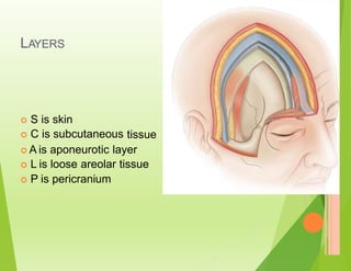
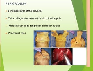
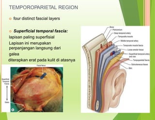
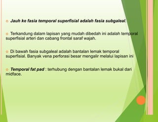
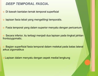
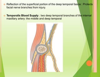
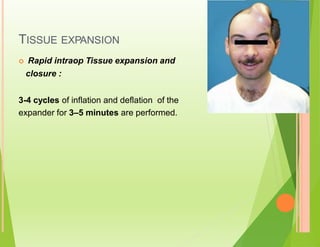
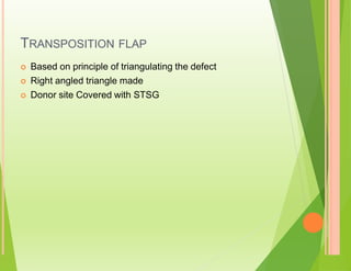
The document discusses scalp anatomy and reconstruction of scalp defects using flap procedures. It describes the layers of the scalp, blood supply, nerve supply and causes of scalp defects. Various reconstruction options are summarized including primary closure, skin grafting, local flaps such as rotation and transposition flaps, regional flaps such as trapezius flaps, and free tissue transfer including latissimus dorsi flaps. Factors to consider for reconstruction include the location and size of the defect, surrounding tissues, and preservation of the native hairline.

![GALEAL FLAP
Based on STA
galeal flap is commonly based on a named scalp vessel or combination of
vessels.
Flap length can often cross the midline
Can be elevated with frontalis muscle of the forehead to reconstruct the
anterior cranial base.
Can be taken with bone [vascularized cranial bone for reconstruction about
the orbit and facial skeleton ]
Subgaleal areolar tissue can be raised with the underlying periosteum as a
turnover flap to provide vascularized coverage for denuded calvaria](https://image.slidesharecdn.com/pptscalpdefectbedahsarafbima-230315124725-9a4f0920/85/PPT-SCALP-DEFECT-BEDAH-SARAF-BIMA-pptx-33-320.jpg)

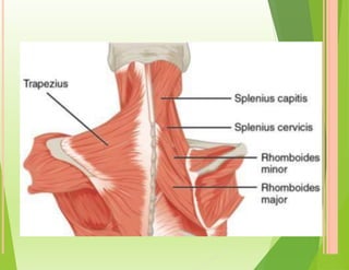
![REGIONAL FLAP
Trapezius flap : type 2
For occipital defects
blood supply :- transverse cervical
dorsal scapular
occipital arteries
Pattern :
Transverse flap : upper fibres [A/w shoulder drop]
Vertical flap : middle and lower fibres
8-10 cm donor defect can be closed primarily](https://image.slidesharecdn.com/pptscalpdefectbedahsarafbima-230315124725-9a4f0920/85/PPT-SCALP-DEFECT-BEDAH-SARAF-BIMA-pptx-37-320.jpg)
![LD FLAP [PEDICLED /FREE]
By passage of the muscle through the axilla,
defects in the orbit and temporal bone can be
repaired.](https://image.slidesharecdn.com/pptscalpdefectbedahsarafbima-230315124725-9a4f0920/85/PPT-SCALP-DEFECT-BEDAH-SARAF-BIMA-pptx-39-320.jpg)
![FREE TISSUE TRANSFER
LD [Flap of choice for large
RFF
ALT
Omental Flap with STSG
defects]
Free temporo-occipital scalp flap for free hair
baring tissue transfer](https://image.slidesharecdn.com/pptscalpdefectbedahsarafbima-230315124725-9a4f0920/85/PPT-SCALP-DEFECT-BEDAH-SARAF-BIMA-pptx-41-320.jpg)
![CHECKLIST
Named vessel included?
Native hairline preserved ?
I.
II.
III. Mode of
IV. Inherent
occipital
injury?
inelasticity of galea and mobile parietal and
region [neck]
V. Donor site : less sensitive cosmetically?](https://image.slidesharecdn.com/pptscalpdefectbedahsarafbima-230315124725-9a4f0920/85/PPT-SCALP-DEFECT-BEDAH-SARAF-BIMA-pptx-42-320.jpg)