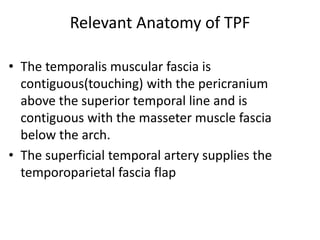
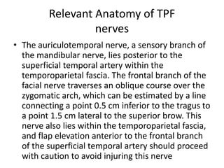
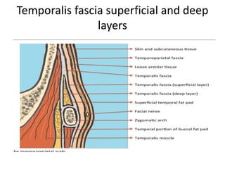
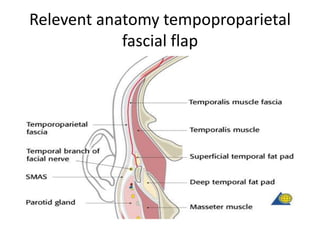
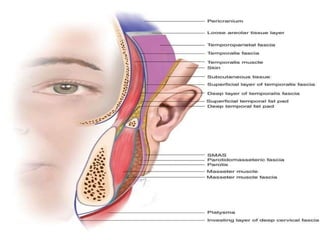
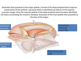
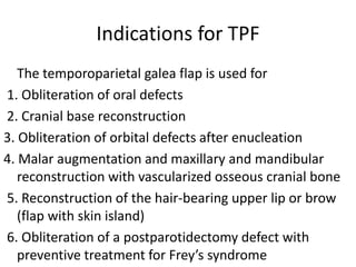
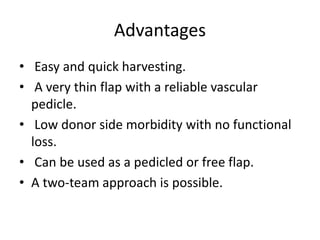
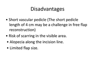
Temporoparietal Fascial Flap
Dr Jameel Kifayatullah
Senior lecturer Khyber College of
dentistry Peshawar ,Pakistan
Relevant Anatomy of TPF
• The first layer consists of the skin and subcutaneous
tissue.
• Immediately deep and firmly bound to this layer is
the temporoparietal (sometimes called superficial
temporal) fascia
• The temporoparietal fascia (TPF) is an extension of
the subcutaneous musculoaponeurotic system
(SMAS) inferiorly and the galea aponeurotica
superiorly.
Relevant Anatomy of TPF
• Beneath the temporoparietal fascia lies a loose
areolar and avascular tissue layer that separates the
fascia from the temporalis muscular fascia
(sometimes termed the deep temporal fascia).
• division of the temporalis muscular fascia as it
splits into a superficial and deep layer (of the deep
temporal fascia) surrounding a fatty tissue pad at
the temporal line of fusion, approximately 2 cm
above the zygomatic arch
Relevant Anatomy of TPF
• The temporalis muscular fascia is
contiguous(touching) with the pericranium
above the superior temporal line and is
contiguous with the masseter muscle fascia
below the arch.
• The superficial temporal artery supplies the
temporoparietal fascia flap
Relevant Anatomy of TPF
nerves
• The auriculotemporal nerve, a sensory branch of
the mandibular nerve, lies posterior to the
superficial temporal artery within the
temporoparietal fascia. The frontal branch of the
facial nerve traverses an oblique course over the
zygomatic arch, which can be estimated by a line
connecting a point 0.5 cm inferior to the tragus to
a point 1.5 cm lateral to the superior brow. This
nerve also lies within the temporoparietal fascia,
and flap elevation anterior to the frontal branch
of the superficial temporal artery should proceed
with caution to avoid injuring this nerve
Temporalis fascia superficial and deep
layers
Relevent anatomy tempoproparietal
fascial flap
SMAS
• The Superficial Muscular Aponeurotic System
(SMAS) is a fibrous network that invests the
facial muscles and connects them with the
dermis. It is continuous with the platysma
inferiorly; superiorly it attaches to the
zygomatic arch. In the lower face, the facial
nerve courses deep to the SMAS and the
platysma.
Fascia
• A fascia (/ˈfæʃ(i)ə/; plural fasciae /ˈfæʃii/;
adjective fascial; from Latin: "band") is a band or
sheet of connective tissue, primarily collagen,
beneath the skin that attaches, stabilizes, encloses,
and separates muscles and other
internal organs. Fascia is classified by layer,
as superficial fascia, deep fascia,
and visceral or parietal fascia, or by its function
and anatomical location.
• fascia is made up of fibrous connective
tissue containing closely packed bundles of
collagen fibers oriented in a wavy pattern parallel
to the direction of pull.
Landmarks
• The important topography of the
temporoparietal flap to mark on the scalp
includes the
• superior temporal line,
• the course of the superficial temporal artery
trunk and the frontal and parietal branches,
• approximate course of the frontal branch of
the facial nerve
TOPOGRAPHY
The proposed scalp incision has been marked. Note dotted line over
lateral brow marking the course of the temporal branch of the facial
nerve
A
incision
• The TPF is initially exposed at the level of the
ear through a pre-auricular facelift incision
which is carried into the hairline. This may be
extended through the scalp in a T or Y pattern;
a zigzag pattern has also been described
Identification of superficial temporal
vessels
• The TPF lies immediately deep to the hair
follicles in the pre-auricular area. The
superficial temporal vessels are identified and
the surgeon verifies that the caliber of the
vessels is adequate for flap elevation.
Identification of the Superficial
temporal artery and vein
C
Epinephrine infiltration
A 1:200,000 epinephrine solution is now
infiltrated into the area of the scalp to control
bleeding from the scalp flaps
Elevation of the scalp flap
• Elevation of the scalp flap begins inferiorly where
the plane between the dermis and superficial
temporal fascia is more easily identified
• As dissection proceeds cephalad(cranially),
separation becomes increasingly difficult
secondary to the fibrous connections and the
perforating vessels from the superficial temporal
artery to the overlying scalp. Meticulous
hemostasis is required. Countertraction of the
scalp by an assistant significantly facilitates
dissection
Flap size estimation
• After the scalp flaps have been elevated, a
template may be made of the recipient site
defect, and the template may be applied to
the TPFF
Extent of dissection
• Anterior dissection is not carried below the
level of the anterior branch of the superficial
temporal artery. Care is also taken to stay far
enough posterior to avoid injuring the
branches of the temporal nerve.
Once the overlying skin has been freed from the tpF, the tpF is incised at its superior
margin and elevated along with its pedicle in retrograde fashion. the loose areolar
plane deep to the tpFF is raised along with the flap rather expediently with
visualization of the deep temporal fascia below.
D
Raising of TPFF
Flap elevation
• The TPF is then incised and elevated along
with its pedicle in retrograde fashion. The
loose areolar plane deep to the TPFF is raised
along with the flap rather expediently with
visualization of the deep temporal fascia
below
Dissection
• Dissection then proceeds to the major pedicle.
Dissection is usually stopped at the base of
the tragus. Further dissection into the parotid
gland is not recommended as injury to the
facial nerve may occur. Flap splitting may be
carried out between the major vascular
branches of the STA, usually between the
central and posterior branch or anterior and
posterior branch if two smaller areas require
coverage
Dissection then proceeds to the major pedicle. harvest of the deep temporal fascia requires
preservation of the perforat- ing vessel which is identified just inferior to the superior
auricular margin. Once the vascular pedicle of the deep temporal fascia has been identified,
the fascia surrounding the muscle is elevated. Dissection of the main pedicle then proceeds to
the base of the tragus.
Superficial
temporal
fascia flap
Flap can be split
here
ep temporal
fascia
oral branch
acial nerve
Indications for TPF
The temporoparietal galea flap is used for
1. Obliteration of oral defects
2. Cranial base reconstruction
3. Obliteration of orbital defects after enucleation
4. Malar augmentation and maxillary and mandibular
reconstruction with vascularized osseous cranial bone
5. Reconstruction of the hair-bearing upper lip or brow
(flap with skin island)
6. Obliteration of a postparotidectomy defect with
preventive treatment for Frey’s syndrome
Indications
As a pedicled flap: – Ear (microtia, anotia) and
scalp reconstruction.
• Orbital, maxillary, palatal, and cheek coverage.
• Facial reanimation and lip reconstruction.
• Skull base defects
Advantages
• Easy and quick harvesting.
• A very thin flap with a reliable vascular
pedicle.
• Low donor side morbidity with no functional
loss.
• Can be used as a pedicled or free flap.
• A two-team approach is possible.
Disadvantages
• Short vascular pedicle (The short pedicle
length of 4 cm may be a challenge in free flap
reconstruction)
• Risk of scarring in the visible area.
• Alopecia along the incision line.
• Limited flap size.

temporoparietal flaps

  • 1.
    Temporoparietal Fascial Flap DrJameel Kifayatullah Senior lecturer Khyber College of dentistry Peshawar ,Pakistan
  • 3.
    Relevant Anatomy ofTPF • The first layer consists of the skin and subcutaneous tissue. • Immediately deep and firmly bound to this layer is the temporoparietal (sometimes called superficial temporal) fascia • The temporoparietal fascia (TPF) is an extension of the subcutaneous musculoaponeurotic system (SMAS) inferiorly and the galea aponeurotica superiorly.
  • 4.
    Relevant Anatomy ofTPF • Beneath the temporoparietal fascia lies a loose areolar and avascular tissue layer that separates the fascia from the temporalis muscular fascia (sometimes termed the deep temporal fascia). • division of the temporalis muscular fascia as it splits into a superficial and deep layer (of the deep temporal fascia) surrounding a fatty tissue pad at the temporal line of fusion, approximately 2 cm above the zygomatic arch
  • 5.
    Relevant Anatomy ofTPF • The temporalis muscular fascia is contiguous(touching) with the pericranium above the superior temporal line and is contiguous with the masseter muscle fascia below the arch. • The superficial temporal artery supplies the temporoparietal fascia flap
  • 6.
    Relevant Anatomy ofTPF nerves • The auriculotemporal nerve, a sensory branch of the mandibular nerve, lies posterior to the superficial temporal artery within the temporoparietal fascia. The frontal branch of the facial nerve traverses an oblique course over the zygomatic arch, which can be estimated by a line connecting a point 0.5 cm inferior to the tragus to a point 1.5 cm lateral to the superior brow. This nerve also lies within the temporoparietal fascia, and flap elevation anterior to the frontal branch of the superficial temporal artery should proceed with caution to avoid injuring this nerve
  • 7.
  • 8.
  • 10.
    SMAS • The SuperficialMuscular Aponeurotic System (SMAS) is a fibrous network that invests the facial muscles and connects them with the dermis. It is continuous with the platysma inferiorly; superiorly it attaches to the zygomatic arch. In the lower face, the facial nerve courses deep to the SMAS and the platysma.
  • 11.
    Fascia • A fascia(/ˈfæʃ(i)ə/; plural fasciae /ˈfæʃii/; adjective fascial; from Latin: "band") is a band or sheet of connective tissue, primarily collagen, beneath the skin that attaches, stabilizes, encloses, and separates muscles and other internal organs. Fascia is classified by layer, as superficial fascia, deep fascia, and visceral or parietal fascia, or by its function and anatomical location. • fascia is made up of fibrous connective tissue containing closely packed bundles of collagen fibers oriented in a wavy pattern parallel to the direction of pull.
  • 12.
    Landmarks • The importanttopography of the temporoparietal flap to mark on the scalp includes the • superior temporal line, • the course of the superficial temporal artery trunk and the frontal and parietal branches, • approximate course of the frontal branch of the facial nerve
  • 13.
  • 14.
    The proposed scalpincision has been marked. Note dotted line over lateral brow marking the course of the temporal branch of the facial nerve A
  • 15.
    incision • The TPFis initially exposed at the level of the ear through a pre-auricular facelift incision which is carried into the hairline. This may be extended through the scalp in a T or Y pattern; a zigzag pattern has also been described
  • 16.
    Identification of superficialtemporal vessels • The TPF lies immediately deep to the hair follicles in the pre-auricular area. The superficial temporal vessels are identified and the surgeon verifies that the caliber of the vessels is adequate for flap elevation.
  • 17.
    Identification of theSuperficial temporal artery and vein C
  • 18.
    Epinephrine infiltration A 1:200,000epinephrine solution is now infiltrated into the area of the scalp to control bleeding from the scalp flaps
  • 19.
    Elevation of thescalp flap • Elevation of the scalp flap begins inferiorly where the plane between the dermis and superficial temporal fascia is more easily identified • As dissection proceeds cephalad(cranially), separation becomes increasingly difficult secondary to the fibrous connections and the perforating vessels from the superficial temporal artery to the overlying scalp. Meticulous hemostasis is required. Countertraction of the scalp by an assistant significantly facilitates dissection
  • 20.
    Flap size estimation •After the scalp flaps have been elevated, a template may be made of the recipient site defect, and the template may be applied to the TPFF
  • 21.
    Extent of dissection •Anterior dissection is not carried below the level of the anterior branch of the superficial temporal artery. Care is also taken to stay far enough posterior to avoid injuring the branches of the temporal nerve.
  • 22.
    Once the overlyingskin has been freed from the tpF, the tpF is incised at its superior margin and elevated along with its pedicle in retrograde fashion. the loose areolar plane deep to the tpFF is raised along with the flap rather expediently with visualization of the deep temporal fascia below. D
  • 23.
  • 24.
    Flap elevation • TheTPF is then incised and elevated along with its pedicle in retrograde fashion. The loose areolar plane deep to the TPFF is raised along with the flap rather expediently with visualization of the deep temporal fascia below
  • 25.
    Dissection • Dissection thenproceeds to the major pedicle. Dissection is usually stopped at the base of the tragus. Further dissection into the parotid gland is not recommended as injury to the facial nerve may occur. Flap splitting may be carried out between the major vascular branches of the STA, usually between the central and posterior branch or anterior and posterior branch if two smaller areas require coverage
  • 26.
    Dissection then proceedsto the major pedicle. harvest of the deep temporal fascia requires preservation of the perforat- ing vessel which is identified just inferior to the superior auricular margin. Once the vascular pedicle of the deep temporal fascia has been identified, the fascia surrounding the muscle is elevated. Dissection of the main pedicle then proceeds to the base of the tragus. Superficial temporal fascia flap Flap can be split here ep temporal fascia oral branch acial nerve
  • 27.
    Indications for TPF Thetemporoparietal galea flap is used for 1. Obliteration of oral defects 2. Cranial base reconstruction 3. Obliteration of orbital defects after enucleation 4. Malar augmentation and maxillary and mandibular reconstruction with vascularized osseous cranial bone 5. Reconstruction of the hair-bearing upper lip or brow (flap with skin island) 6. Obliteration of a postparotidectomy defect with preventive treatment for Frey’s syndrome
  • 28.
    Indications As a pedicledflap: – Ear (microtia, anotia) and scalp reconstruction. • Orbital, maxillary, palatal, and cheek coverage. • Facial reanimation and lip reconstruction. • Skull base defects
  • 29.
    Advantages • Easy andquick harvesting. • A very thin flap with a reliable vascular pedicle. • Low donor side morbidity with no functional loss. • Can be used as a pedicled or free flap. • A two-team approach is possible.
  • 30.
    Disadvantages • Short vascularpedicle (The short pedicle length of 4 cm may be a challenge in free flap reconstruction) • Risk of scarring in the visible area. • Alopecia along the incision line. • Limited flap size.