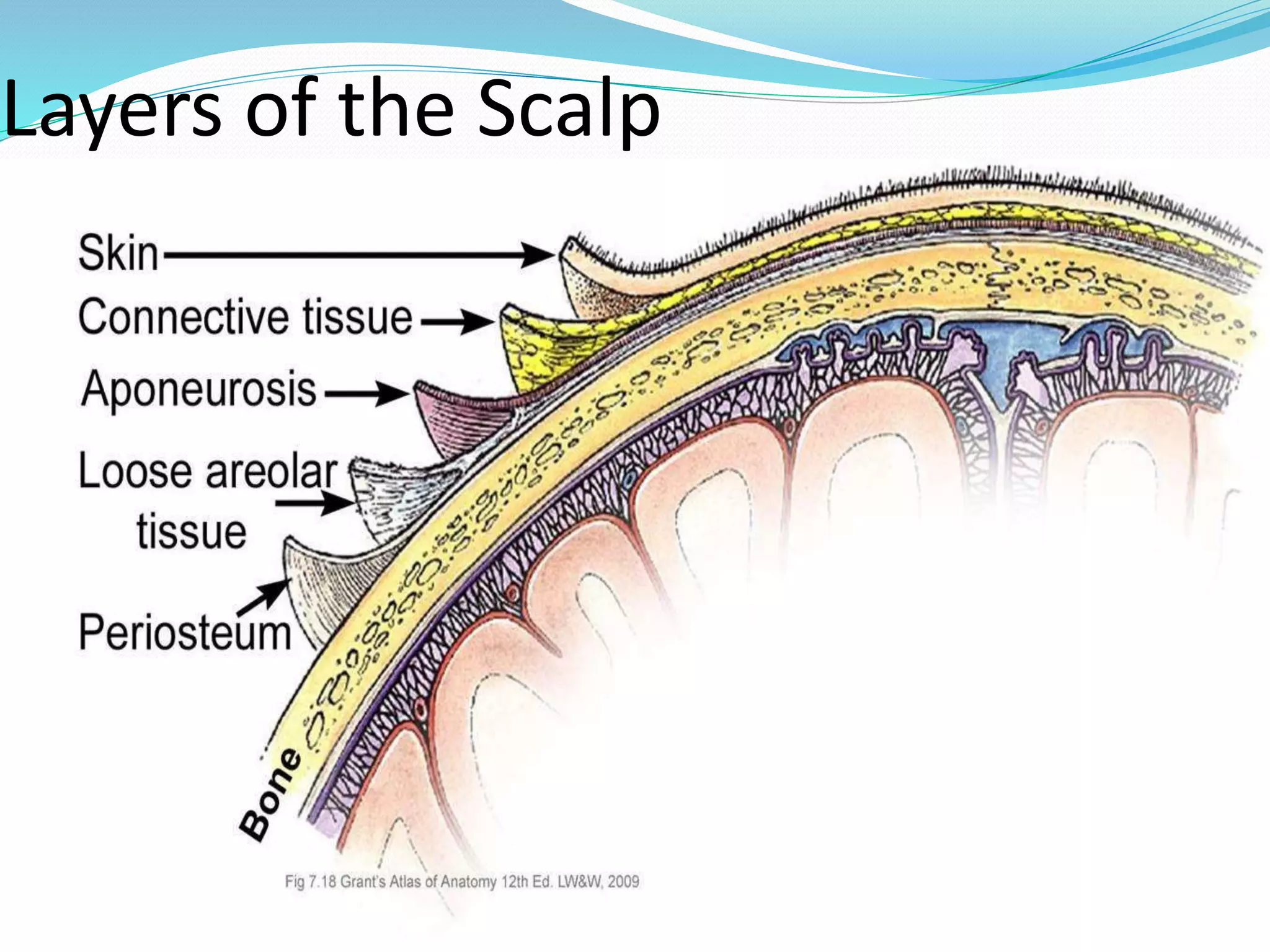
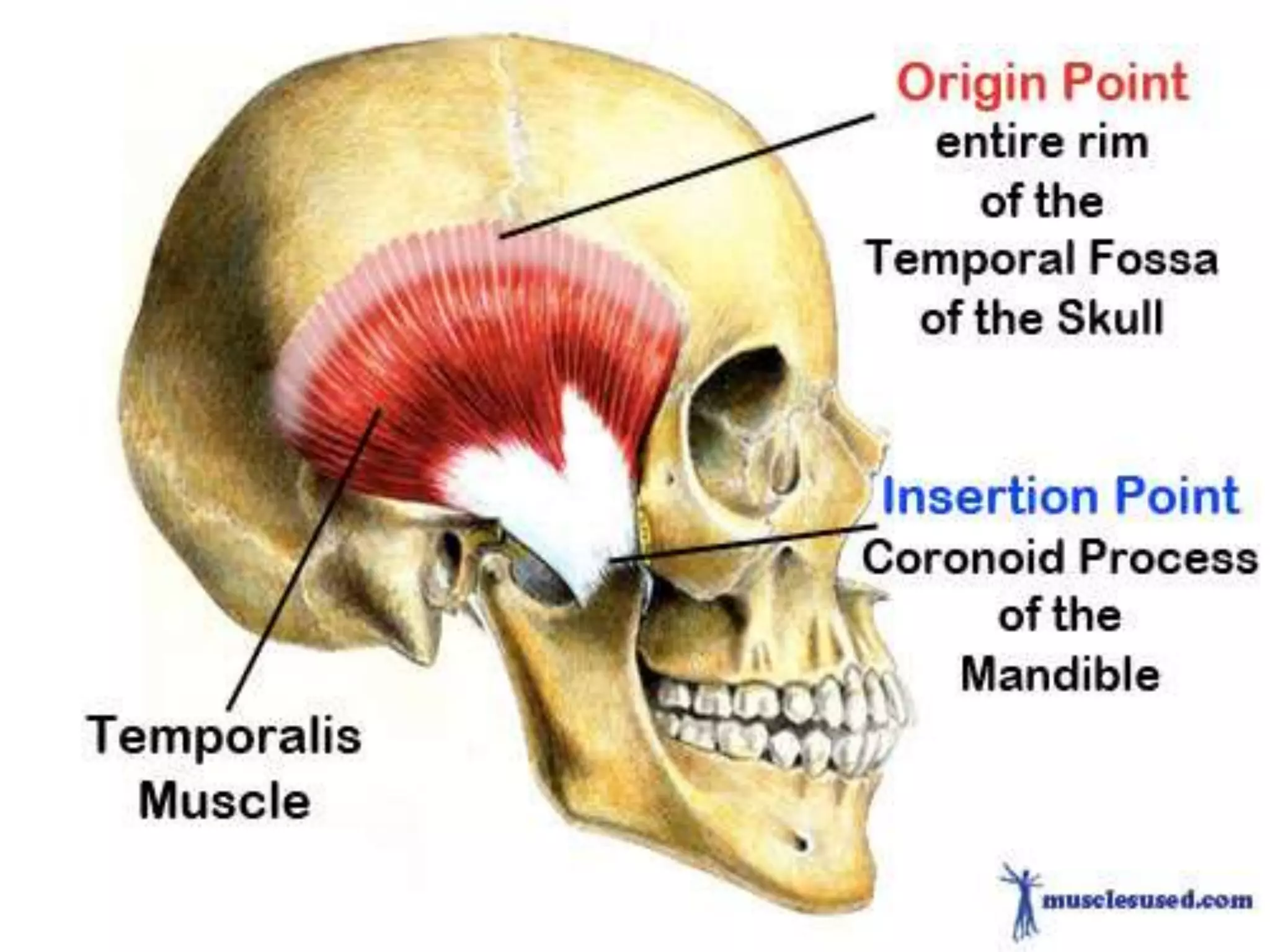
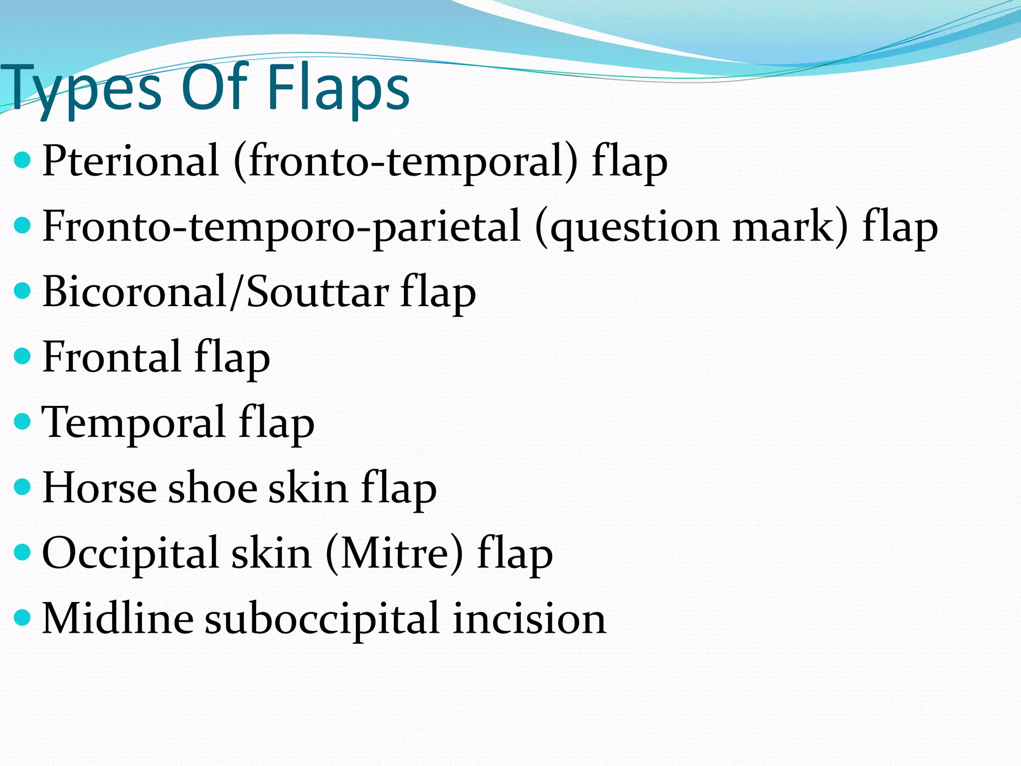
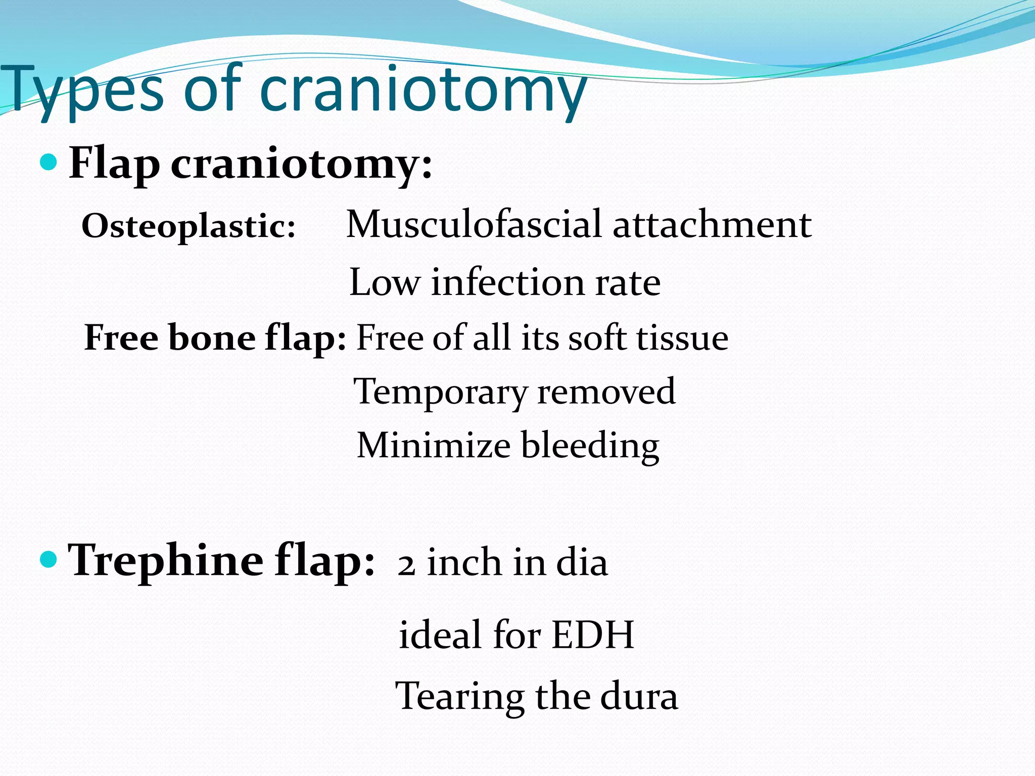
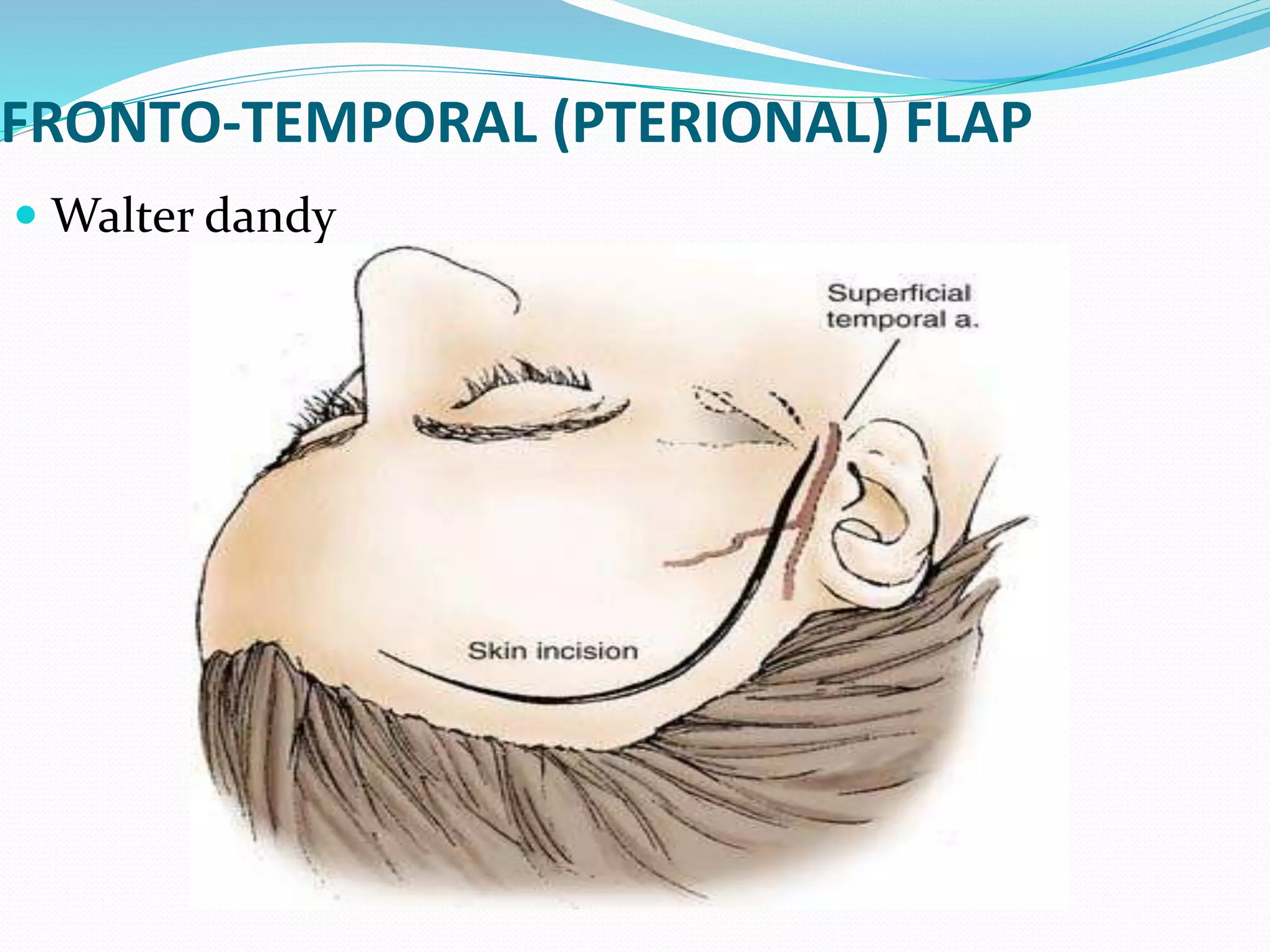
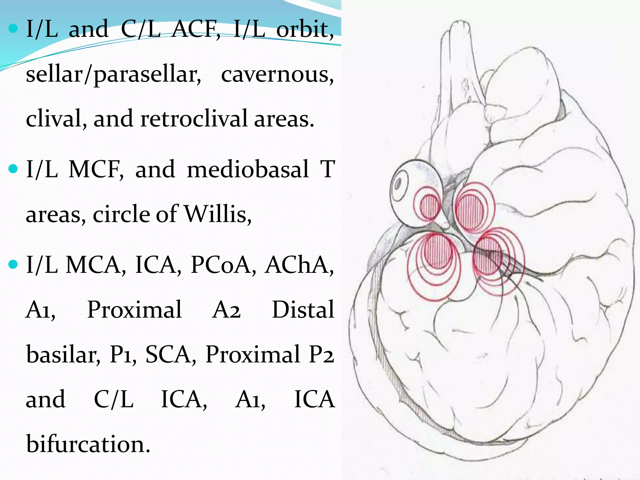
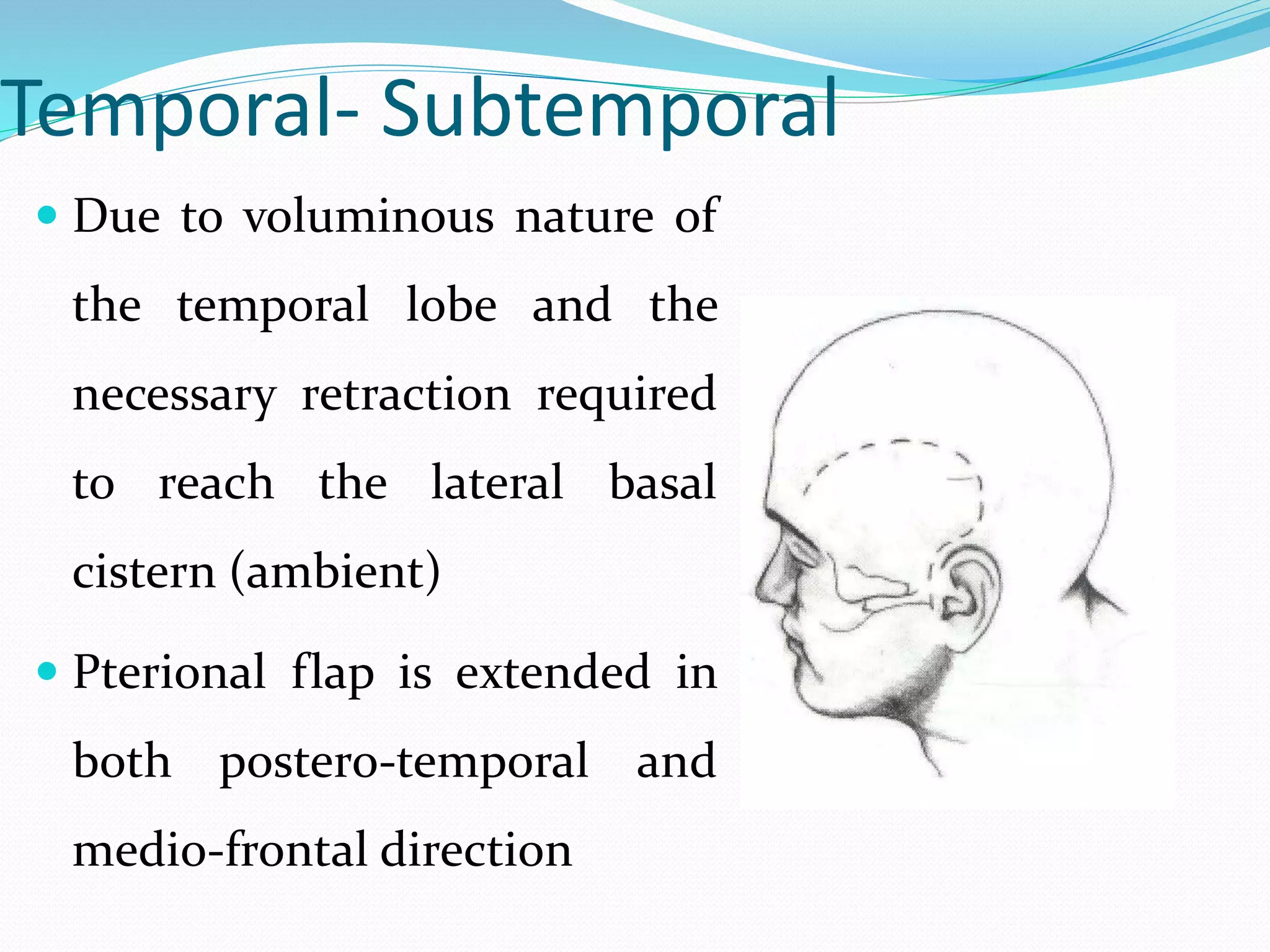
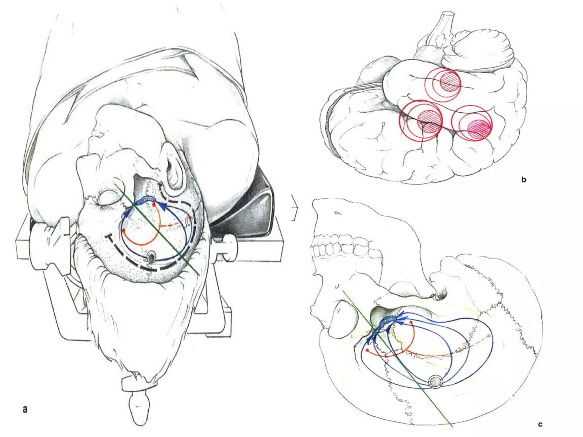
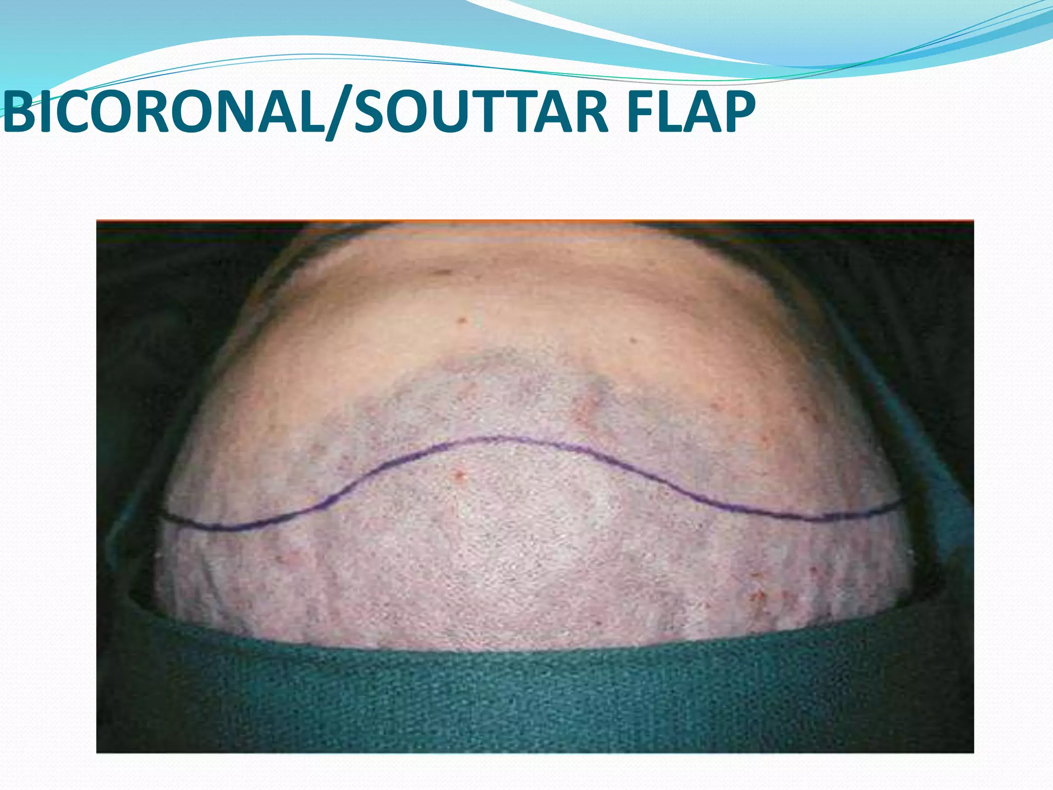
SlideShare a Scribd company logo
1 of 65
DR. AJAY KUMAR SINGH
DNB NEUROSURGERY
VPIMS
Layers of the Scalp
 The 1st three, regarded as a single layer, contains fibro-
adipose tissue, nerve and vessels.
 The epicranius divided into Occipitofrontalis and
Temporoparietalis part.
 Laterally the superfacial facia continue into the temporal
facia and posteriorly as superfacial facia of neck.
The temporoparietalis lies
below the frontal part of
the Occipitofronatlis and
anterior and posterior
auricle muscle.
Temporalis muscle:
 T’ fossa and from deep T’ fascia
 Pass below the zygomatic arch and attached to coronoid
process and the anterior wall of mandibular ramus.
 T’ fascia contains T’ muscle, it is attached above along
the Supr T’ line and below it has two layers- one is
attached to lateral margin of upper border and the other
to the medial margin of zygomatic arch
 Deep T br of ant trunk of Mandibualr N.
 The Frontal br of the Facial n is contained in the
superficial layer of T’ fascia (as it crossed the
zygomatic arch into the temporal region, 1.5 -3 cm
above the arch and 1-1.5 cm posterior to lateral orbital
rim, where it crossed from the facia to run within the
superficial temporal fascia.
 Recommended that the superior layer is cut from its
attacement on the inferior Temporal line till near to
its attachment on the zygoma and it is separated
from the inferior layer and reflected along the flap.
Vascular Supply
 Branches of external carotid:
Occipital.
Posterior auricular.
Superficial temporal.
 Branches of internal carotid:
Supratrochlear.
Supraorbital.
Venous supply
 Freely anastomse with one another
 Connected to the diploic veins of the skull bones and the
intracranial dural sinuses through several emissary veins.
 Supratrochlear and supraorbital: facial vein
 Superficial temporal vein: in front of auricle: IJV
 Posterior auricular vein: EJV
 Occipital vein : suboccipital plexus, EJV
Landmarks
 Nasion
 Bregma : 13 cm from Nasion
 Inion: EOP
 Lambda: 6.5 cm. above the inion.
 Pterion: Middle meningeal artery, 3.5 cm behind, and
1.2 cm above, the level of the frontozygomatic suture.
 Asterion: Transverse sigmoid junction, 4 cm behind
and 1.2 cm above the level of the auricular point.
PRINCIPLES OF SCALP FLAPS
 Position of lesion in imaging in axial coronal and
sagittal plane.
 Relationship with the external landmark like, Ext ear
canal, margin of the ear, glabella, EOP.
 Deep refrence point, anatomical midline, tentorium,
formen of monro, the lateral ventricle.
 Position of important structures
 Plan for enlarging incision, obtain adequate closure
 Adequate exposure of the lesion
 Based on at least two named arteries.
 Length not more than 1.5 times base of flap
 Burr hole depend on size and location of craniotomy,
nature of lesion, patient age, and degree of adherence
of dura to the inner table.
 Incisions within the hairline
 No cross incisions
TAYLOR HAUGHTON LINES
 Taylor Haughton line
In 1900 Taylor and Haughton described a technique to
define a line on the scalp directly above the central
fissure.
 1. Draw a Nasion-Inion line ( Nasion - Just below
Glabella and Inion -External Occipital protruberance)
 2. Divide the Nasion-Inion line in to 25%, 50% and 75%
 3. Bregma is the point between the 25% and 50% points
and Lambda is at 75% point
 4. Sylvian fissure is drawn from the orbito-temporal
angle (A point of depressin where eyebrow ends) to the
75% point on naso-inion line.
 5. Draw a line perpendicular to the root of the zygoma
starting at preauricular point
 6. Central sulcus is drawn from 50% point on naso-inion
line to the point where the sylvian line cuts the
perpendicular line
The Incisions
Supratentorial
 Anterior incision
 Frontal
 Bicoronal/Souttar flap
 Unilateral frontal flap
 Fronto-temporal
(Pterional)
 Temporal
 ? Mark incision
 Horse shoe shaped
 Cushing’s linear
temporal
 Parietal
 Occipital
 The Mitre
 Horse shoe shaped
occipital
Infratentorial
 Retromastoid
 Paramedian
 Median
 Far lateral
 Combined
 Petrosal
Types Of Flaps
 Pterional (fronto-temporal) flap
 Fronto-temporo-parietal (question mark) flap
 Bicoronal/Souttar flap
 Frontal flap
 Temporal flap
 Horse shoe skin flap
 Occipital skin (Mitre) flap
 Midline suboccipital incision
Types of craniotomy
 Flap craniotomy:
Osteoplastic: Musculofascial attachment
Low infection rate
Free bone flap: Free of all its soft tissue
Temporary removed
Minimize bleeding
 Trephine flap: 2 inch in dia
ideal for EDH
Tearing the dura
FRONTO-TEMPORAL (PTERIONAL) FLAP
 Walter dandy
 I/L and C/L ACF, I/L orbit,
sellar/parasellar, cavernous,
clival, and retroclival areas.
 I/L MCF, and mediobasal T
areas, circle of Willis,
 I/L MCA, ICA, PCoA, AChA,
A1, Proximal A2 Distal
basilar, P1, SCA, Proximal P2
and C/L ICA, A1, ICA
bifurcation.
The Pterional craniotomy
 Position: 20-30 degree in opp direction, inclined about
20 degree towards the floor, and slightly towards the
C/L shoulder.
 Highest point: Malar eminence
 Incison : 1 cm anterior to the tragus or 1 cm superior to
the anterior aspect of the auricle and extend to the
temporal crest becoming perpendicular to the zygoma
and then it curves sharply ending at the hairline 2 cm
away from the midline. (Supr temporal art)
Upon retraction of the skin
flap, pericranial lines of
incision are made through
the fascia and periosteum
underneath the
frontozygomatic process, 0.5
cm superiorly to the
temporal line, and diagonally
along the frontal bone.
 The loose areolar tissue is
dissected with the periosteum
and a thin layer from the
temporalis muscle and reflected
back over the aponeurosis.
 The fat layer between the two
facial layer should be dissected
separately to avoid injury to the
frontal branch of the facial
nerve.
 The pericranium of the
temporalis muscle is
separated from the
inferior surface of the
frontozygomatic
process and from the
temporal fossa and
reflected.
 A single burr hole is placed in the parietal bone,
approx 2-3 cm posterior to the pterion below the
temporal line in the shallow depression that runs
obliquely over the lateral pterional wing.
 This shallow depression approximate the course of
MMA.
 No pressure over the I/L eye.(Gauze).
 Flap raise upto supraorbital ridge.
 Key burr hole (6.8 mm superior and 4.5 mm posterior to
the frontozygomatic suture)
 2nd burr hole 2 cm medially and 4-5 cm above key hole.
 3rd burr hole, 5-7 cm lateral to the 2nd.
 4th if necessary, in the temporal squama, just above the
root of zygoma.
 In connecting key hole with temporal hole, resistance
will be met where greater wing of sphenoid meet the
temporal squama.
 MMA may bleed after elevation of bone flap.
Temporal- Subtemporal
 Due to voluminous nature of
the temporal lobe and the
necessary retraction required
to reach the lateral basal
cistern (ambient)
 Pterional flap is extended in
both postero-temporal and
medio-frontal direction
 Middle cranial fossa
 Lesion in Meckel’s Cave, trigeminal neuromas
 Inferior temporal and lateral temporo-occipital
gyrus
 Tentorial edge
 Petroclival and petrous tumors
 Basilar terminus aneurysm
 Posterior to the
mastoid process and is
carried upward the
superior temporal line
before decending to
inscribe a sqaure flap
based on ear and
terminates at mid
zygoma
BICORONAL/SOUTTAR FLAP
 Indication : Frontal lobe, frontobasal lesion
anterior to chiasma, genu of CC, circle of willis,
and sella.
 B/L side Fronto-temporal lesions and cranial base.
 Position: Straight, 10-20 degree towards floor
 Incision: Superior to zygomatic arch, 1 cm anterior to tragus-
extends over the bregma to the corresponding site on the opposite
side. Reflect up to orbit rim.
 Artery: Supraorbital/trochlear vessels.
 Burr hole:
1. Fronto-temporal line and the zygomatic process of the frontal
bone
2. Glabella, either removed as single piece or two separate frontal
flaps
3. Two burr hole on either side of SSS.
 4. 5-7 cm lateral to the SSS burr hole.
Drawback:
 Late visualisation of optic
nerves, ICA
 Risk of CSF leak
 Frontal lobe injury
 Risk of injury to SSS.
QUESTION MARK SKIN FLAP (TRAUMA
FLAP) or Falconer’s incision
Indication: Cranial trauma, malignant MCA infarct
Incision:
 Start at the zygomatic arch< 1 cm ant to the tragus
 Proceed superiorly and then curve posteriorly at the level
of top of the pinna
 4-6 cm behind the pinna it is taken superiorly
 1-2 cm I/L to the midline curve anteriorly to end behind
the hairline.
 Arterial supply: superficial temporal and supra orbital
vessels.
MITRE SKIN FLAP
Mitre skin flap
 Mitre hats worn by bishops
 Medial limb begins from the inion to the vertex and
then curves to come over the ear.
 Occipital lobe, posterior falx and superior tentorial
surface.
 Blood supply from the occipital artery
Supratentorial Paramedian
Indication:
 Tumor on the surface
 Within a sulcus or fissure
 Deep within the white matter
 Intraventricular tumor
Frontal paramedian
Paramedian centroparietal
Paramedian parieto-occipital
Median (Parasagittal) interhemispheric
 The medial part of the sup F’, paracentral, and anterior two third
of the cingulate gyrus and corpus callosum.
 Lateral and third ventricle.
 Sellar and suprasellar masses.
Frontal (anterior and posterior)
Central
Pareito-occipital
Frontal anterior :
Indicatoin: parasagittal and falcine lesion, medial
frontal and cigulate gyrus, anterior callosal and lateral
and third ventricle and paraventricular lesion.
 Incision: Bicoronal incision, curvilenear.
 Burr hole: Single burr hole 1-2 mm from midline on
the C/L side of SSS to the lesion.
Anterior and posterior frontal median
craniotomies for parasaggital and
interhemispheric exploration
Frontal posterior:
 Indication: Posterior part of medial frontal gyrus and
paracentral lesion, lateral ventricle and paraventricular
areas.
 Incison: Curvilinear.
 Burr hole: same as Anterior frontal but posterior to
coronal suture.
Pareito-occcipital :
Indication:
 Medial surface of parietal and occipital lobes,
posterior part of cingulate gyrus, superior partof
parahippocampal gyrus, posterior part of CC and the
lateral ventricle.
 Pineal and parapenial areas.
 Burr hole : medially, a few mm across the midline on
the C/L side of SSS
Infratentorial
 Suboccipital
 Lateral suboccipital retrosigmoid
 Paramedian
 Median
THANK YOU

More Related Content

What's hot

Foramen Magnum Meningioma
Foramen Magnum MeningiomaForamen Magnum Meningioma
Foramen Magnum MeningiomaFarrukh Javeed
 
“BASE OF SKULL: ANATOMY and APPLIED ASPECT”
“BASE OF SKULL: ANATOMY and APPLIED ASPECT”“BASE OF SKULL: ANATOMY and APPLIED ASPECT”
“BASE OF SKULL: ANATOMY and APPLIED ASPECT”RAKESHTALUKDAR2
 
Cavernous sinus-ANATOMY AND SURGICAL APPROACHES
Cavernous sinus-ANATOMY AND SURGICAL APPROACHESCavernous sinus-ANATOMY AND SURGICAL APPROACHES
Cavernous sinus-ANATOMY AND SURGICAL APPROACHESSanjeev Sreenivasan
 
Frontotemporal FTOZ craniotomy
Frontotemporal FTOZ craniotomyFrontotemporal FTOZ craniotomy
Frontotemporal FTOZ craniotomyDr. Shahnawaz Alam
 
Skull base anatomy by Dr. Aditya Tiwari
Skull base anatomy by Dr. Aditya TiwariSkull base anatomy by Dr. Aditya Tiwari
Skull base anatomy by Dr. Aditya TiwariAditya Tiwari
 
Craniometrics and ventricular access
Craniometrics and ventricular accessCraniometrics and ventricular access
Craniometrics and ventricular accessDr. Shahnawaz Alam
 
Brain stem surgical anatomy and approaches
Brain stem surgical anatomy and approachesBrain stem surgical anatomy and approaches
Brain stem surgical anatomy and approachesKode Sashanka
 
NEUROSURGICAL TENETS OF PITUITARY GLAND
NEUROSURGICAL TENETS OF PITUITARY GLANDNEUROSURGICAL TENETS OF PITUITARY GLAND
NEUROSURGICAL TENETS OF PITUITARY GLANDsuresh Bishokarma
 
APPROACH TO PETROCLIVAL MENINGIOMA
APPROACH TO PETROCLIVAL MENINGIOMAAPPROACH TO PETROCLIVAL MENINGIOMA
APPROACH TO PETROCLIVAL MENINGIOMADr. Shahnawaz Alam
 
Sch.40 surgical management of petroclival meningioma
Sch.40 surgical management of petroclival meningioma Sch.40 surgical management of petroclival meningioma
Sch.40 surgical management of petroclival meningioma Neurosurgery Vajira
 
4 th ventricle- Anatomical and surgical perspective
4 th ventricle- Anatomical and surgical perspective4 th ventricle- Anatomical and surgical perspective
4 th ventricle- Anatomical and surgical perspectivesuresh Bishokarma
 
016 Transsphenoidal approch microscopic
016 Transsphenoidal approch microscopic016 Transsphenoidal approch microscopic
016 Transsphenoidal approch microscopicNeurosurgery Vajira
 
Anatomy of the anterior cerebral artery (ACA)
Anatomy of the anterior cerebral artery (ACA)Anatomy of the anterior cerebral artery (ACA)
Anatomy of the anterior cerebral artery (ACA)Mohamed M.A. Zaitoun
 
148 Skull tumour & GB 21.4 skull tumors
148 Skull tumour & GB  21.4 skull tumors148 Skull tumour & GB  21.4 skull tumors
148 Skull tumour & GB 21.4 skull tumorsNeurosurgery Vajira
 
The cerebello pontine angle
The cerebello pontine angleThe cerebello pontine angle
The cerebello pontine angleDr Himanshu Soni
 
Microsurgical anatomy of fourth ventricle
Microsurgical anatomy of fourth ventricleMicrosurgical anatomy of fourth ventricle
Microsurgical anatomy of fourth ventricleSHAMEEJ MUHAMED KV
 

What's hot (20)

ICA anatomy
ICA anatomyICA anatomy
ICA anatomy
 
Foramen Magnum Meningioma
Foramen Magnum MeningiomaForamen Magnum Meningioma
Foramen Magnum Meningioma
 
“BASE OF SKULL: ANATOMY and APPLIED ASPECT”
“BASE OF SKULL: ANATOMY and APPLIED ASPECT”“BASE OF SKULL: ANATOMY and APPLIED ASPECT”
“BASE OF SKULL: ANATOMY and APPLIED ASPECT”
 
Cavernous sinus-ANATOMY AND SURGICAL APPROACHES
Cavernous sinus-ANATOMY AND SURGICAL APPROACHESCavernous sinus-ANATOMY AND SURGICAL APPROACHES
Cavernous sinus-ANATOMY AND SURGICAL APPROACHES
 
Frontotemporal FTOZ craniotomy
Frontotemporal FTOZ craniotomyFrontotemporal FTOZ craniotomy
Frontotemporal FTOZ craniotomy
 
Skull base anatomy by Dr. Aditya Tiwari
Skull base anatomy by Dr. Aditya TiwariSkull base anatomy by Dr. Aditya Tiwari
Skull base anatomy by Dr. Aditya Tiwari
 
Craniometrics and ventricular access
Craniometrics and ventricular accessCraniometrics and ventricular access
Craniometrics and ventricular access
 
Brain stem surgical anatomy and approaches
Brain stem surgical anatomy and approachesBrain stem surgical anatomy and approaches
Brain stem surgical anatomy and approaches
 
NEUROSURGICAL TENETS OF PITUITARY GLAND
NEUROSURGICAL TENETS OF PITUITARY GLANDNEUROSURGICAL TENETS OF PITUITARY GLAND
NEUROSURGICAL TENETS OF PITUITARY GLAND
 
APPROACH TO PETROCLIVAL MENINGIOMA
APPROACH TO PETROCLIVAL MENINGIOMAAPPROACH TO PETROCLIVAL MENINGIOMA
APPROACH TO PETROCLIVAL MENINGIOMA
 
Sch.40 surgical management of petroclival meningioma
Sch.40 surgical management of petroclival meningioma Sch.40 surgical management of petroclival meningioma
Sch.40 surgical management of petroclival meningioma
 
4 th ventricle- Anatomical and surgical perspective
4 th ventricle- Anatomical and surgical perspective4 th ventricle- Anatomical and surgical perspective
4 th ventricle- Anatomical and surgical perspective
 
Surgical approach to thalamus
Surgical approach to thalamusSurgical approach to thalamus
Surgical approach to thalamus
 
016 Transsphenoidal approch microscopic
016 Transsphenoidal approch microscopic016 Transsphenoidal approch microscopic
016 Transsphenoidal approch microscopic
 
Anatomy of the anterior cerebral artery (ACA)
Anatomy of the anterior cerebral artery (ACA)Anatomy of the anterior cerebral artery (ACA)
Anatomy of the anterior cerebral artery (ACA)
 
148 Skull tumour & GB 21.4 skull tumors
148 Skull tumour & GB  21.4 skull tumors148 Skull tumour & GB  21.4 skull tumors
148 Skull tumour & GB 21.4 skull tumors
 
Surgical anatomy of neck
Surgical anatomy of neckSurgical anatomy of neck
Surgical anatomy of neck
 
The cerebello pontine angle
The cerebello pontine angleThe cerebello pontine angle
The cerebello pontine angle
 
Craniopharyngiomas
CraniopharyngiomasCraniopharyngiomas
Craniopharyngiomas
 
Microsurgical anatomy of fourth ventricle
Microsurgical anatomy of fourth ventricleMicrosurgical anatomy of fourth ventricle
Microsurgical anatomy of fourth ventricle
 

Similar to Scalp incision and blood supply

Surgical landmarks and sectional anatomy in neurosurgery
Surgical landmarks  and sectional anatomy in neurosurgerySurgical landmarks  and sectional anatomy in neurosurgery
Surgical landmarks and sectional anatomy in neurosurgeryKIST Surgery
 
Anatomy of External Ear and Middle Ear
Anatomy of External Ear and Middle EarAnatomy of External Ear and Middle Ear
Anatomy of External Ear and Middle EarDiptiman Baliarsingh
 
Anatomy of the external and middle ear
Anatomy of the external and middle earAnatomy of the external and middle ear
Anatomy of the external and middle earSalman Syed
 
Ear discharge and otalgia
Ear discharge and otalgiaEar discharge and otalgia
Ear discharge and otalgiaDennis Lee
 
INFRATEMPORAL FOSSA AND PTERYGOPALATINE FOSSA NEW.pptx
INFRATEMPORAL FOSSA AND PTERYGOPALATINE FOSSA NEW.pptxINFRATEMPORAL FOSSA AND PTERYGOPALATINE FOSSA NEW.pptx
INFRATEMPORAL FOSSA AND PTERYGOPALATINE FOSSA NEW.pptxSudin Kayastha
 
Cavernous sinus
Cavernous sinusCavernous sinus
Cavernous sinusserovars
 
Anatomy of the Sellar Region (A.GH)
Anatomy of the Sellar Region (A.GH)Anatomy of the Sellar Region (A.GH)
Anatomy of the Sellar Region (A.GH)Ahmed Ghoul
 
Temporal bone anatomy and surgical significancepptx
Temporal bone anatomy and surgical significancepptxTemporal bone anatomy and surgical significancepptx
Temporal bone anatomy and surgical significancepptxdruttamnepal
 
Surgical anatomy of nose
Surgical anatomy of noseSurgical anatomy of nose
Surgical anatomy of noseAugustine raj
 
Nasal septum and its diseases[1]
Nasal septum and its diseases[1]Nasal septum and its diseases[1]
Nasal septum and its diseases[1]Priyanka Shastri
 
Middle Ear Anatomy.pptx
Middle Ear Anatomy.pptxMiddle Ear Anatomy.pptx
Middle Ear Anatomy.pptxgopikaraj95
 
SURGICAL ANATOMY OF SALIVARY GLANDS
SURGICAL ANATOMY OF SALIVARY GLANDSSURGICAL ANATOMY OF SALIVARY GLANDS
SURGICAL ANATOMY OF SALIVARY GLANDSDr SAHYA S LAL
 
Mucosal folds and ventilation of middle ear
Mucosal folds and ventilation of middle ear  Mucosal folds and ventilation of middle ear
Mucosal folds and ventilation of middle ear AlkaKapil
 
Blood supply of head and neck
Blood supply of head and neck Blood supply of head and neck
Blood supply of head and neck Pratik Warade
 
middle ear spaces an important topic otorhinolaryngology
middle ear spaces an important topic otorhinolaryngologymiddle ear spaces an important topic otorhinolaryngology
middle ear spaces an important topic otorhinolaryngologyDrsiyaMedfriend
 

Similar to Scalp incision and blood supply (20)

Surgical landmarks and sectional anatomy in neurosurgery
Surgical landmarks  and sectional anatomy in neurosurgerySurgical landmarks  and sectional anatomy in neurosurgery
Surgical landmarks and sectional anatomy in neurosurgery
 
Anatomy of External Ear and Middle Ear
Anatomy of External Ear and Middle EarAnatomy of External Ear and Middle Ear
Anatomy of External Ear and Middle Ear
 
Anatomy of the external and middle ear
Anatomy of the external and middle earAnatomy of the external and middle ear
Anatomy of the external and middle ear
 
Ear discharge and otalgia
Ear discharge and otalgiaEar discharge and otalgia
Ear discharge and otalgia
 
INFRATEMPORAL FOSSA AND PTERYGOPALATINE FOSSA NEW.pptx
INFRATEMPORAL FOSSA AND PTERYGOPALATINE FOSSA NEW.pptxINFRATEMPORAL FOSSA AND PTERYGOPALATINE FOSSA NEW.pptx
INFRATEMPORAL FOSSA AND PTERYGOPALATINE FOSSA NEW.pptx
 
Cavernous sinus
Cavernous sinusCavernous sinus
Cavernous sinus
 
Anatomy of the Sellar Region (A.GH)
Anatomy of the Sellar Region (A.GH)Anatomy of the Sellar Region (A.GH)
Anatomy of the Sellar Region (A.GH)
 
Anatomy of orbital cavity
Anatomy of orbital cavityAnatomy of orbital cavity
Anatomy of orbital cavity
 
Imaging in ent
Imaging in entImaging in ent
Imaging in ent
 
Anatomy of middle ear
Anatomy of middle earAnatomy of middle ear
Anatomy of middle ear
 
Temporal bone anatomy and surgical significancepptx
Temporal bone anatomy and surgical significancepptxTemporal bone anatomy and surgical significancepptx
Temporal bone anatomy and surgical significancepptx
 
Surgical anatomy of nose
Surgical anatomy of noseSurgical anatomy of nose
Surgical anatomy of nose
 
Nasal septum and its diseases[1]
Nasal septum and its diseases[1]Nasal septum and its diseases[1]
Nasal septum and its diseases[1]
 
Anatomy of human ear
Anatomy of human earAnatomy of human ear
Anatomy of human ear
 
Middle Ear Anatomy.pptx
Middle Ear Anatomy.pptxMiddle Ear Anatomy.pptx
Middle Ear Anatomy.pptx
 
SURGICAL ANATOMY OF SALIVARY GLANDS
SURGICAL ANATOMY OF SALIVARY GLANDSSURGICAL ANATOMY OF SALIVARY GLANDS
SURGICAL ANATOMY OF SALIVARY GLANDS
 
Mucosal folds and ventilation of middle ear
Mucosal folds and ventilation of middle ear  Mucosal folds and ventilation of middle ear
Mucosal folds and ventilation of middle ear
 
anatomy of ear.pptx
anatomy of ear.pptxanatomy of ear.pptx
anatomy of ear.pptx
 
Blood supply of head and neck
Blood supply of head and neck Blood supply of head and neck
Blood supply of head and neck
 
middle ear spaces an important topic otorhinolaryngology
middle ear spaces an important topic otorhinolaryngologymiddle ear spaces an important topic otorhinolaryngology
middle ear spaces an important topic otorhinolaryngology
 

More from drajay02

Techiniques of clipping in aneurysm &amp; endovascular option
Techiniques of clipping in aneurysm  &amp; endovascular optionTechiniques of clipping in aneurysm  &amp; endovascular option
Techiniques of clipping in aneurysm &amp; endovascular optiondrajay02
 
Lower cranial nerves ajay
Lower cranial nerves ajayLower cranial nerves ajay
Lower cranial nerves ajaydrajay02
 
Bracial plexus injury localization and management
Bracial plexus injury localization and managementBracial plexus injury localization and management
Bracial plexus injury localization and managementdrajay02
 
Blood brain barrier ajay
Blood brain barrier ajayBlood brain barrier ajay
Blood brain barrier ajaydrajay02
 
Intracranial avm
Intracranial avmIntracranial avm
Intracranial avmdrajay02
 
Orbital tumor and surgical approaches
Orbital tumor and surgical approachesOrbital tumor and surgical approaches
Orbital tumor and surgical approachesdrajay02
 
Urinary bladder dysfunction in neurosuregry
Urinary bladder dysfunction in neurosuregryUrinary bladder dysfunction in neurosuregry
Urinary bladder dysfunction in neurosuregrydrajay02
 
Neurocysticercosis
NeurocysticercosisNeurocysticercosis
Neurocysticercosisdrajay02
 

More from drajay02 (8)

Techiniques of clipping in aneurysm &amp; endovascular option
Techiniques of clipping in aneurysm  &amp; endovascular optionTechiniques of clipping in aneurysm  &amp; endovascular option
Techiniques of clipping in aneurysm &amp; endovascular option
 
Lower cranial nerves ajay
Lower cranial nerves ajayLower cranial nerves ajay
Lower cranial nerves ajay
 
Bracial plexus injury localization and management
Bracial plexus injury localization and managementBracial plexus injury localization and management
Bracial plexus injury localization and management
 
Blood brain barrier ajay
Blood brain barrier ajayBlood brain barrier ajay
Blood brain barrier ajay
 
Intracranial avm
Intracranial avmIntracranial avm
Intracranial avm
 
Orbital tumor and surgical approaches
Orbital tumor and surgical approachesOrbital tumor and surgical approaches
Orbital tumor and surgical approaches
 
Urinary bladder dysfunction in neurosuregry
Urinary bladder dysfunction in neurosuregryUrinary bladder dysfunction in neurosuregry
Urinary bladder dysfunction in neurosuregry
 
Neurocysticercosis
NeurocysticercosisNeurocysticercosis
Neurocysticercosis
 

Recently uploaded

Arihant handbook biology for class 11 .pdf
Arihant handbook biology for class 11 .pdfArihant handbook biology for class 11 .pdf
Arihant handbook biology for class 11 .pdfchloefrazer622
 
Hybridoma Technology ( Production , Purification , and Application )
Hybridoma Technology  ( Production , Purification , and Application  ) Hybridoma Technology  ( Production , Purification , and Application  )
Hybridoma Technology ( Production , Purification , and Application ) Sakshi Ghasle
 
Introduction to AI in Higher Education_draft.pptx
Introduction to AI in Higher Education_draft.pptxIntroduction to AI in Higher Education_draft.pptx
Introduction to AI in Higher Education_draft.pptxpboyjonauth
 
POINT- BIOCHEMISTRY SEM 2 ENZYMES UNIT 5.pptx
POINT- BIOCHEMISTRY SEM 2 ENZYMES UNIT 5.pptxPOINT- BIOCHEMISTRY SEM 2 ENZYMES UNIT 5.pptx
POINT- BIOCHEMISTRY SEM 2 ENZYMES UNIT 5.pptxSayali Powar
 
Nutritional Needs Presentation - HLTH 104
Nutritional Needs Presentation - HLTH 104Nutritional Needs Presentation - HLTH 104
Nutritional Needs Presentation - HLTH 104misteraugie
 
Paris 2024 Olympic Geographies - an activity
Paris 2024 Olympic Geographies - an activityParis 2024 Olympic Geographies - an activity
Paris 2024 Olympic Geographies - an activityGeoBlogs
 
Industrial Policy - 1948, 1956, 1973, 1977, 1980, 1991
Industrial Policy - 1948, 1956, 1973, 1977, 1980, 1991Industrial Policy - 1948, 1956, 1973, 1977, 1980, 1991
Industrial Policy - 1948, 1956, 1973, 1977, 1980, 1991RKavithamani
 
Contemporary philippine arts from the regions_PPT_Module_12 [Autosaved] (1).pptx
Contemporary philippine arts from the regions_PPT_Module_12 [Autosaved] (1).pptxContemporary philippine arts from the regions_PPT_Module_12 [Autosaved] (1).pptx
Contemporary philippine arts from the regions_PPT_Module_12 [Autosaved] (1).pptxRoyAbrique
 
SOCIAL AND HISTORICAL CONTEXT - LFTVD.pptx
SOCIAL AND HISTORICAL CONTEXT - LFTVD.pptxSOCIAL AND HISTORICAL CONTEXT - LFTVD.pptx
SOCIAL AND HISTORICAL CONTEXT - LFTVD.pptxiammrhaywood
 
URLs and Routing in the Odoo 17 Website App
URLs and Routing in the Odoo 17 Website AppURLs and Routing in the Odoo 17 Website App
URLs and Routing in the Odoo 17 Website AppCeline George
 
1029 - Danh muc Sach Giao Khoa 10 . pdf
1029 -  Danh muc Sach Giao Khoa 10 . pdf1029 -  Danh muc Sach Giao Khoa 10 . pdf
1029 - Danh muc Sach Giao Khoa 10 . pdfQucHHunhnh
 
Web & Social Media Analytics Previous Year Question Paper.pdf
Web & Social Media Analytics Previous Year Question Paper.pdfWeb & Social Media Analytics Previous Year Question Paper.pdf
Web & Social Media Analytics Previous Year Question Paper.pdfJayanti Pande
 
Organic Name Reactions for the students and aspirants of Chemistry12th.pptx
Organic Name Reactions  for the students and aspirants of Chemistry12th.pptxOrganic Name Reactions  for the students and aspirants of Chemistry12th.pptx
Organic Name Reactions for the students and aspirants of Chemistry12th.pptxVS Mahajan Coaching Centre
 
“Oh GOSH! Reflecting on Hackteria's Collaborative Practices in a Global Do-It...
“Oh GOSH! Reflecting on Hackteria's Collaborative Practices in a Global Do-It...“Oh GOSH! Reflecting on Hackteria's Collaborative Practices in a Global Do-It...
“Oh GOSH! Reflecting on Hackteria's Collaborative Practices in a Global Do-It...Marc Dusseiller Dusjagr
 
18-04-UA_REPORT_MEDIALITERAСY_INDEX-DM_23-1-final-eng.pdf
18-04-UA_REPORT_MEDIALITERAСY_INDEX-DM_23-1-final-eng.pdf18-04-UA_REPORT_MEDIALITERAСY_INDEX-DM_23-1-final-eng.pdf
18-04-UA_REPORT_MEDIALITERAСY_INDEX-DM_23-1-final-eng.pdfssuser54595a
 
Grant Readiness 101 TechSoup and Remy Consulting
Grant Readiness 101 TechSoup and Remy ConsultingGrant Readiness 101 TechSoup and Remy Consulting
Grant Readiness 101 TechSoup and Remy ConsultingTechSoup
 
Z Score,T Score, Percential Rank and Box Plot Graph
Z Score,T Score, Percential Rank and Box Plot GraphZ Score,T Score, Percential Rank and Box Plot Graph
Z Score,T Score, Percential Rank and Box Plot GraphThiyagu K
 

Recently uploaded (20)

Mattingly "AI & Prompt Design: The Basics of Prompt Design"
Mattingly "AI & Prompt Design: The Basics of Prompt Design"Mattingly "AI & Prompt Design: The Basics of Prompt Design"
Mattingly "AI & Prompt Design: The Basics of Prompt Design"
 
INDIA QUIZ 2024 RLAC DELHI UNIVERSITY.pptx
INDIA QUIZ 2024 RLAC DELHI UNIVERSITY.pptxINDIA QUIZ 2024 RLAC DELHI UNIVERSITY.pptx
INDIA QUIZ 2024 RLAC DELHI UNIVERSITY.pptx
 
Arihant handbook biology for class 11 .pdf
Arihant handbook biology for class 11 .pdfArihant handbook biology for class 11 .pdf
Arihant handbook biology for class 11 .pdf
 
Hybridoma Technology ( Production , Purification , and Application )
Hybridoma Technology  ( Production , Purification , and Application  ) Hybridoma Technology  ( Production , Purification , and Application  )
Hybridoma Technology ( Production , Purification , and Application )
 
Introduction to AI in Higher Education_draft.pptx
Introduction to AI in Higher Education_draft.pptxIntroduction to AI in Higher Education_draft.pptx
Introduction to AI in Higher Education_draft.pptx
 
POINT- BIOCHEMISTRY SEM 2 ENZYMES UNIT 5.pptx
POINT- BIOCHEMISTRY SEM 2 ENZYMES UNIT 5.pptxPOINT- BIOCHEMISTRY SEM 2 ENZYMES UNIT 5.pptx
POINT- BIOCHEMISTRY SEM 2 ENZYMES UNIT 5.pptx
 
Nutritional Needs Presentation - HLTH 104
Nutritional Needs Presentation - HLTH 104Nutritional Needs Presentation - HLTH 104
Nutritional Needs Presentation - HLTH 104
 
Paris 2024 Olympic Geographies - an activity
Paris 2024 Olympic Geographies - an activityParis 2024 Olympic Geographies - an activity
Paris 2024 Olympic Geographies - an activity
 
Industrial Policy - 1948, 1956, 1973, 1977, 1980, 1991
Industrial Policy - 1948, 1956, 1973, 1977, 1980, 1991Industrial Policy - 1948, 1956, 1973, 1977, 1980, 1991
Industrial Policy - 1948, 1956, 1973, 1977, 1980, 1991
 
Contemporary philippine arts from the regions_PPT_Module_12 [Autosaved] (1).pptx
Contemporary philippine arts from the regions_PPT_Module_12 [Autosaved] (1).pptxContemporary philippine arts from the regions_PPT_Module_12 [Autosaved] (1).pptx
Contemporary philippine arts from the regions_PPT_Module_12 [Autosaved] (1).pptx
 
SOCIAL AND HISTORICAL CONTEXT - LFTVD.pptx
SOCIAL AND HISTORICAL CONTEXT - LFTVD.pptxSOCIAL AND HISTORICAL CONTEXT - LFTVD.pptx
SOCIAL AND HISTORICAL CONTEXT - LFTVD.pptx
 
URLs and Routing in the Odoo 17 Website App
URLs and Routing in the Odoo 17 Website AppURLs and Routing in the Odoo 17 Website App
URLs and Routing in the Odoo 17 Website App
 
1029 - Danh muc Sach Giao Khoa 10 . pdf
1029 -  Danh muc Sach Giao Khoa 10 . pdf1029 -  Danh muc Sach Giao Khoa 10 . pdf
1029 - Danh muc Sach Giao Khoa 10 . pdf
 
Código Creativo y Arte de Software | Unidad 1
Código Creativo y Arte de Software | Unidad 1Código Creativo y Arte de Software | Unidad 1
Código Creativo y Arte de Software | Unidad 1
 
Web & Social Media Analytics Previous Year Question Paper.pdf
Web & Social Media Analytics Previous Year Question Paper.pdfWeb & Social Media Analytics Previous Year Question Paper.pdf
Web & Social Media Analytics Previous Year Question Paper.pdf
 
Organic Name Reactions for the students and aspirants of Chemistry12th.pptx
Organic Name Reactions  for the students and aspirants of Chemistry12th.pptxOrganic Name Reactions  for the students and aspirants of Chemistry12th.pptx
Organic Name Reactions for the students and aspirants of Chemistry12th.pptx
 
“Oh GOSH! Reflecting on Hackteria's Collaborative Practices in a Global Do-It...
“Oh GOSH! Reflecting on Hackteria's Collaborative Practices in a Global Do-It...“Oh GOSH! Reflecting on Hackteria's Collaborative Practices in a Global Do-It...
“Oh GOSH! Reflecting on Hackteria's Collaborative Practices in a Global Do-It...
 
18-04-UA_REPORT_MEDIALITERAСY_INDEX-DM_23-1-final-eng.pdf
18-04-UA_REPORT_MEDIALITERAСY_INDEX-DM_23-1-final-eng.pdf18-04-UA_REPORT_MEDIALITERAСY_INDEX-DM_23-1-final-eng.pdf
18-04-UA_REPORT_MEDIALITERAСY_INDEX-DM_23-1-final-eng.pdf
 
Grant Readiness 101 TechSoup and Remy Consulting
Grant Readiness 101 TechSoup and Remy ConsultingGrant Readiness 101 TechSoup and Remy Consulting
Grant Readiness 101 TechSoup and Remy Consulting
 
Z Score,T Score, Percential Rank and Box Plot Graph
Z Score,T Score, Percential Rank and Box Plot GraphZ Score,T Score, Percential Rank and Box Plot Graph
Z Score,T Score, Percential Rank and Box Plot Graph
 

Scalp incision and blood supply

  • 1. DR. AJAY KUMAR SINGH DNB NEUROSURGERY VPIMS
  • 3.  The 1st three, regarded as a single layer, contains fibro- adipose tissue, nerve and vessels.  The epicranius divided into Occipitofrontalis and Temporoparietalis part.  Laterally the superfacial facia continue into the temporal facia and posteriorly as superfacial facia of neck.
  • 4.
  • 5. The temporoparietalis lies below the frontal part of the Occipitofronatlis and anterior and posterior auricle muscle.
  • 6.
  • 7. Temporalis muscle:  T’ fossa and from deep T’ fascia  Pass below the zygomatic arch and attached to coronoid process and the anterior wall of mandibular ramus.  T’ fascia contains T’ muscle, it is attached above along the Supr T’ line and below it has two layers- one is attached to lateral margin of upper border and the other to the medial margin of zygomatic arch
  • 8.
  • 9.  Deep T br of ant trunk of Mandibualr N.  The Frontal br of the Facial n is contained in the superficial layer of T’ fascia (as it crossed the zygomatic arch into the temporal region, 1.5 -3 cm above the arch and 1-1.5 cm posterior to lateral orbital rim, where it crossed from the facia to run within the superficial temporal fascia.
  • 10.
  • 11.  Recommended that the superior layer is cut from its attacement on the inferior Temporal line till near to its attachment on the zygoma and it is separated from the inferior layer and reflected along the flap.
  • 12.
  • 13.
  • 14. Vascular Supply  Branches of external carotid: Occipital. Posterior auricular. Superficial temporal.  Branches of internal carotid: Supratrochlear. Supraorbital.
  • 15.
  • 16. Venous supply  Freely anastomse with one another  Connected to the diploic veins of the skull bones and the intracranial dural sinuses through several emissary veins.  Supratrochlear and supraorbital: facial vein  Superficial temporal vein: in front of auricle: IJV  Posterior auricular vein: EJV  Occipital vein : suboccipital plexus, EJV
  • 17.
  • 18. Landmarks  Nasion  Bregma : 13 cm from Nasion  Inion: EOP  Lambda: 6.5 cm. above the inion.  Pterion: Middle meningeal artery, 3.5 cm behind, and 1.2 cm above, the level of the frontozygomatic suture.  Asterion: Transverse sigmoid junction, 4 cm behind and 1.2 cm above the level of the auricular point.
  • 19. PRINCIPLES OF SCALP FLAPS  Position of lesion in imaging in axial coronal and sagittal plane.  Relationship with the external landmark like, Ext ear canal, margin of the ear, glabella, EOP.  Deep refrence point, anatomical midline, tentorium, formen of monro, the lateral ventricle.  Position of important structures
  • 20.  Plan for enlarging incision, obtain adequate closure  Adequate exposure of the lesion  Based on at least two named arteries.  Length not more than 1.5 times base of flap  Burr hole depend on size and location of craniotomy, nature of lesion, patient age, and degree of adherence of dura to the inner table.  Incisions within the hairline  No cross incisions
  • 22.  Taylor Haughton line In 1900 Taylor and Haughton described a technique to define a line on the scalp directly above the central fissure.  1. Draw a Nasion-Inion line ( Nasion - Just below Glabella and Inion -External Occipital protruberance)  2. Divide the Nasion-Inion line in to 25%, 50% and 75%  3. Bregma is the point between the 25% and 50% points and Lambda is at 75% point
  • 23.  4. Sylvian fissure is drawn from the orbito-temporal angle (A point of depressin where eyebrow ends) to the 75% point on naso-inion line.  5. Draw a line perpendicular to the root of the zygoma starting at preauricular point  6. Central sulcus is drawn from 50% point on naso-inion line to the point where the sylvian line cuts the perpendicular line
  • 24. The Incisions Supratentorial  Anterior incision  Frontal  Bicoronal/Souttar flap  Unilateral frontal flap  Fronto-temporal (Pterional)  Temporal  ? Mark incision  Horse shoe shaped  Cushing’s linear temporal  Parietal  Occipital  The Mitre  Horse shoe shaped occipital
  • 25. Infratentorial  Retromastoid  Paramedian  Median  Far lateral  Combined  Petrosal
  • 26. Types Of Flaps  Pterional (fronto-temporal) flap  Fronto-temporo-parietal (question mark) flap  Bicoronal/Souttar flap  Frontal flap  Temporal flap  Horse shoe skin flap  Occipital skin (Mitre) flap  Midline suboccipital incision
  • 27. Types of craniotomy  Flap craniotomy: Osteoplastic: Musculofascial attachment Low infection rate Free bone flap: Free of all its soft tissue Temporary removed Minimize bleeding  Trephine flap: 2 inch in dia ideal for EDH Tearing the dura
  • 29.  I/L and C/L ACF, I/L orbit, sellar/parasellar, cavernous, clival, and retroclival areas.  I/L MCF, and mediobasal T areas, circle of Willis,  I/L MCA, ICA, PCoA, AChA, A1, Proximal A2 Distal basilar, P1, SCA, Proximal P2 and C/L ICA, A1, ICA bifurcation.
  • 30.
  • 31. The Pterional craniotomy  Position: 20-30 degree in opp direction, inclined about 20 degree towards the floor, and slightly towards the C/L shoulder.  Highest point: Malar eminence  Incison : 1 cm anterior to the tragus or 1 cm superior to the anterior aspect of the auricle and extend to the temporal crest becoming perpendicular to the zygoma and then it curves sharply ending at the hairline 2 cm away from the midline. (Supr temporal art)
  • 32.
  • 33. Upon retraction of the skin flap, pericranial lines of incision are made through the fascia and periosteum underneath the frontozygomatic process, 0.5 cm superiorly to the temporal line, and diagonally along the frontal bone.
  • 34.  The loose areolar tissue is dissected with the periosteum and a thin layer from the temporalis muscle and reflected back over the aponeurosis.  The fat layer between the two facial layer should be dissected separately to avoid injury to the frontal branch of the facial nerve.
  • 35.  The pericranium of the temporalis muscle is separated from the inferior surface of the frontozygomatic process and from the temporal fossa and reflected.
  • 36.
  • 37.  A single burr hole is placed in the parietal bone, approx 2-3 cm posterior to the pterion below the temporal line in the shallow depression that runs obliquely over the lateral pterional wing.  This shallow depression approximate the course of MMA.
  • 38.  No pressure over the I/L eye.(Gauze).  Flap raise upto supraorbital ridge.  Key burr hole (6.8 mm superior and 4.5 mm posterior to the frontozygomatic suture)  2nd burr hole 2 cm medially and 4-5 cm above key hole.  3rd burr hole, 5-7 cm lateral to the 2nd.  4th if necessary, in the temporal squama, just above the root of zygoma.
  • 39.
  • 40.  In connecting key hole with temporal hole, resistance will be met where greater wing of sphenoid meet the temporal squama.  MMA may bleed after elevation of bone flap.
  • 41. Temporal- Subtemporal  Due to voluminous nature of the temporal lobe and the necessary retraction required to reach the lateral basal cistern (ambient)  Pterional flap is extended in both postero-temporal and medio-frontal direction
  • 42.  Middle cranial fossa  Lesion in Meckel’s Cave, trigeminal neuromas  Inferior temporal and lateral temporo-occipital gyrus  Tentorial edge  Petroclival and petrous tumors  Basilar terminus aneurysm
  • 43.  Posterior to the mastoid process and is carried upward the superior temporal line before decending to inscribe a sqaure flap based on ear and terminates at mid zygoma
  • 44.
  • 46. BICORONAL/SOUTTAR FLAP  Indication : Frontal lobe, frontobasal lesion anterior to chiasma, genu of CC, circle of willis, and sella.  B/L side Fronto-temporal lesions and cranial base.  Position: Straight, 10-20 degree towards floor
  • 47.  Incision: Superior to zygomatic arch, 1 cm anterior to tragus- extends over the bregma to the corresponding site on the opposite side. Reflect up to orbit rim.  Artery: Supraorbital/trochlear vessels.  Burr hole: 1. Fronto-temporal line and the zygomatic process of the frontal bone 2. Glabella, either removed as single piece or two separate frontal flaps 3. Two burr hole on either side of SSS.  4. 5-7 cm lateral to the SSS burr hole.
  • 48. Drawback:  Late visualisation of optic nerves, ICA  Risk of CSF leak  Frontal lobe injury  Risk of injury to SSS.
  • 49. QUESTION MARK SKIN FLAP (TRAUMA FLAP) or Falconer’s incision
  • 50. Indication: Cranial trauma, malignant MCA infarct Incision:  Start at the zygomatic arch< 1 cm ant to the tragus  Proceed superiorly and then curve posteriorly at the level of top of the pinna  4-6 cm behind the pinna it is taken superiorly  1-2 cm I/L to the midline curve anteriorly to end behind the hairline.  Arterial supply: superficial temporal and supra orbital vessels.
  • 52. Mitre skin flap  Mitre hats worn by bishops  Medial limb begins from the inion to the vertex and then curves to come over the ear.  Occipital lobe, posterior falx and superior tentorial surface.  Blood supply from the occipital artery
  • 53. Supratentorial Paramedian Indication:  Tumor on the surface  Within a sulcus or fissure  Deep within the white matter  Intraventricular tumor Frontal paramedian Paramedian centroparietal Paramedian parieto-occipital
  • 54.
  • 55. Median (Parasagittal) interhemispheric  The medial part of the sup F’, paracentral, and anterior two third of the cingulate gyrus and corpus callosum.  Lateral and third ventricle.  Sellar and suprasellar masses. Frontal (anterior and posterior) Central Pareito-occipital
  • 56. Frontal anterior : Indicatoin: parasagittal and falcine lesion, medial frontal and cigulate gyrus, anterior callosal and lateral and third ventricle and paraventricular lesion.  Incision: Bicoronal incision, curvilenear.  Burr hole: Single burr hole 1-2 mm from midline on the C/L side of SSS to the lesion.
  • 57. Anterior and posterior frontal median craniotomies for parasaggital and interhemispheric exploration
  • 58. Frontal posterior:  Indication: Posterior part of medial frontal gyrus and paracentral lesion, lateral ventricle and paraventricular areas.  Incison: Curvilinear.  Burr hole: same as Anterior frontal but posterior to coronal suture.
  • 59. Pareito-occcipital : Indication:  Medial surface of parietal and occipital lobes, posterior part of cingulate gyrus, superior partof parahippocampal gyrus, posterior part of CC and the lateral ventricle.  Pineal and parapenial areas.  Burr hole : medially, a few mm across the midline on the C/L side of SSS
  • 60.
  • 61. Infratentorial  Suboccipital  Lateral suboccipital retrosigmoid  Paramedian  Median
  • 62.
  • 63.
  • 64.