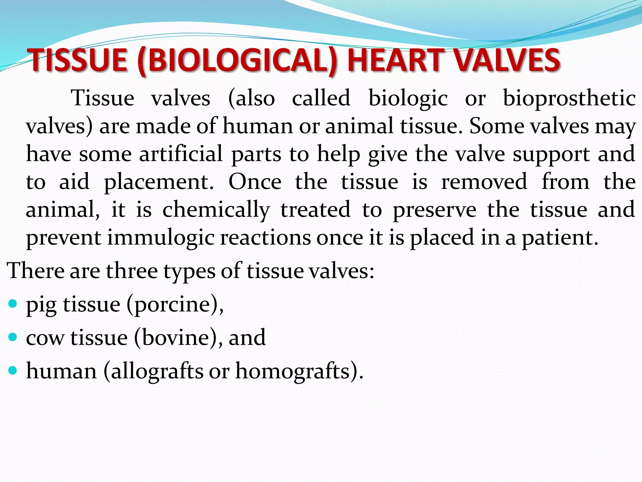
DEFINITION
“Prosthetic heart valve is a device
implanted in the heart of a patient with
valvular heart disease.”
- Brunner & Suddarths (2012)
“A surgical implant used to replace an
abnormal heart valve. Most prosthetic valves
require open heart surgery. The implanted
valves can be porcine (from a pig) or
mechanical (man-made). Also known as
porcine heart valve, heart graft, mechanical
heart valve.”
- Jose Vega (2010)
ANATOMY & PHYSIOLOGY OF HEART
HEART VALVES
TYPES OF PROSTHETIC VALVES
MECHANICAL VALVES
CAGED BALL:-
 The first artificial heart valve was the caged-ball, which
utilizes a metal cage to house a silicone elastomer ball.
 When blood pressure in the chamber of the heart exceeds
that of the pressure on the outside of the chamber the ball
is pushed against the cage and allows blood to flow.
 At the completion of the heart's contraction, the pressure
inside the chamber drops and is lower than beyond the
valve, so the ball moves back against the base of the valve
forming a seal.
Advantages
 Oldest prosthetic valve.
 Durabilty upto 40 yr
Disadvantages
 High profile
 Hemolysis
 High thrombogenecity
 Poor hemodynamics in small sizes
Unique features
 Occluder travels completely out of the orifice, reduces
thrombus & pannus growing from the sewing ring.
 Continuously changing points of contact of the ball
reduces the wear & tear in any one area
 Thrombogenic risk 4-6% per year.
TILTING DISC VALVE
 Tilting disk valves have a single circular occluder controlled
by a metal strut.
 They are made of a metal ring covered by an Eptfe fabric, into
which the suture threads are stitched in order to hold the
valve in place.
 The metal ring holds, by means of two metal supports, a disc
which opens and closes as the heart pumps blood through
the valve.
 The disc is usually made of an extremely hard carbon
material (pyrolytic carbon), in order to allow the valve to
function for years without wearing out.
Advantages
 Low profile
 Good hemodynamics even in small sizes
 Excellent durability
 Permit central laminar flow.
Disadvantages
 Anticoagulation mandatory
 Higher risk of thrombosis than cage ball
 Sudden catastrophic valve thrombosis.
BILEAFLET VALVES
 They have two semicircular leaflets retained
within the ring by hinges. The potential for
impeded leaflet movement due to interference
with cardiac structures is slim, as the open leaflets
are positioned in the middle of the blood stream
and enclosed within the ring in the closed
position.
 Bileaflet valves are the most protected as the
leaflets hardly protrude from the valve ring, even
during maximum opening.
Advantages:-
 Low bulk - flat profile.
 Less thrombogenicy.
 Central laminar flow.
 Two semicircular discs that pivot between open
and closed positions.
 No need for supporting struts.
 Good hemodynamics even in small sizes.
 2 lateral, 1 central minor orifice , no chance of
sudden catastro thrombosis.
Disadvantages:-
 Anticoagulation mandatory risk of thrombosis.
TISSUE (BIOLOGICAL) HEART VALVES
Tissue valves (also called biologic or bioprosthetic
valves) are made of human or animal tissue. Some valves may
have some artificial parts to help give the valve support and
to aid placement. Once the tissue is removed from the
animal, it is chemically treated to preserve the tissue and
prevent immulogic reactions once it is placed in a patient.
There are three types of tissue valves:
 pig tissue (porcine),
 cow tissue (bovine), and
 human (allografts or homografts).
Porcine Stented valves
 The porcine stented valve was the first
generation of porcine tissue valves. They
have been available for more than 30 years.
 The valves are made from natural porcine
aortic valves, but may be used for aortic or
mitral valve replacement. They are trimmed
and then fixed in buffered glutaraldehyde at
high pressure.
Porcine stentless valve
 The porcine stentless valve is used for aortic valve
replacement. The valve is made from a natural
porcine aortic valve and is fixed in buffered
glutaraldehyde solution at a low pressure.
 No stents or synthetic sewing rings are used.
Therefore, these valves are very similar to the
homograft valve (see below).
These valves are technically more difficult to
implant but are useful in patients with small
hypertrophied hearts.
Pericardial valves
The Carpentier-Edwards PERIMOUNT
Pericardial Bioprosthesis
 Pericardial valves include the Perimount series
valves (Edwards LifeSciences). Ionescu-Shiley
pericardial valves have been discontinued. More
recently, stentless porcine valves have been used.
They offer improved hemodynamics with a
decreased transvalvular pressure gradient when
compared with older stented models.
Other types of biological valves
 Xenografts are tissue valves (eg,
bioprostheses, heterografts); most are
from pigs (porcine), but valves from cows
(bovine) may also be used. Their viability is
7 to 10 years.
 They do not generate thrombi, thereby
eliminating the need for longterm
anticoagulation.
 Homografts, or allografts (ie, human
valves), are obtained from cadaver tissue
donations.
 The aortic valve and a portion of the aorta or
the pulmonic valve and a portion of the
pulmonary artery are harvested and stored
cryogenically.
 Homografts are not always available and are
very expensive.
 Autografts (ie, autologous valves) are obtained by
excising the p atient’s own pulmonic valve and a
portion of the pulmonary artery for use as the
aortic valve.
 Anticoagulation is unnecessary because the valve
is the patient’s own tissue and is not
thrombogenic.
 The autograft is an alternative for children (it may
grow as the child grows), women of childbearing
age, young adults, patients with a history of peptic
ulcer disease, and those who cannot tolerate
anticoagulation.
Radiologic Identification
Starr-Edwards caged ball valve :
Radiopaque base ring
 Radiopaque cage
Three struts for the aortic valve; 4
struts for the mitral or tricuspid valve
Cinefluoroscopy:
Structural integrity
Motion of the disc or poppet
Excessive tilt ("rocking") of the base
ring - partial dehiscence of the valve
Aortic valve prosthesis
Fluoroscopy of a normally functioning CarboMedics
bileaflet prosthesis in mitral position
A=opening angle B=closing angle
MRI:
 Not useful in assessing prosthetic-valve
structure
 Used only when prosthetic-valve
regurgitation or para valvular leakage is
suspected but not adequately visualized by
echocardiography
Cardiac Catheterization:
Measure the transvalvular pressure
gradient, from which the EOA can be
calculated
Can visualize and quantify valvular or
paravalvular regurgitation
Echocardiography of Stentless
Aortic Homografts
 Doppler flow characteristics similar to native valve.
 Only 2-D evidence: Increased Echo intensity, and
Thickness of aortic annulus.
Valve dysfunction
complication example Role of echo
Primary mechanical failure Ball variance
Strut fracture
Visualize structure, assess
gradient & regurgitation
Nonstructural dysfunction Pt- prosthesis mismatch
pannus
Gradient, visualize tissue in
& around the sewing ring
Bleeding event Intracranial hge Source of embolus, presence
& mobility of masses
Endocarditis Vegetation, abcess,
dehiscence
Visualize area around the
sewing ring, echo dense /
lucent area, perivalvular
regurgitation
Thrombosis Thrombus impedes opening
&closing of occluder
mechanism
Localize mass, assess
gradient, detect
regurgitation
Embolism stroke Identify & characterize the
source of emboli
Patient-prosthesis mismatch
 When the effective prosthetic valve area, after
insertion into the patient less than that of a normal
valve (Rahimtoola in 1978)
 EOA indexed to BSA is less than 0.85 cm2/m2
 EOA (echo) differs from geometric orifice area
(measured directly)
 EOA for each prostheses type & size obtained in
literature from pts normally functioning prostheses
 Average if > 1 value
-- mild (0.9 - 1 cm² /m²
-- moderate (0.6 - 0.9 cm2/m²
-- severe (iEOA < 0.6cm²/m² (Rahimtoola)
Three-step algorithm
 Step 1: Calculation of the patient BSA.
 Step 2: Reference to the specific table for
identification of the adequate valvular EOA
according to the patient BSA.
 Step 3: Selection of the most appropriate
type and size of valve prosthesis according
to the target iEOA
Valve thrombosis
Incidence of 0.1 to 5.7 % per patient/year
<0.2% per year for mechanical valves
<0.1% bioprosthetic valves
 small thrombus, at the hinge portion of a bileaflet valve
obstruct the mechanism
 tilting disk -- a much larger thrombus to prevent function
 Ball and cage valves – less susceptible  occluder has no
contact at all with the valve housing for a portion of every
cycle
Clinical
 Non obstructive- incidental/embolic phenomenon
 Partial obstruction- dyspnea,systemic embolism , fever
 Severe obstruction- overt heart failure
Fibrinolytic therapy
Rt sided thrombosis 80-100% success rate
• Surgery for fibrinolysis failure/symptoms >
3 wk
 Surgery – Lt sided thrombosis, large clot
burden
 2 types of protocol
-rescue fibrinolysis (short protocol for unstable pt)
- long protocol for stable pt
 Short protocol
- r tPA 10 mg bolus + 90 mg in 90 min or
- SK 15lac in 60 min
 Long protocol
-- SK- 5lac u in 20 min f/b 15lac u for 10 hr
-- rtPA -- 10 mgbolus f/b 90mg/hr for 9 hrs
 Urokinase
 High dose: 4,500 IU/kg/h for 12 h without heparin
 Low dose: 2,000 IU/kg/h with heparin for 24 h
Embolization
 Cerebral embolization  CT normal/infarctwarf
& heparin – 72 hrs APTT lower therapeutic level
till the desired INR
 Anticoagulantion delayed for at least 7 to 14 days -
ICH, extensive cerebral infarction  OAC
Excessive Anticoagulation
 Vit K 2.5 mg daily until the INR is acceptable
 Fresh frozen plasma
 Human recombinant factor VIIa, 15 to 19 g/kg
(INR >10.0 with bleeding)
Paravalvular Regurgitation
 Mild or moderate paravalvular leakage -
asymptomatic , may have only a mild hemolytic
anemia
- can be observed carefully with serial echo
 Severe paravalvular leakage - usually have
symptoms of heart failure or severe anemia
- should be treated with surgical repair or
replacement of the valve
PROSTHETIC VALVES

PROSTHETIC VALVES

  • 2.
    DEFINITION “Prosthetic heart valveis a device implanted in the heart of a patient with valvular heart disease.” - Brunner & Suddarths (2012)
  • 3.
    “A surgical implantused to replace an abnormal heart valve. Most prosthetic valves require open heart surgery. The implanted valves can be porcine (from a pig) or mechanical (man-made). Also known as porcine heart valve, heart graft, mechanical heart valve.” - Jose Vega (2010)
  • 4.
  • 5.
  • 6.
  • 7.
    MECHANICAL VALVES CAGED BALL:- The first artificial heart valve was the caged-ball, which utilizes a metal cage to house a silicone elastomer ball.  When blood pressure in the chamber of the heart exceeds that of the pressure on the outside of the chamber the ball is pushed against the cage and allows blood to flow.  At the completion of the heart's contraction, the pressure inside the chamber drops and is lower than beyond the valve, so the ball moves back against the base of the valve forming a seal.
  • 9.
    Advantages  Oldest prostheticvalve.  Durabilty upto 40 yr Disadvantages  High profile  Hemolysis  High thrombogenecity  Poor hemodynamics in small sizes Unique features  Occluder travels completely out of the orifice, reduces thrombus & pannus growing from the sewing ring.  Continuously changing points of contact of the ball reduces the wear & tear in any one area  Thrombogenic risk 4-6% per year.
  • 10.
    TILTING DISC VALVE Tilting disk valves have a single circular occluder controlled by a metal strut.  They are made of a metal ring covered by an Eptfe fabric, into which the suture threads are stitched in order to hold the valve in place.  The metal ring holds, by means of two metal supports, a disc which opens and closes as the heart pumps blood through the valve.  The disc is usually made of an extremely hard carbon material (pyrolytic carbon), in order to allow the valve to function for years without wearing out.
  • 12.
    Advantages  Low profile Good hemodynamics even in small sizes  Excellent durability  Permit central laminar flow. Disadvantages  Anticoagulation mandatory  Higher risk of thrombosis than cage ball  Sudden catastrophic valve thrombosis.
  • 13.
    BILEAFLET VALVES  Theyhave two semicircular leaflets retained within the ring by hinges. The potential for impeded leaflet movement due to interference with cardiac structures is slim, as the open leaflets are positioned in the middle of the blood stream and enclosed within the ring in the closed position.  Bileaflet valves are the most protected as the leaflets hardly protrude from the valve ring, even during maximum opening.
  • 15.
    Advantages:-  Low bulk- flat profile.  Less thrombogenicy.  Central laminar flow.  Two semicircular discs that pivot between open and closed positions.  No need for supporting struts.  Good hemodynamics even in small sizes.  2 lateral, 1 central minor orifice , no chance of sudden catastro thrombosis. Disadvantages:-  Anticoagulation mandatory risk of thrombosis.
  • 16.
    TISSUE (BIOLOGICAL) HEARTVALVES Tissue valves (also called biologic or bioprosthetic valves) are made of human or animal tissue. Some valves may have some artificial parts to help give the valve support and to aid placement. Once the tissue is removed from the animal, it is chemically treated to preserve the tissue and prevent immulogic reactions once it is placed in a patient. There are three types of tissue valves:  pig tissue (porcine),  cow tissue (bovine), and  human (allografts or homografts).
  • 17.
    Porcine Stented valves The porcine stented valve was the first generation of porcine tissue valves. They have been available for more than 30 years.  The valves are made from natural porcine aortic valves, but may be used for aortic or mitral valve replacement. They are trimmed and then fixed in buffered glutaraldehyde at high pressure.
  • 19.
    Porcine stentless valve The porcine stentless valve is used for aortic valve replacement. The valve is made from a natural porcine aortic valve and is fixed in buffered glutaraldehyde solution at a low pressure.  No stents or synthetic sewing rings are used. Therefore, these valves are very similar to the homograft valve (see below). These valves are technically more difficult to implant but are useful in patients with small hypertrophied hearts.
  • 21.
    Pericardial valves The Carpentier-EdwardsPERIMOUNT Pericardial Bioprosthesis  Pericardial valves include the Perimount series valves (Edwards LifeSciences). Ionescu-Shiley pericardial valves have been discontinued. More recently, stentless porcine valves have been used. They offer improved hemodynamics with a decreased transvalvular pressure gradient when compared with older stented models.
  • 23.
    Other types ofbiological valves  Xenografts are tissue valves (eg, bioprostheses, heterografts); most are from pigs (porcine), but valves from cows (bovine) may also be used. Their viability is 7 to 10 years.  They do not generate thrombi, thereby eliminating the need for longterm anticoagulation.
  • 24.
     Homografts, orallografts (ie, human valves), are obtained from cadaver tissue donations.  The aortic valve and a portion of the aorta or the pulmonic valve and a portion of the pulmonary artery are harvested and stored cryogenically.  Homografts are not always available and are very expensive.
  • 25.
     Autografts (ie,autologous valves) are obtained by excising the p atient’s own pulmonic valve and a portion of the pulmonary artery for use as the aortic valve.  Anticoagulation is unnecessary because the valve is the patient’s own tissue and is not thrombogenic.  The autograft is an alternative for children (it may grow as the child grows), women of childbearing age, young adults, patients with a history of peptic ulcer disease, and those who cannot tolerate anticoagulation.
  • 26.
    Radiologic Identification Starr-Edwards cagedball valve : Radiopaque base ring  Radiopaque cage Three struts for the aortic valve; 4 struts for the mitral or tricuspid valve
  • 28.
    Cinefluoroscopy: Structural integrity Motion ofthe disc or poppet Excessive tilt ("rocking") of the base ring - partial dehiscence of the valve Aortic valve prosthesis
  • 29.
    Fluoroscopy of anormally functioning CarboMedics bileaflet prosthesis in mitral position A=opening angle B=closing angle
  • 30.
    MRI:  Not usefulin assessing prosthetic-valve structure  Used only when prosthetic-valve regurgitation or para valvular leakage is suspected but not adequately visualized by echocardiography
  • 31.
    Cardiac Catheterization: Measure thetransvalvular pressure gradient, from which the EOA can be calculated Can visualize and quantify valvular or paravalvular regurgitation
  • 32.
    Echocardiography of Stentless AorticHomografts  Doppler flow characteristics similar to native valve.  Only 2-D evidence: Increased Echo intensity, and Thickness of aortic annulus.
  • 33.
    Valve dysfunction complication exampleRole of echo Primary mechanical failure Ball variance Strut fracture Visualize structure, assess gradient & regurgitation Nonstructural dysfunction Pt- prosthesis mismatch pannus Gradient, visualize tissue in & around the sewing ring Bleeding event Intracranial hge Source of embolus, presence & mobility of masses Endocarditis Vegetation, abcess, dehiscence Visualize area around the sewing ring, echo dense / lucent area, perivalvular regurgitation Thrombosis Thrombus impedes opening &closing of occluder mechanism Localize mass, assess gradient, detect regurgitation Embolism stroke Identify & characterize the source of emboli
  • 34.
    Patient-prosthesis mismatch  Whenthe effective prosthetic valve area, after insertion into the patient less than that of a normal valve (Rahimtoola in 1978)  EOA indexed to BSA is less than 0.85 cm2/m2  EOA (echo) differs from geometric orifice area (measured directly)  EOA for each prostheses type & size obtained in literature from pts normally functioning prostheses  Average if > 1 value -- mild (0.9 - 1 cm² /m² -- moderate (0.6 - 0.9 cm2/m² -- severe (iEOA < 0.6cm²/m² (Rahimtoola)
  • 35.
    Three-step algorithm  Step1: Calculation of the patient BSA.  Step 2: Reference to the specific table for identification of the adequate valvular EOA according to the patient BSA.  Step 3: Selection of the most appropriate type and size of valve prosthesis according to the target iEOA
  • 37.
    Valve thrombosis Incidence of0.1 to 5.7 % per patient/year <0.2% per year for mechanical valves <0.1% bioprosthetic valves  small thrombus, at the hinge portion of a bileaflet valve obstruct the mechanism  tilting disk -- a much larger thrombus to prevent function  Ball and cage valves – less susceptible  occluder has no contact at all with the valve housing for a portion of every cycle Clinical  Non obstructive- incidental/embolic phenomenon  Partial obstruction- dyspnea,systemic embolism , fever  Severe obstruction- overt heart failure
  • 38.
    Fibrinolytic therapy Rt sidedthrombosis 80-100% success rate • Surgery for fibrinolysis failure/symptoms > 3 wk  Surgery – Lt sided thrombosis, large clot burden
  • 39.
     2 typesof protocol -rescue fibrinolysis (short protocol for unstable pt) - long protocol for stable pt  Short protocol - r tPA 10 mg bolus + 90 mg in 90 min or - SK 15lac in 60 min  Long protocol -- SK- 5lac u in 20 min f/b 15lac u for 10 hr -- rtPA -- 10 mgbolus f/b 90mg/hr for 9 hrs  Urokinase  High dose: 4,500 IU/kg/h for 12 h without heparin  Low dose: 2,000 IU/kg/h with heparin for 24 h
  • 40.
    Embolization  Cerebral embolization CT normal/infarctwarf & heparin – 72 hrs APTT lower therapeutic level till the desired INR  Anticoagulantion delayed for at least 7 to 14 days - ICH, extensive cerebral infarction  OAC
  • 41.
    Excessive Anticoagulation  VitK 2.5 mg daily until the INR is acceptable  Fresh frozen plasma  Human recombinant factor VIIa, 15 to 19 g/kg (INR >10.0 with bleeding)
  • 43.
    Paravalvular Regurgitation  Mildor moderate paravalvular leakage - asymptomatic , may have only a mild hemolytic anemia - can be observed carefully with serial echo  Severe paravalvular leakage - usually have symptoms of heart failure or severe anemia - should be treated with surgical repair or replacement of the valve