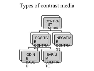
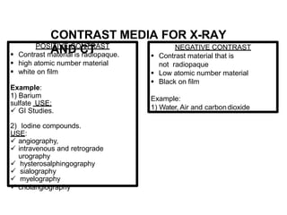
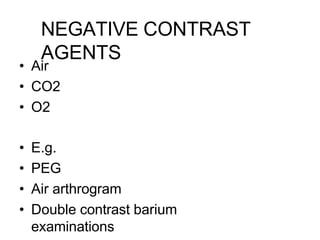
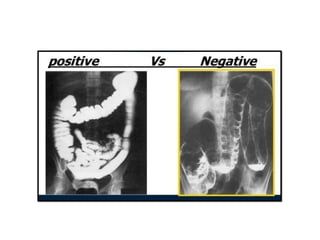
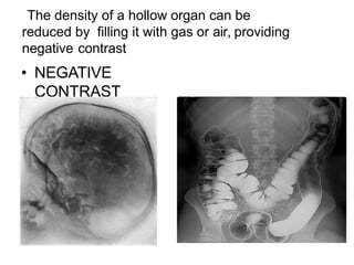
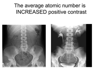
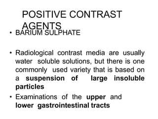
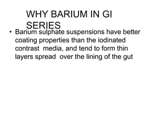
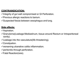
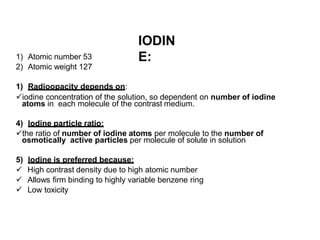
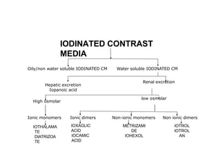
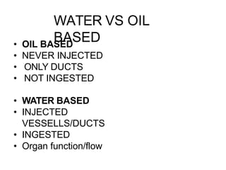
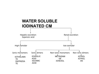
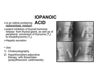
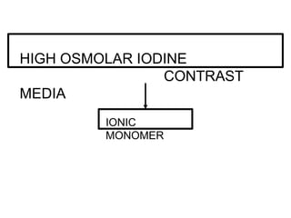
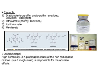
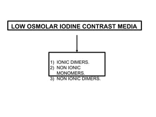
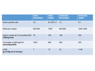
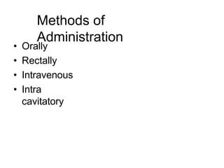
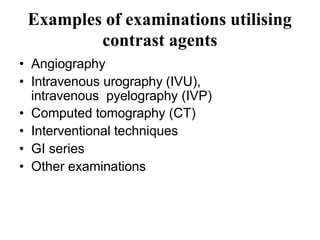
CONTRAST
MEDIA
Dr. PRITHVIRAJ JADHAV
Assisstant Professor
Dept . Of Radiodiagnosis
Aims of this session
• Why contrast agents are used
• What are the desirable features of a
contrast agent
• Types of contrast agents used in clinical
settings
• Methods of administrating contrast agents
• Examples of examinations utilising contrast
agents
• Problems/issues associated with
administrating contrast agents
Why contrast agents are used
• Different tissues within the body
attenuate the beam of X-rays to
different degrees.
• The degree of attenuation of an X-ray
beam by an element is complex, but one
of the major variables is the number of
electrons in the path of the beam with
which it can interact.
• The number of electrons in the path of
the beam is dependent upon three
factors:
• The thickness of the substance being
studied
• Its density
• The number of electrons per atom of the
element (which is equal to its atomic
number)
Where there is inherent
considerable difference between
the densities of two organs
• However, if the two organs have similar
densities and similar average atomic
numbers, then it is not possible to
distinguish them on a radiograph,
because no natural contrast exists.
• For example, it is not possible to identify
blood vessels within an organ, or to
demonstrate the internal structure of the
kidney, without artificially altering one of
the factors mentioned earlier
2)CONTRAST MEDIUM:
It is a chemical substance of very high or very low atomic
number or weight, therefor it increase or decrease the density
of the organ under examination.
OR
A substance which when introduced into the body will
increase the radiographic contrast in an area where it was
absent or low before.
Two of the factors important in
organ contrast can be artificially
altered,
• the density of an organ, and,
• more usefully, the average atomic number
of a structure.
What are the desirable features of a
contrast agent
• Easy to administer
• No toxicity/carcinogenecity
• Stable compound
• Concentrates in area of interest
• Proper demonstration of the organ
system
• Should have rapid elimination
• Minimal distress to patients (viscosity)
• Cost effective
MODE OF
ADMINISTRATION
1) Orally.
2) Rectally.
3) Intravenously – (injection/
infusion).
4) Mechanically – Filling of a
body cavity or potential
space.
5) Intra-muscularly
CLASSIFICATION OF CONTRAST
MEDIA
Oily/non water soluble IODINATED CM Water soluble IODINATED CM
Renal excretion
Hepatic excretion
Iopanoic acid
High osmolar low osmolar
Ionic dimers Non-ionic monomers
Ionic monomers
IOTHALAMA
TE
DIATRIZOAT
E
IOXAGLIC
ACID
IOCAMIC
ACID
METRIZAMI
DE
IOHEXOL
Non ionic dimers
IOTRO
L
IOTROL
AN
X-RAY & CT ULTRASOUND MRI
Positive CM Negective CM
water,air,CO2
Non water soluble BaSO4 IODINATED CM
Types of contrast media
CONTRA
ST
MEDIA
POSITIV
E
CONTRA
ST
IODIN
E
BASE
D
BARIU
M
SULPHA
TE
NEGATIV
E
CONTRA
ST
POSITIVE CONTRAST
 Contrast material is radiopaque.
 high atomic number material
 white on film
Example:
1) Barium
sulfate USE:
 GI Studies.
2) Iodine compounds.
USE:
 angiography,
 intravenous and retrograde
urography
 hysterosalphingography
 sialography
 myelography
 cholangiography
NEGATIVE CONTRAST
 Contrast material that is
not radiopaque
 Low atomic number material
 Black on film
Example:
1) Water, Air and carbondioxide
CONTRAST MEDIA FOR X-RAY
AND CT
NEGATIVE CONTRAST
AGENTS
• Air
• CO2
• O2
• E.g.
• PEG
• Air arthrogram
• Double contrast barium
examinations
The density of a hollow organ can be
reduced by filling it with gas or air, providing
negative contrast
• NEGATIVE
CONTRAST
The average atomic number is
INCREASED positive contrast
POSITIVE CONTRAST
AGENTS
• BARIUM SULPHATE
• Radiological contrast media are usually
water soluble solutions, but there is one
commonly used variety that is based on
a suspension of large insoluble
particles
• Examinations of the upper and
lower gastrointestinal tracts
• Barium sulphate suspensions have better
coating properties than the iodinated
contrast media, and tend to form thin
layers spread over the lining of the gut
WHY BARIUM IN GI
SERIES
BARIUM
SULPHATE
1) Atomic number:56
2) Highly radiopaque.
3) Non absorbable.
4) Non toxic.
5) Insoluble in water/lipid.
6) Inert to tissues.
7) Can be used for double contrast studies.
8) Route: Orally Or Rectally (aqueous
suspension with 0.3 to 1 g dry weight per
milliliter)
9) Uses:
 barium swallow
 barium meal
 barium meal follow through
 Enteroclysis
 barium enema
BARIUM
SWALLOW
BARIUM
MEAL
BARIUM MEAL FOLLOW
THROUGH
ENTEROCL
YSIS
BARIUM
ENEMA
CONTRAINDICATION:
Integrity of gut wall compromised or GI Perforation.
Previous allergic reactions to barium.
Suspected fistula between oesophagus and lung.
Side effects:
Aspiration.
Granuloma(Leakage:Mediastinum, tissue around Rectum or Intraperitoneal
cavity).
Leakage into the vasculature(life threatening).
Constipation.
worsening ulcerative colitis inflammation.
peritonitis through perforation.
Fatal Reaction(rare).
IODIN
E:
1) Atomic number 53
2) Atomic weight 127
1) Radioopacity depends on:
iodine concentration of the solution, so dependent on number of iodine
atoms in each molecule of the contrast medium.
4) Iodine particle ratio:
the ratio of number of iodine atoms per molecule to the number of
osmotically active particles per molecule of solute in solution
5) Iodine is preferred because:
 High contrast density due to high atomic number
 Allows firm binding to highly variable benzene ring
 Low toxicity
IODINATED CONTRAST
MEDIA
Oily/non water soluble IODINATED CM
Hepatic excretion
Iopanoic acid
High osmolar
Water soluble IODINATED CM
Renal excretion
low osmolar
Ionic monomers Non-ionic monomers Non ionic dimers
IOTHALAMA
TE
DIATRIZOA
TE
Ionic dimers
IOXAGLIC
ACID
IOCAMIC
ACID
METRIZAMI
DE
IOHEXOL
IOTROL
IOTROL
AN
WATER VS OIL
BASED
• OIL BASED
• NEVER INJECTED
• ONLY DUCTS
• NOT INGESTED
• WATER BASED
• INJECTED
VESSELLS/DUCTS
• INGESTED
• Organ function/flow
OILY/NON WATER SOLUBLE
IODINATED CM
Fatty Acids
Insoluble in water
White on the radiograph
Examples:
1) Iophendylate (Myodil,
Pantopaque)- myelographic
agent
2) Lipiodrol Ultrafluide
(Ethiodol)-
lymphangiographic agent.
 COMPLICATION:
1) Fat
Embolism
LYMPHANGIOGRA
PHY
USE:
1) Sialography
2) Hysterosalpingograp
hy
3) Myelography
4) Lymphangiography
5) Dacryocystography
6) Galactography
7) Broncography
SIALOGRAP
HY
HYSTROSALPHINGOGR
APHY
MYELOGRAP
HY
DACROCYSTOGR
APHY
GALACTOGRA
PHY
BRANCHOGRA
PHY
Renal excretion
Hepatic excretion
Iopanoic acid
High osmolar low osmolar
Ionic monomers Non-ionic monomers Non ionic dimers
IOTHALAMA
TE
DIATRIZOA
TE
Ionic dimers
IOXAGLIC
ACID
IOCAMIC
ACID
METRIZAMI
DE
IOHEXOL
IOTROL
IOTROL
AN
WATER SOLUBLE
IODINATED CM
IOPANOIC
ACID
is an iodine-containing
radiocontrast medium.
potent inhibitors of thyroid hormone
release from thyroid gland, as well as of
peripheral conversion of thyroxine (T4)
to triiodothyronine (T3)
Hepatic excretion
• Use:
1) Cholecystography
2) Hyperthyroidism:adjunctive
therapy with thioamides
(propylthiouracil, carbimazole).
HIGH OSMOLAR IODINE
CONTRAST
MEDIA
IONIC
MONOMER
Example:
1) Diatrizoate(urograffin, angiograffin , urovideo,
urovision, trazograff)
2) Iothalamate(conray, Triovideo)
3) Ioxithalamate
4) Metrizoate
Disadvantage:
High osmolality (8 X plasma) because of the non radiopaque
cations (Na & meglumine) is responsible for the adverse
effects.
LOW OSMOLAR IODINE CONTRAST MEDIA
1) IONIC DIMERS.
2) NON IONIC
MONOMERS.
3) NON IONIC DIMERS.
IONIC
MONOMER
IONIC
DIMER
NONIONIC
MONOMER
NONIONIC
DIMER
Iodine particle ratio 3:2 6:2 OR 3:1 3:1 6:1
Molecular weight 600-800 1269 600-800 1500-1626
Iodine content at 0.3 osmol/kg H2O
(300mg I/ml)
70 150 150 300
Osmolality at 280mgI2/ml
(osmol/kg H2O)
1500 560 600 300
LD50
(g of I/kg wt of mouse)
7 12 22 >>26
Methods of
Administration
• Orally
• Rectally
• Intravenous
• Intra
cavitatory
Examples of examinations utilising
contrast agents
• Angiography
• Intravenous urography (IVU),
intravenous pyelography (IVP)
• Computed tomography (CT)
• Interventional techniques
• GI series
• Other examinations
ADDITIVES USED IN CONTRAST
MEDIA
1) Stabilizer:
Ca or Na EDTA
2) Buffers:
Stabilizes pH during storage, Na acid
phosphates
3) Preservatives:
Generally not disclosed by the
manufacturers.
IDEAL CONTRAST
MEDIA
1) High water solubility.
2) Heat & chemical stability(shelf life) ideally- 3 to
5yrs.
3) Biological inertness( non antigenic).
4) Low viscosity.
5) Low or isoosmolar to plasma.
6) Selective excretion, like excretion by kidney is
favorable.
7) Safety: LD50 (lethal dose) should be high.
8) Reasonable cost.
POINTS TO
REMEMBER
Contrast media used for myelography- non-ionic CM.
CM used for cerebral angiography- only meglumine salt.
Least osmolar- Ioxaglate (Hexabrix).
Most hyperosmolar- Iohexol.
Max nausea & vomiting- Ioxaglate (Hexabrix).
Bronchospasm- Meglumine salts.
Viscosity:
• Increase with concentration
• Higher for dimers(big size)
• High viscosity interferes with mixing of contrast media with plasma & body fluids.
• Least viscosity- Omnipaque240
Meticulous heparinization is required during angiography as incidence of
thromboembolic phenomenon is high when CM is mixed with blood.
Choice of
contrast
• Infants (introduction of hyperosmolar fluid into the
bodies of very young children can cause
• problems of fluid balance)
• The elderly (for the same reason)
• Diabetics
• Patients with cardiac impairment
• Patients with renal impairment
• Asthmatics
• Patients who have previously reacted adversely
to a contrast medium
• Patients with a history of allergy
• Patients who are unduly anxious
Adverse effects
VASCULAR TOXICITY --VENOUS
--1. pain at injection site –result of perivenous injection
--2. pain extending up the arm –due to stasis of contrast in arm – relieved by
abduction of arm
--3. delayed limb pain –due to thrombophlebitis as a result of toxic effect on
endothelium
ARTERIAL –arterial endothelial damage & vasodilation –related to hyperosmolarity
SOFT TISSUE TOXICITY – pain ,swelling , erythema even sloughing of skin due to
extravasated contrast medium
--increased risk when pumps are used to inject large amt
of contrast media
CARDIOVASCULAR TOXICITY --
1. Intracoronary injection – cardiac rhythm disturbances
2. Increased vagal activity – depression of SA & AV node –bradycardia , asystole
3. Injection of hypertonic contrast medium – significant fluid & ion shift
e.g. – hypervolemia – due to diffusion of extracellular fluid through capillary walls
HEMATOLOGICAL CHANGES –
1. Erythrocyte damage – injection of HOCM  loss of water from RBC 
dehydrated shrunken RBC  Increased internal viscocity
with loss of ability of RBC to deform to traverse capillary
obstruction of important capillary beds (cerebral , renal,
coronary, pulmonary )
2. Red cell aggregation & coagulation may occur
3. Thrombus formation may occur – commoner with LOCM
4.Sickle cell crisis may be precipitate
NEPHROTOXICITY -- CONTRAST INDUCED NEPHROPATHY –sr. creat conc .
starts to rise within first 24 hrs , reaches peak by 2-3 days &
returns to baseline by 3- 7 days
PREDISPOSING FACTORS – preexisting impairment of renal function
-- diabetes mellitus
--dehydration
--large doses of contrast
--age
--multiple myeloma
--concurrent use of nephrotoxic drug
NEUROTOXICITY – if BBB is impaired , nerve cells are exposed to toxic effects of
contrast media
THYROID FUNCTION – thyrotoxicosis may be exacerbated in preexisting thyrotoxic
symptoms
Idiosyncratic reaction
CLASSIFIED AS –
1. MINOR REACTION -- do not interfere with examination but require patient
reassurance
--e.g. –nausea, vomiting, mild rash , light headache
2. INTERMEDIATE REACTION –interfere with examination
--e.g.- urticaria, facial edema, dyspnea, hypotension
3. SEVERE REACTION -- warrant urgent tretment & hospitalisation
e.g. –circulatory collapse, pulmonary edema, severe angina
convulsions,
Mechanism
HISTAMINE RELEASE – histamine – primary mediator of anaphylaxis –released by
mast cells due to contrast media
COMPLEMENT ACTIVATION – contrast media may activate complement system
which induce histamine release & other biological
mediators
PROTEIN BINDING & ENZYME INHIBITION –contrast media are weakly protein
bound & thus inhibit enzyme acetylcholinesterase
--contrast media side effects are cholinergic effects –
e.g.—vasodilation, bradycardia, bronchospasm
CHEMOTOXICITY – due to cations- especially Na+, effects are seen in neurons,
myocardial cells , capillary endothelium, RBC, kidney
ANXIETY – most contrast reaction result of patients fear & apprehension
--high autonomic nervous system activity in anxious patient is stimulated
further after contrast administration
{
Prophylaxis
PROPHYLAXIS OF RENAL ADVERSE REACTION
IDENTIFICATION OF PATIENTS AT INCREASED RISK-
1. Referring clinician should identify patients with preexisting renal impairment
& inform radiology department
2. sr.creat should be measured within 1 week before administration of contrast
PRECAUTIONS FOR PATIENTS WITH SIGNIFICANT RENAL IMPAIRMENT
(sr.creat > 130 micromol/ l)
1.Consider alternative imaging method
2. 48 hr before stop any treatment with metformin . It should be withheld for further
48 hrs after procedure & renal function should be reassessed before restarting
metformin
3. 24 hr before stop all nephrotoxic drugs, mannitol, & loop diuretics
4. 6 hr before start hydrating patient either orally or intravenously
5. Use smallest possible dose of contrast medium
6. Use low or iso-ismolar contrast media
--no definite evidence that hemodialysis protect patients with impaired renal function
From contrast medium induced nephrotoxicity
PROPHYLAXIS OF NON RENAL ADVERSE EFFECTS
IDENTIFICATION OF PATIENTS AT INCREASED RISK OF ANAPHYLACTOID
REACTION– patients should be asked for history of—
1. previous contrast reaction
2. asthma
3. previous allergic reaction requiring medical treatment
PRECAUTIONS FOR PATIENTS AT INCREASED RISK OF ANAPHYLACTOID
CONTRAST REACTION –
1. Consider an alternative test
2. If contrast is necessary –
a. use non ionic contrast media
b. for previous reactors use a different non ionic contrast than used previously
c. maintain close supervision
d. leave canula in place & observe for 30 min.
e. be ready to treat promptly any adverse reaction & ensure that emergency drugs &
equipment are ready
3. Premedication may be used e.g.- prednisolone 30 mg orally given 12 hr & 2hr
before contrast administration
{
Treatment
OXYGEN – high dose oxygen ( upto 100% ) at rate of of 10- 12 lit /min via face mask
EPINEPHRINE – available in 2 dilutions –
--1 in 1000 – 1mg in 1 ml – s. c. or i.m. use
--1 in 10000 –1mg in 10 ml– for i.v. administration
Epinephrine is administered –
--sc. In a dose 0.1 – 0.3 ml ( 0.1 – 0.3 mg) can be repeated every 10-15 min until a total
dose of 1 mg is administered
--i.v. in same dose of 0.1- 0.3 mg , so due to greater dilution 1-3 ml is injected
COMPLICATIONS- hypertensive crisis , myocardial ischemia , infarction
Administered carefully in following pts- with cardiac disease , with hypertension
on beta blockers
CORTICOSTEROIDS – effective in reducing late reactions , which can be observed
upto 48 hrs
-- i.v. doses of 100- 1000 mg of hydrocortisone recommended
--can be followed by continuous infusion of 300-500 mg in 250 ml saline at rate
60 ml/ hr
PRINCIPLES OF TREATMENT
MILD REACTION
-- reassure patient
--loosen tight clothing, tell patient to take few deep breath & relax
--stay with patient & watch until symptoms subside
Moderate-severe reaction
SKIN REACTION – usually no treatment is needed
-- if pruritus is severe use diphenhydramine ( 50 mg )
--if patient develops severe erythema or angioedema use H1 & H2
blockers ( cimetidine 300 mg in 20 ml ) – if no response use
epinephrine 0.1-0.3 ml
RESPIRATION REACTION – causes of respiratory decompensation
1. airway & laryngeal edema
2. bronchospasm
3. pulmonary edema
1. LARYNGEAL EDEMA MANAGEMENT – O2
--epinephrine
--intubation if necessary
2.BRONCHOSPASM – MILD – O2
--metered dose inhaler albuterol 2-3 inhalation
MODERATE –epinephrine
-- aminophylline – 5 mg/kg i.v. slowly over 10-20
min
SEVERE – epinephrine i.v.
3. PULMONARY EDEMA – elevate head end of bed
--O2 10 L/ min
--furosemide 40 mg i.v. slowly
--morphine 1-3 mg i.v.
--hydrocortisone 100 mg i.v. slowly
--shift to ICU
HYPOTENSION – MILD – 1. release any abdominal compression
2. elevate legs
3. O2
4. isotonic i.v. fluids – administered rapidly
SEVERE – BRADYCARDIA – atropine 0.6 mg i.v. slowly , repeat 3- 5 min,
max upto 3mg
--TACYCARDIA – epinephrine 1- 3ml ( 1: 10000) i.v. upto 10 ml or dopamine
SEIZURES OR CONVULSIONS –
MILD –1. turn patient to one side to avoid aspiration . Be sure airway is clear & open
2. O2- 10 L/min
SEVERE – diazepam 5 mg i.v. slowly
HYPERTENSIVE CRISES –1. O2 10 L/ min
--nitroglycerine 0.4 mg tablet sublingually
-- if no response , nifedipine --10 mg capsule sublingually
-- ECG
-- if pheochromocytoma , phentolamine 5 mg i. v.
-- frusemide 40 mg i.v. slowly
EXTRAVASATION OF CONTRAST MATERIAL –
1.eLevation of affected extremity above heart level
2. Ice packs ( 20 min t.i.d. for 2-3 days )
3. Plastic surgery consultation if – large volume extravasation
-- skin ulceration or blistering
-- worsening symptoms after 2-4 hrs
4. Close follow up until resolution
MRI CONTRAST MEDIA
MECHANISM OF ACTION – act indirectly by altering magnetic properties of
Hydrogen ions (protons ) in water & lipid . They alter rate of relaxation of protons
FERROMAGNETIC – retain magnetism even when applied field is removed .
Interfere with cell function – unsafe for clinical use
PARAMAGNETIC – e.g. gadolinium . Have magnetic moments which align to applied
field , but once gradient field is turned off, thermal energy within overcome alignment.
Their maximum effect is on protons – shortening of T1 relaxation time & hence
increased signal intensity ( white ) on T1 images .
SUPERPARAMAGNETIC – particles of iron oxide ( Fe3O4), small particles of iron
Oxide ( SPIO ), ultrasmall particles of iron oxide ( USPIO )
--cause abrupt changes in local magnetic field – results in rapid proton dephasing –
Reduction in T2 relaxation – decreased signal intensity ( black ) on T2 images
THANK YOU

Contrast agents

  • 1.
    CONTRAST MEDIA Dr. PRITHVIRAJ JADHAV AssisstantProfessor Dept . Of Radiodiagnosis
  • 2.
    Aims of thissession • Why contrast agents are used • What are the desirable features of a contrast agent • Types of contrast agents used in clinical settings • Methods of administrating contrast agents • Examples of examinations utilising contrast agents • Problems/issues associated with administrating contrast agents
  • 3.
    Why contrast agentsare used • Different tissues within the body attenuate the beam of X-rays to different degrees. • The degree of attenuation of an X-ray beam by an element is complex, but one of the major variables is the number of electrons in the path of the beam with which it can interact.
  • 4.
    • The numberof electrons in the path of the beam is dependent upon three factors: • The thickness of the substance being studied • Its density • The number of electrons per atom of the element (which is equal to its atomic number)
  • 5.
    Where there isinherent considerable difference between the densities of two organs
  • 6.
    • However, ifthe two organs have similar densities and similar average atomic numbers, then it is not possible to distinguish them on a radiograph, because no natural contrast exists. • For example, it is not possible to identify blood vessels within an organ, or to demonstrate the internal structure of the kidney, without artificially altering one of the factors mentioned earlier
  • 8.
    2)CONTRAST MEDIUM: It isa chemical substance of very high or very low atomic number or weight, therefor it increase or decrease the density of the organ under examination. OR A substance which when introduced into the body will increase the radiographic contrast in an area where it was absent or low before.
  • 9.
    Two of thefactors important in organ contrast can be artificially altered, • the density of an organ, and, • more usefully, the average atomic number of a structure.
  • 10.
    What are thedesirable features of a contrast agent • Easy to administer • No toxicity/carcinogenecity • Stable compound • Concentrates in area of interest • Proper demonstration of the organ system • Should have rapid elimination • Minimal distress to patients (viscosity) • Cost effective
  • 11.
    MODE OF ADMINISTRATION 1) Orally. 2)Rectally. 3) Intravenously – (injection/ infusion). 4) Mechanically – Filling of a body cavity or potential space. 5) Intra-muscularly
  • 12.
    CLASSIFICATION OF CONTRAST MEDIA Oily/nonwater soluble IODINATED CM Water soluble IODINATED CM Renal excretion Hepatic excretion Iopanoic acid High osmolar low osmolar Ionic dimers Non-ionic monomers Ionic monomers IOTHALAMA TE DIATRIZOAT E IOXAGLIC ACID IOCAMIC ACID METRIZAMI DE IOHEXOL Non ionic dimers IOTRO L IOTROL AN X-RAY & CT ULTRASOUND MRI Positive CM Negective CM water,air,CO2 Non water soluble BaSO4 IODINATED CM
  • 13.
    Types of contrastmedia CONTRA ST MEDIA POSITIV E CONTRA ST IODIN E BASE D BARIU M SULPHA TE NEGATIV E CONTRA ST
  • 14.
    POSITIVE CONTRAST  Contrastmaterial is radiopaque.  high atomic number material  white on film Example: 1) Barium sulfate USE:  GI Studies. 2) Iodine compounds. USE:  angiography,  intravenous and retrograde urography  hysterosalphingography  sialography  myelography  cholangiography NEGATIVE CONTRAST  Contrast material that is not radiopaque  Low atomic number material  Black on film Example: 1) Water, Air and carbondioxide CONTRAST MEDIA FOR X-RAY AND CT
  • 15.
    NEGATIVE CONTRAST AGENTS • Air •CO2 • O2 • E.g. • PEG • Air arthrogram • Double contrast barium examinations
  • 17.
    The density ofa hollow organ can be reduced by filling it with gas or air, providing negative contrast • NEGATIVE CONTRAST
  • 18.
    The average atomicnumber is INCREASED positive contrast
  • 19.
    POSITIVE CONTRAST AGENTS • BARIUMSULPHATE • Radiological contrast media are usually water soluble solutions, but there is one commonly used variety that is based on a suspension of large insoluble particles • Examinations of the upper and lower gastrointestinal tracts
  • 20.
    • Barium sulphatesuspensions have better coating properties than the iodinated contrast media, and tend to form thin layers spread over the lining of the gut WHY BARIUM IN GI SERIES
  • 21.
    BARIUM SULPHATE 1) Atomic number:56 2)Highly radiopaque. 3) Non absorbable. 4) Non toxic. 5) Insoluble in water/lipid. 6) Inert to tissues. 7) Can be used for double contrast studies. 8) Route: Orally Or Rectally (aqueous suspension with 0.3 to 1 g dry weight per milliliter) 9) Uses:  barium swallow  barium meal  barium meal follow through  Enteroclysis  barium enema
  • 22.
  • 23.
  • 25.
    CONTRAINDICATION: Integrity of gutwall compromised or GI Perforation. Previous allergic reactions to barium. Suspected fistula between oesophagus and lung. Side effects: Aspiration. Granuloma(Leakage:Mediastinum, tissue around Rectum or Intraperitoneal cavity). Leakage into the vasculature(life threatening). Constipation. worsening ulcerative colitis inflammation. peritonitis through perforation. Fatal Reaction(rare).
  • 26.
    IODIN E: 1) Atomic number53 2) Atomic weight 127 1) Radioopacity depends on: iodine concentration of the solution, so dependent on number of iodine atoms in each molecule of the contrast medium. 4) Iodine particle ratio: the ratio of number of iodine atoms per molecule to the number of osmotically active particles per molecule of solute in solution 5) Iodine is preferred because:  High contrast density due to high atomic number  Allows firm binding to highly variable benzene ring  Low toxicity
  • 27.
    IODINATED CONTRAST MEDIA Oily/non watersoluble IODINATED CM Hepatic excretion Iopanoic acid High osmolar Water soluble IODINATED CM Renal excretion low osmolar Ionic monomers Non-ionic monomers Non ionic dimers IOTHALAMA TE DIATRIZOA TE Ionic dimers IOXAGLIC ACID IOCAMIC ACID METRIZAMI DE IOHEXOL IOTROL IOTROL AN
  • 28.
    WATER VS OIL BASED •OIL BASED • NEVER INJECTED • ONLY DUCTS • NOT INGESTED • WATER BASED • INJECTED VESSELLS/DUCTS • INGESTED • Organ function/flow
  • 29.
    OILY/NON WATER SOLUBLE IODINATEDCM Fatty Acids Insoluble in water White on the radiograph Examples: 1) Iophendylate (Myodil, Pantopaque)- myelographic agent 2) Lipiodrol Ultrafluide (Ethiodol)- lymphangiographic agent.  COMPLICATION: 1) Fat Embolism LYMPHANGIOGRA PHY
  • 30.
    USE: 1) Sialography 2) Hysterosalpingograp hy 3)Myelography 4) Lymphangiography 5) Dacryocystography 6) Galactography 7) Broncography
  • 31.
  • 32.
  • 33.
    Renal excretion Hepatic excretion Iopanoicacid High osmolar low osmolar Ionic monomers Non-ionic monomers Non ionic dimers IOTHALAMA TE DIATRIZOA TE Ionic dimers IOXAGLIC ACID IOCAMIC ACID METRIZAMI DE IOHEXOL IOTROL IOTROL AN WATER SOLUBLE IODINATED CM
  • 34.
    IOPANOIC ACID is an iodine-containing radiocontrastmedium. potent inhibitors of thyroid hormone release from thyroid gland, as well as of peripheral conversion of thyroxine (T4) to triiodothyronine (T3) Hepatic excretion • Use: 1) Cholecystography 2) Hyperthyroidism:adjunctive therapy with thioamides (propylthiouracil, carbimazole).
  • 35.
  • 36.
    Example: 1) Diatrizoate(urograffin, angiograffin, urovideo, urovision, trazograff) 2) Iothalamate(conray, Triovideo) 3) Ioxithalamate 4) Metrizoate Disadvantage: High osmolality (8 X plasma) because of the non radiopaque cations (Na & meglumine) is responsible for the adverse effects.
  • 37.
    LOW OSMOLAR IODINECONTRAST MEDIA 1) IONIC DIMERS. 2) NON IONIC MONOMERS. 3) NON IONIC DIMERS.
  • 38.
    IONIC MONOMER IONIC DIMER NONIONIC MONOMER NONIONIC DIMER Iodine particle ratio3:2 6:2 OR 3:1 3:1 6:1 Molecular weight 600-800 1269 600-800 1500-1626 Iodine content at 0.3 osmol/kg H2O (300mg I/ml) 70 150 150 300 Osmolality at 280mgI2/ml (osmol/kg H2O) 1500 560 600 300 LD50 (g of I/kg wt of mouse) 7 12 22 >>26
  • 39.
    Methods of Administration • Orally •Rectally • Intravenous • Intra cavitatory
  • 40.
    Examples of examinationsutilising contrast agents • Angiography • Intravenous urography (IVU), intravenous pyelography (IVP) • Computed tomography (CT) • Interventional techniques • GI series • Other examinations
  • 41.
    ADDITIVES USED INCONTRAST MEDIA 1) Stabilizer: Ca or Na EDTA 2) Buffers: Stabilizes pH during storage, Na acid phosphates 3) Preservatives: Generally not disclosed by the manufacturers.
  • 42.
    IDEAL CONTRAST MEDIA 1) Highwater solubility. 2) Heat & chemical stability(shelf life) ideally- 3 to 5yrs. 3) Biological inertness( non antigenic). 4) Low viscosity. 5) Low or isoosmolar to plasma. 6) Selective excretion, like excretion by kidney is favorable. 7) Safety: LD50 (lethal dose) should be high. 8) Reasonable cost.
  • 43.
    POINTS TO REMEMBER Contrast mediaused for myelography- non-ionic CM. CM used for cerebral angiography- only meglumine salt. Least osmolar- Ioxaglate (Hexabrix). Most hyperosmolar- Iohexol. Max nausea & vomiting- Ioxaglate (Hexabrix). Bronchospasm- Meglumine salts. Viscosity: • Increase with concentration • Higher for dimers(big size) • High viscosity interferes with mixing of contrast media with plasma & body fluids. • Least viscosity- Omnipaque240 Meticulous heparinization is required during angiography as incidence of thromboembolic phenomenon is high when CM is mixed with blood.
  • 44.
    Choice of contrast • Infants(introduction of hyperosmolar fluid into the bodies of very young children can cause • problems of fluid balance) • The elderly (for the same reason) • Diabetics • Patients with cardiac impairment • Patients with renal impairment • Asthmatics • Patients who have previously reacted adversely to a contrast medium • Patients with a history of allergy • Patients who are unduly anxious
  • 45.
    Adverse effects VASCULAR TOXICITY--VENOUS --1. pain at injection site –result of perivenous injection --2. pain extending up the arm –due to stasis of contrast in arm – relieved by abduction of arm --3. delayed limb pain –due to thrombophlebitis as a result of toxic effect on endothelium ARTERIAL –arterial endothelial damage & vasodilation –related to hyperosmolarity SOFT TISSUE TOXICITY – pain ,swelling , erythema even sloughing of skin due to extravasated contrast medium --increased risk when pumps are used to inject large amt of contrast media
  • 46.
    CARDIOVASCULAR TOXICITY -- 1.Intracoronary injection – cardiac rhythm disturbances 2. Increased vagal activity – depression of SA & AV node –bradycardia , asystole 3. Injection of hypertonic contrast medium – significant fluid & ion shift e.g. – hypervolemia – due to diffusion of extracellular fluid through capillary walls HEMATOLOGICAL CHANGES – 1. Erythrocyte damage – injection of HOCM  loss of water from RBC  dehydrated shrunken RBC  Increased internal viscocity with loss of ability of RBC to deform to traverse capillary obstruction of important capillary beds (cerebral , renal, coronary, pulmonary ) 2. Red cell aggregation & coagulation may occur 3. Thrombus formation may occur – commoner with LOCM 4.Sickle cell crisis may be precipitate
  • 47.
    NEPHROTOXICITY -- CONTRASTINDUCED NEPHROPATHY –sr. creat conc . starts to rise within first 24 hrs , reaches peak by 2-3 days & returns to baseline by 3- 7 days PREDISPOSING FACTORS – preexisting impairment of renal function -- diabetes mellitus --dehydration --large doses of contrast --age --multiple myeloma --concurrent use of nephrotoxic drug NEUROTOXICITY – if BBB is impaired , nerve cells are exposed to toxic effects of contrast media THYROID FUNCTION – thyrotoxicosis may be exacerbated in preexisting thyrotoxic symptoms
  • 48.
    Idiosyncratic reaction CLASSIFIED AS– 1. MINOR REACTION -- do not interfere with examination but require patient reassurance --e.g. –nausea, vomiting, mild rash , light headache 2. INTERMEDIATE REACTION –interfere with examination --e.g.- urticaria, facial edema, dyspnea, hypotension 3. SEVERE REACTION -- warrant urgent tretment & hospitalisation e.g. –circulatory collapse, pulmonary edema, severe angina convulsions,
  • 49.
    Mechanism HISTAMINE RELEASE –histamine – primary mediator of anaphylaxis –released by mast cells due to contrast media COMPLEMENT ACTIVATION – contrast media may activate complement system which induce histamine release & other biological mediators PROTEIN BINDING & ENZYME INHIBITION –contrast media are weakly protein bound & thus inhibit enzyme acetylcholinesterase --contrast media side effects are cholinergic effects – e.g.—vasodilation, bradycardia, bronchospasm CHEMOTOXICITY – due to cations- especially Na+, effects are seen in neurons, myocardial cells , capillary endothelium, RBC, kidney ANXIETY – most contrast reaction result of patients fear & apprehension --high autonomic nervous system activity in anxious patient is stimulated further after contrast administration
  • 50.
  • 51.
    PROPHYLAXIS OF RENALADVERSE REACTION IDENTIFICATION OF PATIENTS AT INCREASED RISK- 1. Referring clinician should identify patients with preexisting renal impairment & inform radiology department 2. sr.creat should be measured within 1 week before administration of contrast PRECAUTIONS FOR PATIENTS WITH SIGNIFICANT RENAL IMPAIRMENT (sr.creat > 130 micromol/ l) 1.Consider alternative imaging method 2. 48 hr before stop any treatment with metformin . It should be withheld for further 48 hrs after procedure & renal function should be reassessed before restarting metformin 3. 24 hr before stop all nephrotoxic drugs, mannitol, & loop diuretics 4. 6 hr before start hydrating patient either orally or intravenously 5. Use smallest possible dose of contrast medium 6. Use low or iso-ismolar contrast media --no definite evidence that hemodialysis protect patients with impaired renal function From contrast medium induced nephrotoxicity
  • 52.
    PROPHYLAXIS OF NONRENAL ADVERSE EFFECTS IDENTIFICATION OF PATIENTS AT INCREASED RISK OF ANAPHYLACTOID REACTION– patients should be asked for history of— 1. previous contrast reaction 2. asthma 3. previous allergic reaction requiring medical treatment PRECAUTIONS FOR PATIENTS AT INCREASED RISK OF ANAPHYLACTOID CONTRAST REACTION – 1. Consider an alternative test 2. If contrast is necessary – a. use non ionic contrast media b. for previous reactors use a different non ionic contrast than used previously c. maintain close supervision d. leave canula in place & observe for 30 min. e. be ready to treat promptly any adverse reaction & ensure that emergency drugs & equipment are ready 3. Premedication may be used e.g.- prednisolone 30 mg orally given 12 hr & 2hr before contrast administration
  • 53.
  • 54.
    OXYGEN – highdose oxygen ( upto 100% ) at rate of of 10- 12 lit /min via face mask EPINEPHRINE – available in 2 dilutions – --1 in 1000 – 1mg in 1 ml – s. c. or i.m. use --1 in 10000 –1mg in 10 ml– for i.v. administration Epinephrine is administered – --sc. In a dose 0.1 – 0.3 ml ( 0.1 – 0.3 mg) can be repeated every 10-15 min until a total dose of 1 mg is administered --i.v. in same dose of 0.1- 0.3 mg , so due to greater dilution 1-3 ml is injected COMPLICATIONS- hypertensive crisis , myocardial ischemia , infarction Administered carefully in following pts- with cardiac disease , with hypertension on beta blockers CORTICOSTEROIDS – effective in reducing late reactions , which can be observed upto 48 hrs -- i.v. doses of 100- 1000 mg of hydrocortisone recommended --can be followed by continuous infusion of 300-500 mg in 250 ml saline at rate 60 ml/ hr PRINCIPLES OF TREATMENT
  • 55.
    MILD REACTION -- reassurepatient --loosen tight clothing, tell patient to take few deep breath & relax --stay with patient & watch until symptoms subside
  • 56.
    Moderate-severe reaction SKIN REACTION– usually no treatment is needed -- if pruritus is severe use diphenhydramine ( 50 mg ) --if patient develops severe erythema or angioedema use H1 & H2 blockers ( cimetidine 300 mg in 20 ml ) – if no response use epinephrine 0.1-0.3 ml RESPIRATION REACTION – causes of respiratory decompensation 1. airway & laryngeal edema 2. bronchospasm 3. pulmonary edema 1. LARYNGEAL EDEMA MANAGEMENT – O2 --epinephrine --intubation if necessary
  • 57.
    2.BRONCHOSPASM – MILD– O2 --metered dose inhaler albuterol 2-3 inhalation MODERATE –epinephrine -- aminophylline – 5 mg/kg i.v. slowly over 10-20 min SEVERE – epinephrine i.v. 3. PULMONARY EDEMA – elevate head end of bed --O2 10 L/ min --furosemide 40 mg i.v. slowly --morphine 1-3 mg i.v. --hydrocortisone 100 mg i.v. slowly --shift to ICU HYPOTENSION – MILD – 1. release any abdominal compression 2. elevate legs 3. O2 4. isotonic i.v. fluids – administered rapidly SEVERE – BRADYCARDIA – atropine 0.6 mg i.v. slowly , repeat 3- 5 min, max upto 3mg --TACYCARDIA – epinephrine 1- 3ml ( 1: 10000) i.v. upto 10 ml or dopamine
  • 58.
    SEIZURES OR CONVULSIONS– MILD –1. turn patient to one side to avoid aspiration . Be sure airway is clear & open 2. O2- 10 L/min SEVERE – diazepam 5 mg i.v. slowly HYPERTENSIVE CRISES –1. O2 10 L/ min --nitroglycerine 0.4 mg tablet sublingually -- if no response , nifedipine --10 mg capsule sublingually -- ECG -- if pheochromocytoma , phentolamine 5 mg i. v. -- frusemide 40 mg i.v. slowly EXTRAVASATION OF CONTRAST MATERIAL – 1.eLevation of affected extremity above heart level 2. Ice packs ( 20 min t.i.d. for 2-3 days ) 3. Plastic surgery consultation if – large volume extravasation -- skin ulceration or blistering -- worsening symptoms after 2-4 hrs 4. Close follow up until resolution
  • 59.
    MRI CONTRAST MEDIA MECHANISMOF ACTION – act indirectly by altering magnetic properties of Hydrogen ions (protons ) in water & lipid . They alter rate of relaxation of protons FERROMAGNETIC – retain magnetism even when applied field is removed . Interfere with cell function – unsafe for clinical use PARAMAGNETIC – e.g. gadolinium . Have magnetic moments which align to applied field , but once gradient field is turned off, thermal energy within overcome alignment. Their maximum effect is on protons – shortening of T1 relaxation time & hence increased signal intensity ( white ) on T1 images . SUPERPARAMAGNETIC – particles of iron oxide ( Fe3O4), small particles of iron Oxide ( SPIO ), ultrasmall particles of iron oxide ( USPIO ) --cause abrupt changes in local magnetic field – results in rapid proton dephasing – Reduction in T2 relaxation – decreased signal intensity ( black ) on T2 images
  • 60.