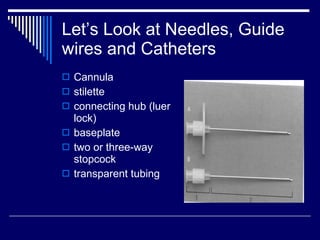
Downloaded 1,492 times


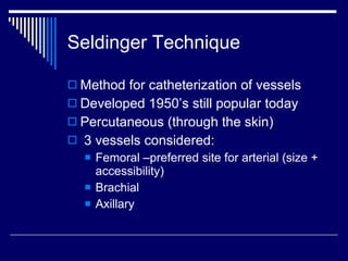
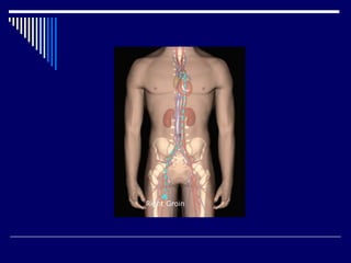


The document discusses angiography procedures and equipment. It describes the personnel involved including radiologists, nurses, and technologists. It outlines the angiography room setup and various equipment used, including x-ray generators, tubes, injectors, and digital imaging systems. Key steps of the Seldinger technique for vascular catheter insertion are provided. Post-procedure care and risks are also summarized.


![Invasive_Cardio-Devices_procedures[1].pdf](https://cdn.slidesharecdn.com/ss_thumbnails/invasivecardio-devicesprocedures1-240129085722-eb86cfb0-thumbnail.jpg?width=640&height=640&fit=bounds)


