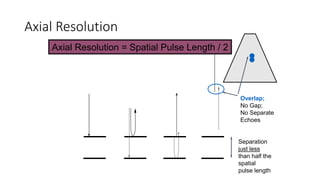
Downloaded 65 times
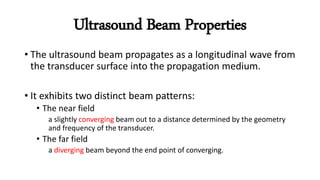
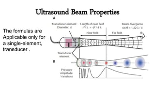
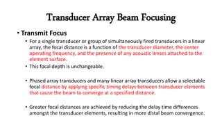
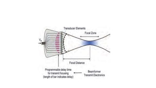
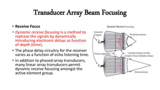
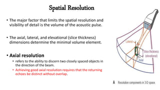
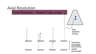
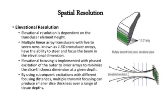

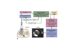


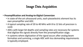
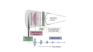
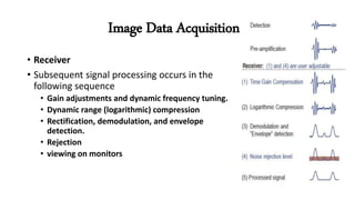
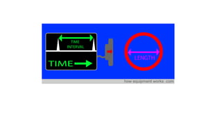

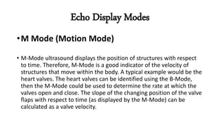
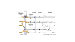

Ultrasound imaging uses transducers to emit sound waves into the body and receive echoes. There are three main types of ultrasound displays: A-mode shows amplitude of echoes over time. B-mode uses brightness to display 2D cross-sections. M-mode images motion over time, useful for evaluating moving structures like heart valves. The ultrasound beam has near and far field regions. Arrays allow focusing at different depths through phased delays. Spatial resolution depends on pulse length and beam width. Modern systems digitally process echo signals in parallel from each transducer to form high-quality images.


