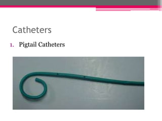
LEFT
VENTRICULOGRAPHY
Nomusa Sibanda
Bsc CVT
4th sem
What is Ventriculography
• Left heart ventricular angiography is a
procedure to look at the left-sided heart
chambers and the function of the left-sided
valves. It is sometimes combined with coronary
angiography.
• A ventriculogram is a test that shows images
of your heart. The images show how well your
heart is pumping.
•
• Cardiac ventriculography involves injecting contrast
media into the heart's ventricle(s) to measure the
volume of blood pumped. Cardiac ventriculography
can be performed with a
radionuclide in radionuclide ventriculography or
with an iodine-based contrast in cardiac chamber
catheterization.
• A radionuclide is an unstable form of a
chemical element that releases radiation as
it breaks down and becomes more stable.
Radionuclides may occur in nature or be made in a
laboratory. In medicine, they are used in imaging
tests and in treatment.
Indications
• Assessment of left ventricular function
including left ventricular ejection fraction, wall
motion abnormalities, ventricular size and mass.
• Identification and assessment of mitral
regurgitation.
• Identification and assessment of ventricular
septal defects.
How the test is done
• A doctor inserts a thin, flexible catheter into
your heart. The doctor uses the catheter to
inject dye into your heart. This dye makes the
inside of your heart show up on an X-ray. Then the
doctor can see how well your heart is pumping.
• The 3 major measurements obtained by cardiac
ventriculography are:
• Ejection fraction
• Stroke Volume
• Cardiac Output
Catheters
1. Pigtail Catheters
• One of the most commonly used catheters
• The loop shape keeps the end-hole away from
direct contact with endocardium.
• Multiple side-holes stabilize the catheter within
the left ventricle during contrast injection and
reducing the magnitude of catheter recoil.
2. Sones catheters
• The Sones catheter is used widely for left
ventriculography when catheterization is
performed from brachial approach.
• The Sones catheter should be positioned in an
axial orientation with its tip midway between the
aortic valve and left ventricular apex.
• Operator should hold the catheter during
injection.
3. NIH and Eppendorf Catheters
• The NIH and Eppendorf catheters have multiple
side holes and no end holes.
• Multiple side holes allow for rapid injection of
large volumes of contrast material.
4. Lehman Catheter
• The Lehman catheter has multiple side holes
and a tapered closed tip.
• The tapered tip helps in negotiating tortous
vessels and in crossing stenosis aortic valves.
• The long tip reduces the chances of endocardial
staining.
Views
• The 2 standard views for ventriculography are:
Right Anterior Oblique (RAO)
Left Anterior Oblique (LAO)
RAO
• In general the RAO view demonstrates Anterior,
Apical and Inferior ventricular walls.
• Cranial RAO view best shows Left Anterior
Descending Artery (LAD).
• RAO caudal view is the best overall view and the
best view for LCx. In addition, it allows good
assessment of the distal LM and the
proximal LAD.
LAO
• LAO cranial view allows for better imaging of
lateral and septal ventricular walls.
• This view is particularly useful in patients with
lateral ischemia especially circumflex ischemia,
VSD and mitral regurgitation.
• LAO caudal view allows a good assessment of
the left main artery, proximal LAD and LCx and
proximal branches.
Use of the Injector
• An injector is used to deliver contrast material to
the ventricles to assess cardiac structure and
function.
• Power injected left ventriculography (Power LV)
using standard volume (36 ml over 3 seconds) of
contrast through a 5 Fr. angled pigtail catheter
in the left ventricle is a common routine clinical
practice during left heart catheterization.
• Cardiologists have commonly used power
contrast media injectors to achieve opacification
for these diagnostic procedures, which require
high flow, high volume, fixed rate injections
delivered with relatively high pressures. Typical
applications of this sort are ventriculograms,
aortograms and researches.
• Before the left ventriculographic run, a test
injection of small amount of contrast material is
performed:
1. Assess catheter and patient position
2. Confirm that ventricular ectopy does not occur
3. Exclude a reaction to contrast media
Complications of Injection
• Arrhythmias- ventricular extrasystoles occur
frequently during ventriculography
• Intramyocardial injection- usually caused by
improper position of catheter
• Fascicular block- catheter induced fascicular
block usually resolves within 12 to 48 hours.
Thank you

LEFT VENTRICULOGRAPHY.pptx

  • 1.
  • 2.
    What is Ventriculography •Left heart ventricular angiography is a procedure to look at the left-sided heart chambers and the function of the left-sided valves. It is sometimes combined with coronary angiography. • A ventriculogram is a test that shows images of your heart. The images show how well your heart is pumping. •
  • 3.
    • Cardiac ventriculographyinvolves injecting contrast media into the heart's ventricle(s) to measure the volume of blood pumped. Cardiac ventriculography can be performed with a radionuclide in radionuclide ventriculography or with an iodine-based contrast in cardiac chamber catheterization. • A radionuclide is an unstable form of a chemical element that releases radiation as it breaks down and becomes more stable. Radionuclides may occur in nature or be made in a laboratory. In medicine, they are used in imaging tests and in treatment.
  • 4.
    Indications • Assessment ofleft ventricular function including left ventricular ejection fraction, wall motion abnormalities, ventricular size and mass. • Identification and assessment of mitral regurgitation. • Identification and assessment of ventricular septal defects.
  • 5.
    How the testis done • A doctor inserts a thin, flexible catheter into your heart. The doctor uses the catheter to inject dye into your heart. This dye makes the inside of your heart show up on an X-ray. Then the doctor can see how well your heart is pumping. • The 3 major measurements obtained by cardiac ventriculography are: • Ejection fraction • Stroke Volume • Cardiac Output
  • 6.
  • 7.
    • One ofthe most commonly used catheters • The loop shape keeps the end-hole away from direct contact with endocardium. • Multiple side-holes stabilize the catheter within the left ventricle during contrast injection and reducing the magnitude of catheter recoil.
  • 8.
  • 9.
    • The Sonescatheter is used widely for left ventriculography when catheterization is performed from brachial approach. • The Sones catheter should be positioned in an axial orientation with its tip midway between the aortic valve and left ventricular apex. • Operator should hold the catheter during injection.
  • 10.
    3. NIH andEppendorf Catheters
  • 11.
    • The NIHand Eppendorf catheters have multiple side holes and no end holes. • Multiple side holes allow for rapid injection of large volumes of contrast material.
  • 12.
    4. Lehman Catheter •The Lehman catheter has multiple side holes and a tapered closed tip. • The tapered tip helps in negotiating tortous vessels and in crossing stenosis aortic valves. • The long tip reduces the chances of endocardial staining.
  • 13.
    Views • The 2standard views for ventriculography are: Right Anterior Oblique (RAO) Left Anterior Oblique (LAO)
  • 14.
    RAO • In generalthe RAO view demonstrates Anterior, Apical and Inferior ventricular walls. • Cranial RAO view best shows Left Anterior Descending Artery (LAD). • RAO caudal view is the best overall view and the best view for LCx. In addition, it allows good assessment of the distal LM and the proximal LAD.
  • 15.
    LAO • LAO cranialview allows for better imaging of lateral and septal ventricular walls. • This view is particularly useful in patients with lateral ischemia especially circumflex ischemia, VSD and mitral regurgitation. • LAO caudal view allows a good assessment of the left main artery, proximal LAD and LCx and proximal branches.
  • 16.
    Use of theInjector • An injector is used to deliver contrast material to the ventricles to assess cardiac structure and function. • Power injected left ventriculography (Power LV) using standard volume (36 ml over 3 seconds) of contrast through a 5 Fr. angled pigtail catheter in the left ventricle is a common routine clinical practice during left heart catheterization.
  • 17.
    • Cardiologists havecommonly used power contrast media injectors to achieve opacification for these diagnostic procedures, which require high flow, high volume, fixed rate injections delivered with relatively high pressures. Typical applications of this sort are ventriculograms, aortograms and researches.
  • 18.
    • Before theleft ventriculographic run, a test injection of small amount of contrast material is performed: 1. Assess catheter and patient position 2. Confirm that ventricular ectopy does not occur 3. Exclude a reaction to contrast media
  • 19.
    Complications of Injection •Arrhythmias- ventricular extrasystoles occur frequently during ventriculography • Intramyocardial injection- usually caused by improper position of catheter • Fascicular block- catheter induced fascicular block usually resolves within 12 to 48 hours.
  • 20.

Editor's Notes